. 2014 Dec 22;207(6):717-33.
doi: 10.1083/jcb.201405077.

Opposing ISWI- and CHD-class chromatin remodeling activities orchestrate heterochromatic DNA repair

Affiliations

Opposing ISWI- and CHD-class chromatin remodeling activities orchestrate heterochromatic DNA repair

Karolin Klement et al. J Cell Biol. .

Abstract

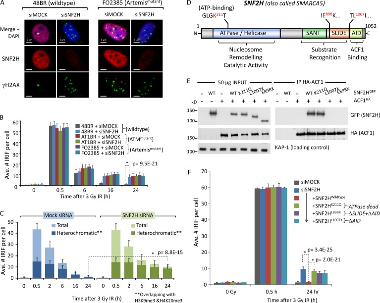
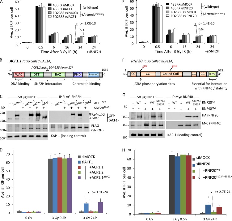
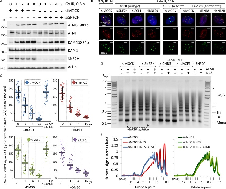
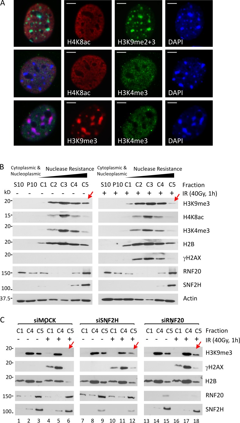
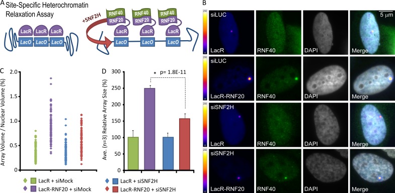
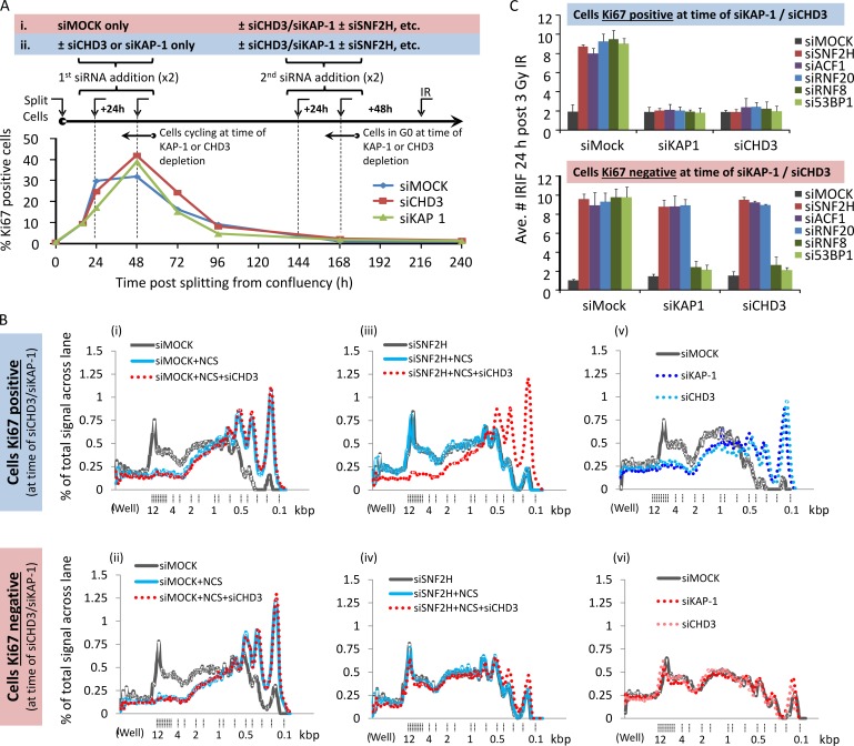
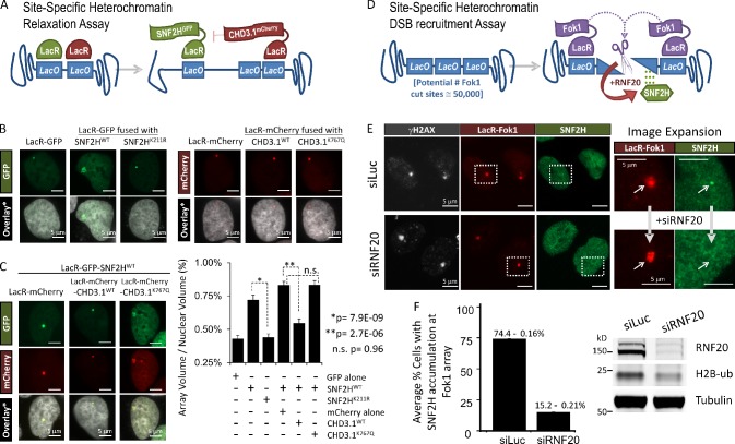
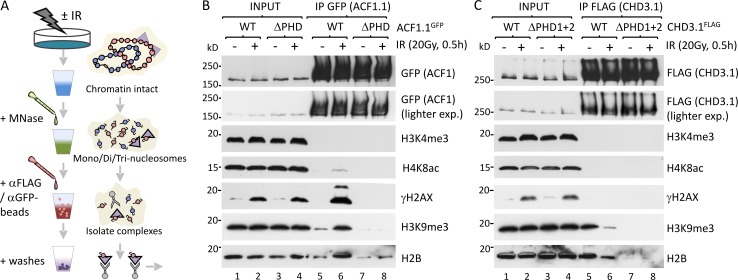
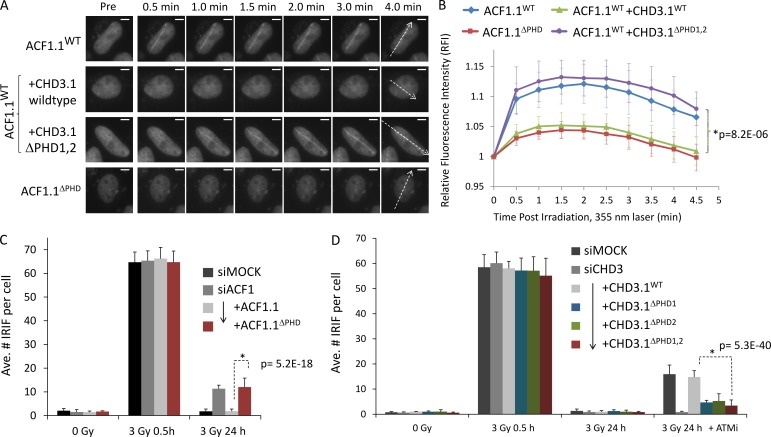
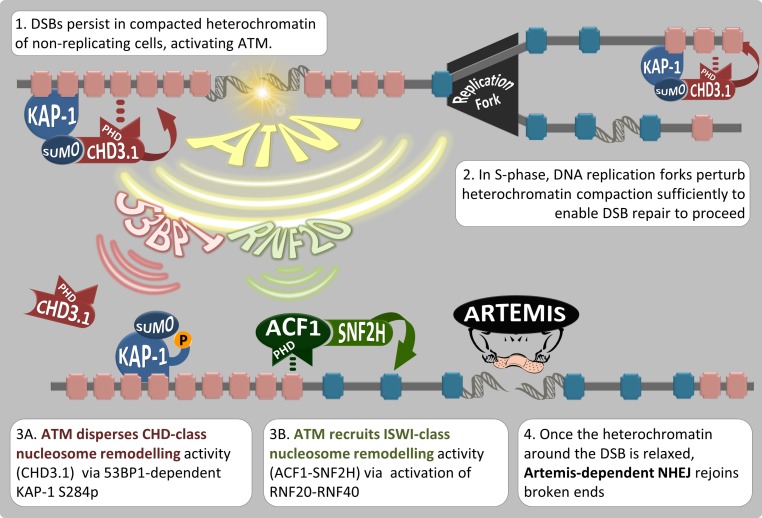
Heterochromatin is a barrier to DNA repair that correlates strongly with elevated somatic mutation in cancer. CHD class II nucleosome remodeling activity (specifically CHD3.1) retained by KAP-1 increases heterochromatin compaction and impedes DNA double-strand break (DSB) repair requiring Artemis. This obstruction is alleviated by chromatin relaxation via ATM-dependent KAP-1S824 phosphorylation (pKAP-1) and CHD3.1 dispersal from heterochromatic DSBs; however, how heterochromatin compaction is actually adjusted after CHD3.1 dispersal is unknown. In this paper, we demonstrate that Artemis-dependent DSB repair in heterochromatin requires ISWI (imitation switch)-class ACF1-SNF2H nucleosome remodeling. Compacted chromatin generated by CHD3.1 after DNA replication necessitates ACF1-SNF2H-mediated relaxation for DSB repair. ACF1-SNF2H requires RNF20 to bind heterochromatic DSBs, underlies RNF20-mediated chromatin relaxation, and functions downstream of pKAP-1-mediated CHD3.1 dispersal to enable DSB repair. CHD3.1 and ACF1-SNF2H display counteractive activities but similar histone affinities (via the plant homeodomains of CHD3.1 and ACF1), which we suggest necessitates a two-step dispersal and recruitment system regulating these opposing chromatin remodeling activities during DSB repair.


Figures

Figure 1.
Figure 1.
SNF2H chromatin remodeling activity is required for ATM- and Artemis-dependent DSB repair in heterochromatin. (A) SNF2H was depleted by siRNA (mock = scrambled siRNA) in wild-type (48BR) or Artemis mutant (FO2385) quiescent primary human fibroblasts. Cells were irradiated with 3 Gy IR and immunostained 24 h later for SNF2H (red), γ-H2AX (green), and DAPI (blue). Bars, 5 µm. (B) The mean number of γ-H2AX per nucleus from cells prepared as in A and harvested at 0.5, 6, 16, and 24 h after 3 Gy IR was enumerated for three independent experiments. (C) Quiescent 48BR cells were treated with scrambled (mock) or SNF2H siRNA, irradiated, and harvested as indicated. Cells were then stained with γ-H2AX (green) and H3K9me3 + H4K20me3 (red) and imaged by confocal microscopy. Regions of green- and red-positive signal were identified by software, isolating overlap of γ-H2AX foci in an H3K9me3 + H4K20me3–positive (heterochromatic) region. The heterochromatic foci were enumerated relative to total number. (D) A schematic diagram of human SNF2H. N, N terminus; C, C terminus. (E) The indicated GFP-tagged SNF2H or HA-tagged ACF1 constructs were transfected into 1BRhTERT cells; 24 h later, whole cell extracts were prepared and incubated with anti–HA-agarose for 4 h at 4°C. Washed immunoprecipitates and 50 µg of input lysate were then immunoblotted for the indicated proteins. (F) Confluent 1BRhTERT cells were transfected with SNF2H-B siRNA and, 48 h later, then split to 75% confluency and transfected with the indicated SNF2HGFP constructs. 16 h later, cells were irradiated as indicated, harvested, and immunostained for γ-H2AX and either SNF2H or FLAG. The mean number of γ-H2AX per nucleus was scored in cells with confirmed knockdown or construct expression for three independent experiments. P-values (standard two-tailed Student’s t test) are indicated for significance of relevant data points. Error bars show SD. Ave., average.
Figure 2.
Figure 2.
ACF1 and RNF20 enable SNF2H and Artemis-dependent DSB repair. (A) ACF1 was depleted by siRNA (mock = scrambled siRNA) in wild-type (48BR) or Artemis mutant (FO2385) quiescent primary human fibroblasts before being treated, immunostained, and analyzed as in Fig. 1 A. (B) Schematic diagram of human ACF1. (C) The indicated FLAG-tagged SNF2H or GFP-tagged ACF1 constructs were transfected into 1BRhTERT cells; 24 h later, whole cell extracts were prepared and incubated with anti–FLAG-Sepharose for 3 h at 4°C. Washed GFP immunoprecipitates and 50 µg of input lysate were then immunoblotted for the indicated proteins. (D) HeLa cells were transfected with ACF1-B siRNA and, 48 h later, then split to 75% confluency and transfected with the indicated ACF1GFP constructs. 16 h later, cells were irradiated as indicated, harvested, and immunostained for γ-H2AX; the mean number of γ-H2AX per nucleus was scored in cells with confirmed construct expression for three independent experiments. (E) RNF20 was depleted by siRNA (mock = scrambled siRNA) in wild-type (48BR) or Artemis mutant (FO2385) quiescent primary human fibroblasts before being treated, immunostained, and analyzed as in Fig. 1 A. (F) Schematic diagram of human RNF20. BromoD, bromodomain; CC, coiled coil. (G) The indicated HA-tagged RNF20 constructs were transfected into 1BRhTERT cells alongside wild-type Myc-tagged RNF40; 24 h later, whole cell extracts were prepared and immunoblotted for the indicated proteins. (H) HeLa cells were transfected with RNF20-A siRNA and, 48 h later, then split to 75% confluency and transfected with the indicated RNF20HA constructs alongside wild-type RNF40Myc. 16 h later, cells were irradiated as indicated, harvested, and immunostained for γ-H2AX and either RNF20 or HA; the mean number of γ-H2AX per nucleus was scored in cells with confirmed knockdown or construct expression for three independent experiments. P-values (standard two-tailed Student’s t test) are indicated. Error bars show SD. Ave., average.
Figure 3.
Figure 3.
SNF2H-ACF1 and RNF20 enable DSB-induced chromatin relaxation downstream of KAP-1 phosphorylation and CHD3.1 dispersal. (A) SNF2H was depleted by siRNA (mock = scrambled siRNA) in wild-type 48BR cells and irradiated with 0, 1, 2, 4, and 8 Gy IR before being harvested 0.5 h later and immunoblotted for the indicated proteins. (B) Wild-type (48BR), ATM mutant (AT1BR), or Artemis mutant (FO2385) quiescent primary human fibroblasts were treated with SNF2H, ACF1, RNF20, or RNF8 siRNA (as indicated) before being irradiated and harvested as indicated. Cells were immunostained for pKAP-1 (red) and γ-H2AX (green). Bars, 5 µm. (C) 48BR cells were treated with siRNA as in B and were irradiated with 0, 1, 4, and 16 Gy IR ± DMSO or 10 µM Ku55933 ATM inhibitor (ATMi). Cells were incubated with 0.1% (vol/vol) Triton X-100 in PBS for 30 s before being fixed and immunostained for CHD3.1 (red) or γ-H2AX (green). Nuclear CHD3.1 signal was quantified as in Goodarzi et al. (2011); data from three independent experiments were plotted together. Solid bars indicate means with SD. A.U., arbitrary unit. (D) HeLa cells were treated with siRNA as indicated. 48 h later, cells were treated with 200 ng/ml neocarzinostatin (NCS) ± ATM inhibitor (as indicated). 0.5 after NCS treatment, nuclei were purified and treated with MNase, and DNA were isolated as described in Goodarzi et al. (2011). 2.5 µg DNA was resolved by 1.2% agarose gel and visualized by ethidium bromide staining. (E) The quantification of signal in each lane of the gel in D. Data are expressed as the percentages of the total signal (for a given lane) across the distance from the well to end of the gel. Calibrated kilobase pair sizes are indicated. The experimental dataset shown in this figure is representative of four independent repeat experiments, all showing the same result/trends.
Figure 4.
Figure 4.
SNF2H and RNF20 enable specifically heterochromatic relaxation after DSB induction. (A) Confluence-arrested NIH3T3 cells immunostained for H3K9me2/3, H3K9me3, H4K8ac, and/or H3K4me3, as indicated. Bars, 5 µm. (B) Confluence-arrested NIH3T3 cells were exposed to 0 or 40 Gy IR and, 1 h later, fractionated into extracts (for a full explanation of each fraction, see Materials and methods) and immunoblotted for the indicated proteins. (C) Confluence-arrested NIH3T3 cells were treated with scrambled (Mock), SNF2H, or RNF20 siRNA and, 48 h later, were irradiated, fractionated and immunoblotted as in B. Red arrows highlight the H3K9me3 signal in the C5 fraction undergoing a dynamic change in response to stimuli.
Figure 5.
Figure 5.
RNF20 promotes site-specific heterochromatin relaxation in an SNF2H-dependent manner. (A) Schematic for site-specific chromatin relaxation assay. (B) Cells containing an integrated LacO array within a region of heterochromatin were treated ± siRNA toward luciferase (siLuc) or SNF2H (siSNF2H) and transfected with RNF40Myc and either LacR or LacR-RNF20, as indicated. Color scales indicate false-colored lookup table of pixel intensities (8 bit, pixel range = 1–256). (C and D) The nuclear volume of the LacR-signal (array) as a percentage of the overall nuclear volume was plotted for each condition, either as individual data points (C) or a mean of three independent experiments (D). P-values (standard two-tailed Student’s t test) are indicated. Error bars show SD.
Figure 6.
Figure 6.
SNF2H–ACF1 and RNF20 remain essential for heterochromatic relaxation and DSB repair in the absence of CHD3 in nonproliferating cells. (A) Quiescent, confluence-arrested 48BR cells were split to ∼50% confluency and monitored over the course of 10 d (240 h) for proliferation as measured by Ki67 signal by IF. Two different schemes of siRNA treatment during this time were used: in the first (i) scenario, cells were subject only to mock siRNA while in a proliferative state (24–72 h after splitting); in the second (ii), either KAP-1 or CHD3 siRNA were added to cells while they were proliferating. In both cases, after 7 d (144 h), cells achieved confluency, and they were subjected to further CHD3/KAP-1 siRNA in combination with siRNA targeting SNF2H, ACF1, RNF20, RNF8, or 53BP1. At 9 d (216 h) after splitting, once complete knockdown of all targets was achieved, cells were irradiated with 3 Gy IR harvested a day later. The percentage of cells positive for Ki67 was plotted over time (>250 cells monitored per condition, per experiment). (B) Cells obtained from either treatment schemes i or ii as outlined in A were treated with NCS and processed for the chromatin relaxation assay as in Fig. 3 (D and E). (C) Cells obtained from either treatment schemes i or ii as outlined in A were immunostained for γ-H2AX (green) and the relevant target of siRNA (e.g., in SNF2H-depleted cells, cells were counterstained with SNF2H in the red channel to confirm knockdown). The mean number of IRIF per nucleus was scored in cells with confirmed knockdown for three independent experiments. Error bars show SD. Ave., average.
Figure 7.
Figure 7.
Active CHD3.1 opposes SNF2H chromatin remodeling activity in heterochromatin, which requires RNF20 to localize to heterochromatic DSBs. (A) Schematic of the site-specific heterochromatin relaxation assay. (B) Representative images of cells containing a LacO array integrated within a region of heterochromatin and transfected with LacR-GFP, LacR-GFP-SNF2H (wild type or K211R), LacR-mCherry, or LacR-mCherry-CHD3.1 (wild type or K767Q), as indicated. Bars, 5 µm. (C) Cells from B were transfected with LacR-GFP-SNF2H wild type and either LacR-mCherry or LacR-mCherry-CHD3.1 (wild type or K767Q), as indicated. The nuclear volume of the LacR-GFP signal, as a percentage of the overall nuclear volume was plotted for each condition (from B and C), either as a mean of three independent experiments (∼75 cells for each condition). P-values (standard two-tailed Student’s t test) are indicated. Bars, 5 µm. Error bars show SD. (D) Schematic of the site-specific heterochromatin DSB recruitment assay. (E) U2OS 2-6-3 cells treated with either RNF20 (siRNF20) or luciferase (siLuc) siRNA and stably expressing ER-Fok1-mCherry-LacR-DD were induced with 300 nM 4-OHT and 1 µM Shield-I for 5 h. Subsequently, cells were preextracted using 0.25% Triton X-100 in CSK buffer for 10 min, fixed with formaldehyde, and immunostained with SNF2H (green) and γ-H2AX (greyscale). Boxes are enlarged in the right images. Arrows point to the site of DSB induction at the array. Bars, 5 µm. (F) The mean percentages of cells with SNF2H foci present at Fok1/γ-H2AX foci were quantified (130 cells for each condition from two independent experiments). siRNA efficiency was assessed by immunoblotting.
Figure 8.
Figure 8.
The PHD fingers of ACF1 and CHD3.1 confer similar binding specificity for nucleosomes. (A) A schematic for experiments in B and C. (B) HeLa cells were transfected with wild-type ACF1.1 or ACF1.1 with C/H→A mutations in both Zn-3 and Zn-4 of the PHD finger (ACF1.1ΔPHD). 24 h later, cells were irradiated as indicated, and nucleosome-solubilized (MNase digested) whole cell extracts were prepared, immunoprecipitated with GFP-agarose, and immunoblotted for the indicated proteins. exp., exposure. (C) HeLa cells were transfected with wild-type CHD3.1 or CHD3.1 with C/H→A mutations in both Zn-1 and Zn-2 of the PHD finger 1 and both Zn-3 and Zn-4 of the PHD finger 2 (CHD3.1ΔPHD1+2). 24 h later, cells were irradiated as indicated, and nucleosome-solubilized (MNase digested) whole cell extracts were prepared, immunoprecipitated with GFP-agarose, and immunoblotted for indicated proteins.
Figure 9.
Figure 9.
The PHD fingers of ACF1 and CHD3.1 confer similar binding specificity for nucleosomes. (A) HeLa cells were transfected as in Fig. 8 (B and C) with the indicated constructs and incubated with BrdU for 16–24 h before irradiation with a directed 355-nm laser. GFP signal was imaged live over time. The dotted arrows indicate path of laser microirradiation through nuclei. Bars, 5 µm. (B) The relative fluorescence intensity increase for GFP signal obtained in A was quantified. P-values (standard two-tailed Student’s t test) are indicated. (C) HeLa cells were transfected with ACF1-B siRNA and, 48 h later, then split to 75% confluency and transfected with the indicated ACF1.1GFP constructs. 16 h later, cells were irradiated as indicated, harvested, and immunostained for γ-H2AX; the mean number of γ-H2AX per GFP-positive nucleus was scored for three independent experiments. (D) HeLa cells were transfected with CHD3 siRNA and, 48 h later, then split to 75% confluency and transfected with the indicated CHD3.1FLAG constructs. 16 h later, cells were incubated ± ATM inhibitor (ATMi), irradiated as indicated, harvested, and immunostained for 53BP1 and FLAG; the mean number of 53BP1 per FLAG-positive nucleus was scored for three independent experiments. Ave., average. Error bars show SD. P-values (standard two-tailed Student’s t test) are indicated.
Figure 10.
Figure 10.
Model for heterochromatin nucleosome relaxation and Artemis-dependent DSB repair. (1) Artemis-dependent DSB repair stalls within the KAP-1– and CHD3.1-rich heterochromatin of nondividing cells. (2) During DNA replication, the heterochromatic superstructure is perturbed, and newly synthesized strands remain in an open configuration, bypassing the need for additional chromatin relaxation during repair of DSBs incurred during S phase. (3) ATM protein kinase activity triggers two signaling axes. The first (3A) enables the dispersal of class II CHD chromatin remodeling activity (CHD3.1) via the IRIF mediator proteins (such as 53BP1) and densely localized pKAP-1. The other (3B) promotes the gain of ISWI-class chromatin remodeling activity via RNF20-RNF40 activation and the recruitment/activation of ACF1–SNF2H, which we propose occupies PHD finger binding sites vacated by CHD3.1. (4) These mechanisms converge to enable heterochromatin relaxation, which is favorable for the repair of DSBs via Artemis. P, phosphorylation.

References

    1. Andrin C., McDonald D., Attwood K.M., Rodrigue A., Ghosh S., Mirzayans R., Masson J.Y., Dellaire G., and Hendzel M.J.. 2012. A requirement for polymerized actin in DNA double-strand break repair. Nucleus. 3:384–395 10.4161/nucl.21055 - DOI - PubMed
    1. Bekker-Jensen S., Lukas C., Kitagawa R., Melander F., Kastan M.B., Bartek J., and Lukas J.. 2006. Spatial organization of the mammalian genome surveillance machinery in response to DNA strand breaks. J. Cell Biol. 173:195–206 10.1083/jcb.200510130 - DOI - PMC - PubMed
    1. Beucher A., Birraux J., Tchouandong L., Barton O., Shibata A., Conrad S., Goodarzi A.A., Krempler A., Jeggo P.A., and Löbrich M.. 2009. ATM and Artemis promote homologous recombination of radiation-induced DNA double-strand breaks in G2. EMBO J. 28:3413–3427 10.1038/emboj.2009.276 - DOI - PMC - PubMed
    1. Brehm A., Längst G., Kehle J., Clapier C.R., Imhof A., Eberharter A., Müller J., and Becker P.B.. 2000. dMi-2 and ISWI chromatin remodelling factors have distinct nucleosome binding and mobilization properties. EMBO J. 19:4332–4341 10.1093/emboj/19.16.4332 - DOI - PMC - PubMed
    1. Brunton H., Goodarzi A.A., Noon A.T., Shrikhande A., Hansen R.S., Jeggo P.A., and Shibata A.. 2011. Analysis of human syndromes with disordered chromatin reveals the impact of heterochromatin on the efficacy of ATM-dependent G2/M checkpoint arrest. Mol. Cell. Biol. 31:4022–4035 10.1128/MCB.05289-11 - DOI - PMC - PubMed

Publication types

MeSH terms

Associated data