. 2020 Jun;24(11):6350-6361.
doi: 10.1111/jcmm.15277. Epub 2020 Apr 19.

Molecular mechanisms involved in high glucose-induced valve calcification in a 3D valve model with human valvular cells

Affiliations

Molecular mechanisms involved in high glucose-induced valve calcification in a 3D valve model with human valvular cells

Mihaela Vadana et al. J Cell Mol Med. 2020 Jun.

Abstract

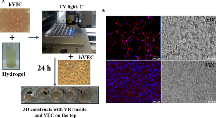
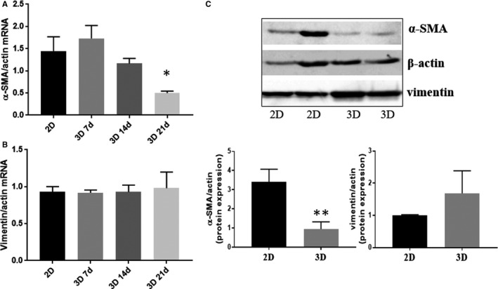
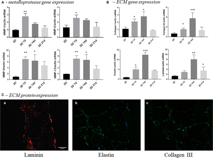
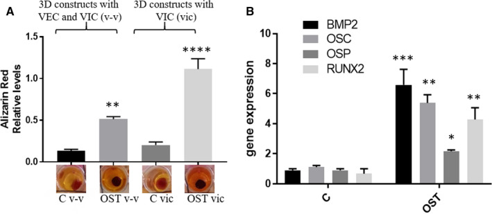
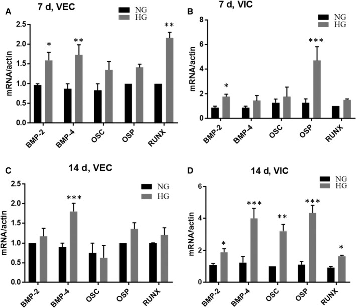
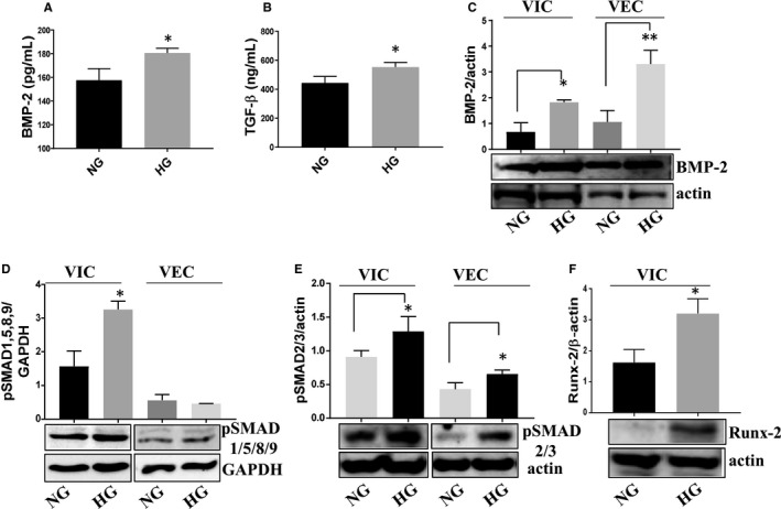
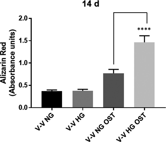
Calcific aortic valve disease (CAVD)-the most common valvular heart disease-is accelerated in diabetes and has no pharmacotherapy. Although it is known that early CAVD is associated with inflammation and osteogenesis, the molecular mechanisms involved in diabetes-associated CAVD still need to be uncovered. In this context, we have developed a 3D construct based on gelatin populated with human valvular endothelial cells (VEC) and valvular interstitial cells (VIC) and evaluated the effect of high glucose (HG) concentration on osteogenic molecules expression and on calcification mechanisms. First, we characterized the 3D model and assessed VIC remodelling properties at different time-points. Then, we exposed it to normal glucose (NG) or high glucose (HG) for 7, 14 and 21 days after which the cells were isolated, separated and investigated individually. Our results showed that encapsulated VIC actively remodel the hydrogel, as demonstrated by an increased expression of extracellular matrix (ECM) proteins and matrix metalloproteinases (MMPs). Moreover, exposure of the construct to HG triggered bone morphogenetic protein (BMP) and TGF-β signalling pathways, up-regulating expression of osteogenic molecules-BMP-2/-4, osteocalcin, osteopontin, SMADs and Runt-related transcription factor (Runx-2)-and increased calcium deposits in an osteogenic environment. These findings underline the potential of the developed 3D model as a suitable system to investigate the mechanisms of human CAVD and may help to better understand the calcification mechanisms in CAVD associated to diabetes.

Keywords: calcific aortic valve disease; high glucose levels; human valvular cells; osteogenic molecules; valve tissue engineering.


Conflict of interest statement

The authors confirm that there are no conflicts of interest.

Figures

FIGURE 1
FIGURE 1
Obtaining the 3D construct, morphology of valvular cells from the 3D construct. A, 3D‐construct development: human VIC at a density of 2 million cells/mL were re‐suspended in G‐MA hydrogel solution (10% porcine gelatin methacrylate, 1% alginate and 0.5% Photo‐initiator—Irgacure 2595). 100 μL of VIC‐laden pre‐polymer solution was dropwise added on a 48‐cavity mould (Ø ‐ 8, 1 mm thickness) and subsequently cross‐linked by exposure for 1 minute to UV light (365 nm). Resulted 3D constructs were removed from the mould and cultured, according to protocol. After 24 h, VEC (5 × 105/cm2) were seeded on top of 3D constructs. B, Valvular cells morphology in 3D construct at day 7 of culture as determined by phalloidin labelled F‐actin (red) and DAPI nuclear staining (blue). Hydrogel formula supports cell network development of VIC (inside the hydrogel) and VEC proliferation as a monolayer on the scaffold surface. Contrast phase images of the surface and inside of hydrogel with VIC encapsulated inside and VEC cultured on top. Scale bar indicates 100 µm
FIGURE 2
FIGURE 2
Phenotype of VIC and VEC from the 3D constructs. A and B, mRNA expression of VIC markers, α‐SMA (A) and vimentin (B), in VIC isolated from 3D constructs cultured for different period of time (7, 14 or 21 days), compared with VIC grown in 2D culture (7 days). n = 3, *P < .05, VIC from 3D compared with VIC from 2D. Data depicted as mean ± SD. C, Evaluation of protein expression for α‐SMA and vimentin in VIC isolated from 3D constructs or from 2D culture by Western blot, n = 3, *P < .05, VIC from 3D compared with VIC from 2D
FIGURE 3
FIGURE 3
Expression of molecules associated with matrix remodelling by VIC cultured in 2‐dimensional conditions compared with VIC isolated from 3D constructs. A, Gene expression of matrix metalloproteases MMP‐1, MMP‐2, MMP‐9 and MMP‐13 in VIC cultured in 2D (7 days) versus VIC from 3D constructs at different periods of time—7, 14 or 21 days, as evaluated by real‐time PCR. B, Gene expression of extracellular matrix proteins by VIC cultured in 2‐dimensional conditions compared with VIC isolated from 3D constructs. The mRNA of matrix proteins was normalized to actin mRNA. Note that VIC isolated from 3D constructs exhibited a time‐dependent increase of collagen I, III, elastin and laminin mRNAs. n = 3, *P < .05, **P < .01, ***P < .001 VIC from 2D versus VIC from 3D. C, Protein expression of ECM as evaluated by immunofluorescent technique. Representative images of laminin (a), elastin (b) and collagen III (c) in sections obtained from 3D constructs with human VEC and VIC. The sections were marked with specific primary antibodies and Alexa‐ or FITC‐coupled secondary antibodies (red or green staining). Nuclei were stained with DAPI (blue staining)
FIGURE 4
FIGURE 4
Osteogenic environment favours the calcium deposits development and induces osteogenic phenotype of VIC in 3D constructs. A, Alizarin Red was used to stain the mineralized nodules formed by cells from 3D constructs with VIC or 3D constructs with VEC and VIC (v‐v) cultured for 14 days. The lower panel shows the representative stained construct from each of the experimental group. The upper graph displays the quantitative measurement of Alizarin Red dye released from the mineralized nodules formed in 3D constructs cultured in normal conditions or exposed to osteogenic media (OST: 10 mM β‐glycerophosphate, 10 ng/mL ascorbic acid and 10−8 mol/l dexamethasone). n = 3, **P < .01, ****P < .0001. B, mRNA expression of osteogenic molecules (BMP‐2, osteocalcin, osteopontin and RUNX2) in VIC isolated from 3D constructs cultured in normal conditions or exposed to OST media for 14 days. Note that VIC isolated from 3D constructs exposed to OST media exhibit a significantly increased expression of osteogenic molecules mRNAs. n = 3, *P < .01, **P < .01, ***P < .001 VIC from 3D‐OST versus VIC from 3D. Data depicted as mean ± SD
FIGURE 5
FIGURE 5
Gene expression of osteogenic molecules expressed by VEC and VIC from 3D constructs cultured in NG or HG conditions. A and C, Gene expression of BMP‐2, BMP‐4, osteocalcin, osteopontin and Runx‐2 in VEC isolated from 3D constructs after 7 (A) and 14 (C) days, as evaluated by real‐time PCR. n = 3. B and D, Gene expression of osteogenic molecules in VIC isolated from 3D constructs after 7 (B) and 14 (D) days, as evaluated by real‐time PCR. The mRNA of osteogenic molecules was normalized to actin mRNA. n = 3, *P < .05, **P < .01, ***P < .001 VIC from 2D versus VIC from 3D
FIGURE 6
FIGURE 6
Protein expression of osteogenic signalling pathway molecules expressed by VEC and VIC from 3D constructs, cultured in NG or HG conditions. A and B, Soluble BMP‐2 and TGF‐β released in the conditioned media by valvular cells from 3D construct exposed to NG or HG conditions, as determined by ELISA assay. n = 3, *P < .05. C‐F, Protein expression of BMP‐2, pSMAD1/5/8/9, pSMAD2/3 and Runx‐2 in VIC and VEC as determined by Western blot. n = 3, *P < .05**P < .01 high glucose vs normal glucose
FIGURE 7
FIGURE 7
Calcium deposits development in 3D constructs exposed to NG or HG concentration in an osteogenic environment. Alizarin Red was used to stain the mineralized nodules formed by cells from 3D constructs with VEC and VIC cultured for 14 days in NG or HG in presence or absence of osteogenic media (OST). n = 3, ****P < .0001

References

    1. Benjamin EJ, Blaha MJ, Chiuve SE, et al. Heart disease and stroke statistics’ 2017 update: A report from the American heart association. Circulation. 2017;135:e146‐e603. - PMC - PubMed
    1. Otto CM, Prendergast B. Aortic‐valve stenosis‐from patients at risk to severe valve obstruction. N Engl J Med. 2014;21:744‐756. - PubMed
    1. Thaden JJ, Nkomo VT, Enriquez‐Sarano M. The global burden of aortic stenosis. Prog Cardiovasc Dis. 2014;56:565‐571. - PubMed
    1. Rossi A, Targher G, Zoppini G, et al. Aortic and mitral annular calcifications are predictive of all‐cause and cardiovascular mortality in patients with type 2 diabetes. Diabetes Care. 2012;35:1781‐1786. - PMC - PubMed
    1. Ginter E, Simko V. Type 2 diabetes mellitus, pandemic in 21st century. Adv Exp Med Biol. 2012;771:42‐50. - PubMed

Publication types

MeSH terms

Supplementary concepts

Associated data