. 2016 Aug;148(4):363-76.
doi: 10.1111/imm.12615.

Distinct expression of interferon-induced protein with tetratricopeptide repeats (IFIT) 1/2/3 and other antiviral genes between subsets of dendritic cells induced by dengue virus 2 infection

Affiliations

Distinct expression of interferon-induced protein with tetratricopeptide repeats (IFIT) 1/2/3 and other antiviral genes between subsets of dendritic cells induced by dengue virus 2 infection

Jingshu Zhang et al. Immunology. 2016 Aug.

Abstract

Dengue virus (DENV) infection is an emerging public health hazard threatening inhabitants of the tropics and sub-tropics. Dendritic cells (DCs) are one of the major targets of DENV and the initiators of the innate immune response against the virus. However, current in vitro research on the DENV-DC interaction is hampered by the low availability of ex vivo DCs and donor variation. In the current study, we attempted to develop a novel in vitro DC model using immature DCs derived from the myeloid leukaemia cell line MUTZ-3 (IMDCs) to investigate the DENV-DC interaction. The IMDCs morphologically and phenotypically resembled human immature monocyte-derived dendritic cells (IMMoDCs). However, the permissiveness of IMDCs to DENV2 was lower than that of IMMoDCs. RT-PCR arrays showed that a group of type I interferon (IFN) -inducible genes, especially IFIT1, IFITM1, and IFI27, were significantly up-regulated in IMMoDCs but not in IMDCs after DENV2 infection. Further investigation revealed that IFIT genes were spontaneously expressed at both transcriptional and protein levels in the naive IMDCs but not in the naive IMMoDCs. It is possible that the poor permissiveness of IMDCs to DENV2 was a result of the high basal levels of IFIT proteins. We conclude that the IMDC model, although less permissive to DENV2, is a useful platform for studying the suppression mechanism of DENV2 and we expand the knowledge of cellular factors that modulate DENV2 infection in the human body.

Keywords: MUTZ-3; dendritic cells; dengue virus.


Figures

Figure 1
Figure 1
Morphologies of primary human monocytes and MUTZ‐3 cells before and after cytokine induction. Both primary human monocytes and MUTZ‐3 cells were round, blast‐like and non‐adherent before cytokine induction (a and c), but both cell types formed dendrites and became adherent after induction (b and d). Magnification: 400 ×.
Figure 2
Figure 2
Surface marker expression profiles of primary human monocytes and MUTZ‐3 cells before and after differentiation. (a) PI− populations of viable cells were gated for detection of CD1a, CD80, CD14, mannose receptor (MR) and DCSIGN. Primary human monocytes and MUTZ‐3 cells were CD14+ CD1a CD80 DCSIGN ; (b) both cell types expressed MR at high levels. (c) After cytokine differentiation, 43·3% of immature MUTZ‐3‐derived dendritic cells (IMDCs) and 55·6% of primary human monocyte‐derived DCs (IMMoDCs) were DCSIGN + MR +; (d) the double positive populations were gated for detection of CD1a, CD80 and CD14. The experiment shown is one representative of three. Black line: non‐stained control; red line: CD1a staining; blue line: CD80 staining; green line: CD14 staining. The details of antibodies for multi‐colour staining are presented in the Supplementary material (Figure S1).
Figure 3
Figure 3
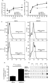
Replication of dengue virus 2 (DENV2) in immature MUTZ‐3‐derived dendritic cells (IMDCs) and primary human monocyte‐derived DCs (IMMoDCs). (a) DENV2 viral genome inside IMDCs and IMMoDCs and (b) in the culture media of IMDCs and IMMoDCs were detected at various time‐points. Grey dashed lines: DENV2‐infected IMDCs; black lines: DENV2‐infected IMMoDCs. The y‐axis represents the logarithm of DENV2 copy number. The results are expressed as mean ± SEM of data from three independent experiments. (c) Infection rates of DENV2 in IMDCs (left panels) and in IMMoDCs (right panels) were detected by flow cytometry at 24 hr post‐infection (p.i.) (upper panel) and at 48 hr p.i. (lower panel), respectively. Black lines: infected cells; grey dashed lines: non‐infected cells. The experiment shown is one representative of three. (d) Replication of DENV2 replicon in IMDCs and IMMoDCs 48 hr post transfection was detected by the TaqMan assay. The reported data are the mean ± SEM of the results of three independent experiments. (e) DENV2 E and NS1 proteins in the culture media of IMDCs and IMMoDCs at various time‐points as determined by dot blotting. Dots from infected samples were considered positive if their integrated densities were higher than the mock controls dots belonging to the same time‐points. Positive dots were indicated with an asterisk. +, infected; −, mock controls.
Figure 4
Figure 4
Effect of dengue virus 2 (DENV2) RNA replication on interferon‐induced protein with tetratricopeptide repeats (IFIT) genes in immature MUTZ‐3‐derived dendritic cells (IMDCs) and primary human monocyte‐derived DCs (IMMoDCs). (a) Expression of three IFIT genes in IMDCs and IMMoDCs at various time‐points post DENV2 infection. Black lines and grey dashed lines show the trends in gene expression in infected IMMoDCs and IMDCs, respectively. The relative gene expression fold‐changes from three independent experiments were calculated and analysed by one‐way analysis of variance plus Tukey's honest significant difference post hoc test. (b) Protein expression time‐courses of IFIT1, IFIT2, and IFIT3 after DENV2 infection in IMDCs (left panel) and in IMMoDCs (right panel). GAPDH was recruited as an internal control. The results shown represent one of three independent experiments. Western blot results were quantified by imageJ software followed by one‐way analysis of variance plus Tukey's honest significant difference post hoc test. + and solid columns: infected samples; − and open columns: mock controls. ***P < 0.001; **P < 0.01; *P < 0.05.
Figure 5
Figure 5
Immunofluorescence assay of IFN‐induced protein with tetratricopeptide repeats (IFIT) proteins in primary human monocyte‐derived dendritic cells (IMMoDC) and immature MUTZ‐3‐derived DC (IMDC) cultures 48 hr after dengue virus 2 (DENV2) infection. After DENV2 infection, DENV antigen E protein was detectable in a small proportion of the IMMoDC culture (a,c,e) and IMDC culture (g,i,k). IFIT1, IFIT2 and IFIT3 proteins had low basal expression levels in the mock IMMoDC culture (b,d,f). Upon DENV2 infection, intensive expression of IFIT proteins was detected in the neighbouring uninfected IMMoDCs but not in infected IMMoDCs (a,c,e). Naive IMDCs had stronger expression of IFIT1, IFIT2 and IFIT3 proteins (h,j,l) compared with naive IMMoDCs (b,d,f). The infected IMDCs had higher expression levels of IFIT1, IFIT2 and IFIT3 proteins (g,i,k) compared with bystander IMDCs in the same culture (h,j,l). Blue: DAPI; green: anti‐DENV E‐FITC; red: anti‐IFIT1/anti‐IFIT2/anti‐IFIT3‐Texas Red; white arrowheads: DENV2‐infected cells; yellow arrowheads: bystander cells.

References

    1. Halstead SB. Dengue. Lancet 2007; 370:1644–52. - PubMed
    1. World Health Organization . Dengue and severe dengue. Geneva: World Health Organization, 2012.
    1. Martina BE, Koraka P, Osterhaus AD. Dengue virus pathogenesis: an integrated view. Clin Microbiol Rev 2009; 22:564–81. - PMC - PubMed
    1. Bente DA, Rico‐Hesse R. Models of dengue virus infection. Drug Discov Today Dis Models 2006; 3:97–103. - PMC - PubMed
    1. Clark KB, Hsiao HM, Noisakran S, Tsai JJ, Perng GC. Role of microparticles in dengue virus infection and its impact on medical intervention strategies. Yale J Biol Med 2012; 85:3–18. - PMC - PubMed

MeSH terms

Associated data