. 2004 Nov;2(11):e355.
doi: 10.1371/journal.pbio.0020355. Epub 2004 Oct 19.

BMP receptor signaling is required for postnatal maintenance of articular cartilage

Affiliations

BMP receptor signaling is required for postnatal maintenance of articular cartilage

Ryan B Rountree et al. PLoS Biol. 2004 Nov.

Abstract

Articular cartilage plays an essential role in health and mobility, but is frequently damaged or lost in millions of people that develop arthritis. The molecular mechanisms that create and maintain this thin layer of cartilage that covers the surface of bones in joint regions are poorly understood, in part because tools to manipulate gene expression specifically in this tissue have not been available. Here we use regulatory information from the mouse Gdf5 gene (a bone morphogenetic protein [BMP] family member) to develop new mouse lines that can be used to either activate or inactivate genes specifically in developing joints. Expression of Cre recombinase from Gdf5 bacterial artificial chromosome clones leads to specific activation or inactivation of floxed target genes in developing joints, including early joint interzones, adult articular cartilage, and the joint capsule. We have used this system to test the role of BMP receptor signaling in joint development. Mice with null mutations in Bmpr1a are known to die early in embryogenesis with multiple defects. However, combining a floxed Bmpr1a allele with the Gdf5-Cre driver bypasses this embryonic lethality, and leads to birth and postnatal development of mice missing the Bmpr1a gene in articular regions. Most joints in the body form normally in the absence of Bmpr1a receptor function. However, articular cartilage within the joints gradually wears away in receptor-deficient mice after birth in a process resembling human osteoarthritis. Gdf5-Cre mice provide a general system that can be used to test the role of genes in articular regions. BMP receptor signaling is required not only for early development and creation of multiple tissues, but also for ongoing maintenance of articular cartilage after birth. Genetic variation in the strength of BMP receptor signaling may be an important risk factor in human osteoarthritis, and treatments that mimic or augment BMP receptor signaling should be investigated as a possible therapeutic strategy for maintaining the health of joint linings.


Conflict of interest statement

The authors have declared that no conflicts of interest exist.

Figures
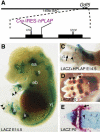
Figure 1
Figure 1. A Genetic System to Drive Gene Recombination in Developing Joints
(A) A 140-kb BAC from the Gdf5 locus was modified by inserting Cre-IRES-hPLAP into the translation start site of Gdf5 and used to make transgenic mice. Not to scale. See Materials and Methods for details. (B–E) Visualization of Gdf5-Cre driven recombination patterns based on activation of lacZ expression from the R26R Cre reporter allele. (B) LACZ activity is visible as blue staining in the ear (ea) and the joints of the shoulder (s), elbow (eb), wrist (w), knee (k), ankle (a), vertebra (vj), and phalanges (black arrowheads) of an E14.5 mouse embryo. (C) E14.5 hindlimb double-stained to show both HPLAP expression from the transgene (grey/purple staining) and LACZ expression from the rearranged R26R allele (blue staining). Note that both markers are visible in the oldest, proximal interphalangeal joint (black arrowhead), only HPLAP activity is visible in the more recently formed medial interphalangeal joint (black arrow), and neither HPLAP nor LACZ expression is visible in the youngest, most distal joint of the digit (white arrowhead). (D) Newborn (P0) forelimb with skin partially removed showing LACZ activity expressed in all phalangeal joints (red Salmon gal staining, black arrowheads) and regions of some tendons (asterisk). (E) Section through the most distal phalangeal joint of a P0 hindlimb stained with Alcian blue to mark cartilage showing LACZ expression (stained red) in all tissues of developing joints: articular cartilage (black arrowhead), precursors of ligaments and synovial membranes (black arrow), and cells where cavitation is occurring (asterisk).
Figure 2
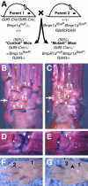
Figure 2. Bmpr1a Is Required for Webbing Regression and Apoptosis in Specific Regions of the Limb
(A and B) Control E14.5 forelimb (A) compared to a, E14.5 mutant forelimb (B) showing webbing between digits 1 and 2 (arrowheads) and extra tissue at the posterior of digit 5 (arrows). (C) Gdf5-Cre induced lacZ expression from R26R in an E13.5 forelimb showing LACZ staining (blue) in metacarpal-phalangeal joints, between digits 1 and 2 (arrowhead), and in a region posterior to digit 5 (arrow). (D and E) Sections of E14.5 hindlimbs showing apoptosis visualized by TUNEL staining (green) and proliferation visualized by staining for histone H3 phosphorylation (red). Controls show strong, uniform TUNEL staining between digits 1 and 2 (D, arrowhead) while mutants show patchy TUNEL staining interspersed with mitotic cells in similar regions (E). Scale bar = 200 μm. (F) Quantitation of TUNEL staining and mitotic cells in the posterior region of the fifth digit shows apoptosis is reduced 30% while proliferation is increased 20% (asterisks indicate statistically significant difference). (G and H) By E15.5, interdigital tissue has regressed in controls (G, arrowhead). In contrast, tissue remains in mutants at this location, primarily derived from cells that have undergone Gdf5-Cre-mediated recombination that inactivates Bmpr1a function and activates expression of LACZ (H). Scale bar = 75 μm.
Figure 3
Figure 3. Gdf5-Cre-Mediated Deletion of Bmpr1a
(A) Breeding strategy simultaneously deletes Bmpr1afloxP and allows visualization of Gdf5-Cre-mediated recombination by lacZ expression from R26R. (B–E) 5-week-old mutant and control mice stained with Alcian blue to mark cartilage and alizarin red to mark bone. (B) Ankle of control with strong blue staining lining each joint (arrowheads). (C) Ankle of mutant showing an absence of blue staining in most regions (arrowheads) and a joint fusion between the central (c) and second (2) tarsals (arrow). (D) Control and (E) mutant metatarsal/phalangeal joint which lacks blue staining in articular regions (arrowheads) but retains staining in the growth plate (asterisks). (F) Control forelimb. (G) Mutant forelimb with webbing between the first and second digit (black arrowhead).
Figure 4
Figure 4. Bmpr1a Is Expressed in Joints and Is Required for Continued Joint Formation in the Ankle Region
(A) Diagram of ankle bones from a wild-type mouse; bones fusing in mutant are colored red. Roman numerals II–IV, metatarsals; 2, 3, and 4/5, distal row of tarsal bones; c, central tarsal bone; ta, talus; ca, calcaneus. (B and C) In situ hybridization at E15.5 showing that Bmpr1a is expressed in ankle joint interzones (B, arrowheads) and in the forming articular regions of the phalangeal joints (C, arrowheads). (D) Near adjacent section to (C) showing Gdf5-Cre induced LACZ expression from R26R in the forming joints of the digits (arrowheads). (E–J) Marker gene expression and R26R LACZ staining patterns on near adjacent sections of control and mutant embryos. In control mice at E15.5 ankle joints are clearly delineated as regions that have down-regulated Col2 (E), express Gdf5 throughout (F), and express LACZ in most cells (G; white arrowheads and black arrows). In mutant embryos at the same stage, joint formation is incomplete. Faint Col2 expression can be seen connecting a medial region of tarsal 2 with metatarsal II (H, white arrowhead), and Gdf5 expression does not extend all the way across the joint at this location (I, white arrowhead). Between tarsals c and 2, mutants express Col2 across the normal joint-forming region (H, black arrow) and lack expression of Gdf5 at sites where skeletal fusions are observed (I, black arrow and bracket). (J) Scale bar = 100 μm.
Figure 5
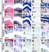
Figure 5. Bmpr1a Is Required to Maintain Expression of ECM Components in Articular Cartilage
In situ hybridization or LACZ staining on near adjacent sections of metacarpal-phalangeal joints (A–C and F–H) and the tarsal 2-metatarsal II joint (D–E and I–J) of P0 mice. At birth, articular cartilage of controls (A–E) and mutants (F–J) appears similar by Safranin O staining (A and F), and Col2 expression (B, G). Mat4 expression confirms that articular cartilage is initially specified in mutants (D andI, brackets). LACZ expression confirms Cre-mediated recombination has occurred in articular cartilage (C, H, E, and J). (K–T) Near adjacent sections of the metacarpal-phalangeal joints of P14 mice. Two weeks after birth, articular cartilage of controls stains with pericellular Safranin O (orange staining, K), and expresses Col2 (L), Agg (M), and SOX9 (N). In contrast, mutant articular cells are smaller and more densely packed, lack pericellular Safranin O staining (P), have reduced expression of Col2 (Q) and Agg (R), but retain normal levels of SOX9 protein (S, brackets; dashed line marks faint edges of articular surfaces). LACZ expression confirms Cre-mediated recombination has occurred in articular cells (O ansd T, brackets). (A and K) Scale bar = 75 μm.
Figure 6
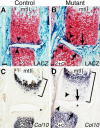
Figure 6. Synovial Membrane Expansion, Articular Surface Erosion, and Accelerated Maturation of Underlying Cartilage in Ankles of Bmpr1a Mutant Mice
Near adjacent sections from the tarsal 2-metatarsal II joint of 7-d-old mice. (A and B) LACZ staining (blue) shows Cre-mediated recombination is largely restricted to articular (arrowheads) and synovial cells (asterisks) in both controls and mutants. (C and D) In situ hybridization shows Col10 expression expands in mutants toward regions of synovial membrane expansion and articular surface erosion (brackets and arrows). This may be a cell nonautonomous effect of joint damage, since the LACZ expressing cells at the articular surface do not show upregulation of Col10 (arrowheads) and the region of expanded Col10 expression is largely made up of cells that have not undergone Cre-mediated recombination. Note the formation of a cartilaginous bridge along the joint capsule of the mutant where joint formation is disrupted at earlier stages (B, white arrowhead, and Figure 3, white arrowheads). (A) Scale bar = 75 μm.
Figure 7
Figure 7. Loss of Bmpr1a Signaling Leads to Articular Cartilage Fibrillation and Degeneration in Digits and Knees of Aging Mice
(A–D) Near adjacent sections of metatarsal-phalangeal joints from 9 month old mice. Articular cartilage of controls is complete and stains strongly with Safranin O (A, orange stain). In contrast, articular cells of mutants are severely fibrillated or absent with much reduced staining of Safranin O (C, arrowheads). LACZ expression confirms Cre-mediated recombination has occurred in articular cells (B and D). (E–P) Sagittal sections through knee joints of 7-wk- (E–J) or 9-mo-old animals (K–P); fe, femur; ti, tibia; gp, growth plate. Seven weeks after birth, the height of the tibial epiphysis is reduced in mutants (E and H, bars), and their articular layer stains poorly with Safranin O, is fibrillated, and is strikingly thinner (F and I, black arrowhead, and brackets). Near adjacent sections with LACZ staining confirm Cre-mediated recombination has occurred in articular cells (G and J). Note that in mutants, LACZ is absent in cells adjacent to those that do stain with Safranin O, suggesting Bmpr1a may act cell autonomously (I and J, white arrowheads). At 9 mo old, the mutant tibial epiphysis is extremely thin (K and N, bars), and the articular layer is completely absent, leaving bone to rub directly on bone (L and O, bracket). LACZ staining shows Cre-mediated recombination occurred in articular cells of controls (M) and in some remaining skeletal tissue of mutants (P). Also note aberrantly formed meniscal cartilage in mutants (E, H, K, and N, arrows), and increased sclerosis in mutant epiphyses (E, H, K, and N, asterisks). (A and K) Scale bar = 50 μm; (I) scale bar = 300 μm.

References

    1. Ahn K, Mishina Y, Hanks MC, Behringer RR, Crenshaw EB, 3rd. BMPR-IA signaling is required for the formation of the apical ectodermal ridge and dorsal-ventral patterning of the limb. Development. 2001;128:4449–4461. - PubMed
    1. Apte SS, Seldin MF, Hayashi M, Olsen BR. Cloning of the human and mouse type X collagen genes and mapping of the mouse type X collagen gene to chromosome 10. Eur J Biochem. 1992;206:217–224. - PubMed
    1. Badley EM. The effect of osteoarthritis on disability and health care use in Canada. J Rheumatol Suppl. 1995;43:19–22. - PubMed
    1. Bau BH, Haag J, Schmid E, Kaiser M, Gebhard PM, Aigner T. Bone morphogenetic protein-mediating receptor-associated Smads as well as common Smad are expressed in human articular chondrocytes but not up-regulated or down-regulated in osteoarthritic cartilage. J Bone Miner Res. 2002;17:2141–2150. - PubMed
    1. Baur ST, Mai JJ, Dymecki SM. Combinatorial signaling through BMP receptor IB and GDF5: Shaping of the distal mouse limb and the genetics of distal limb diversity. Development. 2000;127:605–619. - PubMed

Publication types

MeSH terms

Associated data