Multicenter Study
. 2022 Jul 18;99(3):e258-e269.
doi: 10.1212/WNL.0000000000200351.

Brain Metabolism and Amyloid Load in Individuals With Subjective Cognitive Decline or Pre-Mild Cognitive Impairment

Collaborators, Affiliations
Multicenter Study

Brain Metabolism and Amyloid Load in Individuals With Subjective Cognitive Decline or Pre-Mild Cognitive Impairment

Giacomo Tondo et al. Neurology. .

Abstract

Background and objective: This was a multicenter study aimed at investigating the characteristics of cognitive decline, neuropsychiatric symptoms, and brain imaging in individuals with subjective cognitive decline (SCD) and subtle cognitive decline (pre-mild cognitive impairment [pre-MCI]).

Methods: Data were obtained from the Network-AD project (NET-2011-02346784). The included participants underwent baseline cognitive and neurobehavioral evaluation, FDG-PET, and amyloid PET. We used principal component analysis (PCA) to identify independent neuropsychological and neuropsychiatric dimensions and their association with brain metabolism.

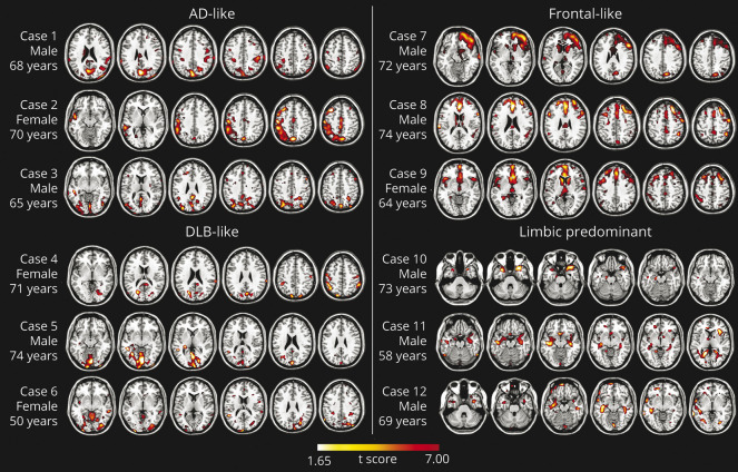
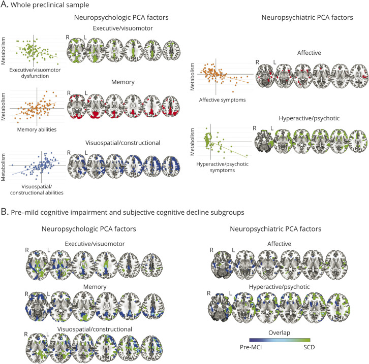
Results: A total of 105 participants (SCD = 49, pre-MCI = 56) were included. FDG-PET was normal in 45% of participants and revealed brain hypometabolism in 55%, with a frontal-like pattern as the most frequent finding (28%). Neuropsychiatric symptoms emerging from the Neuropsychiatric Inventory and the Starkstein Apathy Scale were highly prevalent in the whole sample (78%). An abnormal amyloid load was detected in the 18% of the participants who underwent amyloid PET (n = 60). PCA resulted in 3 neuropsychological factors: (1) executive/visuomotor, correlating with hypometabolism in frontal and occipital cortices and basal ganglia; (2) memory, correlating with hypometabolism in temporoparietal regions; and (3) visuospatial/constructional, correlating with hypometabolism in frontoparietal cortices. Two factors emerged from the neuropsychiatric PCA: (1) affective, correlating with hypometabolism in orbitofrontal and cingulate cortex and insula; (2) hyperactive/psychotic, correlating with hypometabolism in frontal, temporal, and parietal regions.

Discussion: FDG-PET evidence suggests either normal brain function or different patterns of brain hypometabolism in SCD and pre-MCI. These results indicate that SCD and pre-MCI represent heterogeneous populations. Different neuropsychological and neuropsychiatric profiles emerged, which correlated with neuronal dysfunction in specific brain regions. Long-term follow-up studies are needed to assess the risk of progression to dementia in these conditions.


Figures

Figure 1
Figure 1. Examples of Single-Subject FDG-PET Hypometabolic Patterns Resulting From Statistical Parametric Mapping Single-subject analysis vs 112 controls; significance was set at uncorrected p <0.05 at the voxel level with k > 100 voxels
AD = Alzheimer disease; DLB = dementia with Lewy bodies; pre–MCI = pre–mild cognitive impairment; SCD = subjective cognitive decline.
Figure 2
Figure 2. Results of Voxel-wise Multivariate Regression Models Between the Grouped Test in Neuropsychological PCA and Neuropsychiatric PCA Factors and Brain Glucose Metabolism, After Factoring out the Effect of Age (p < 0.05)
(A) Results in the whole preclinical group. On the left of each result, the scatterplots display the correlations between factors and glucose metabolism in the significant clusters in the whole sample. Significant correlations among the 3 neuropsychological principal component analysis (PCA) factors and brain metabolism and the 2 neuropsychiatric PCA factors and brain metabolism are reported on the left and the right, respectively. The executive/visuomotor factor showed a significant negative correlation with metabolism (i.e., linear decrease in glucose metabolism together with impaired performance, thus higher scores) in the superior and the middle frontal gyri, lingual gyrus, cuneus, precuneus, and middle cingulate cortex, plus the caudate nuclei and thalamus, bilaterally. The memory factor showed a significant positive correlation with metabolism (i.e., linear decrease in glucose metabolism together with decreasing scores) in the precuneus, cuneus, superior and inferior parietal lobules, the posterior and middle cingulate cortices, and the superior and the middle frontal gyri. The visuospatial/constructional factor showed a significant positive correlation with right-lateralized metabolism (i.e., linear decrease in glucose metabolism together with decreasing scores of grouped tests), specifically in the angular gyrus, the anterior and middle cingulate cortex, and the dorsolateral frontal cortex. The affective factor showed a significant negative correlation with brain metabolism (i.e., linear decrease in glucose metabolism together with increasing symptoms) mainly in the right hemisphere, insula, temporal pole, anterior cingulate cortex, superior temporal gyrus, and inferior frontal gyrus (pars orbitalis). The hyperactive/psychotic factor showed a significant negative correlation with metabolism (i.e., linear decrease in glucose metabolism together with increasing symptoms) in the orbitofrontal cortex and prefrontal cortex, anterior cingulate cortex, left lateral temporal cortex, insula, and caudate. (B) Results of voxel-wise multivariate regression models (run separately for pre–mild cognitive impairment [pre–MCI] and subjective cognitive decline [SCD] subgroups). Results obtained in pre–MCI and SCD are blue and green, respectively (overlap areas are light blue). The executive/visuomotor factor negatively correlated with metabolism in extended temporoparietal and occipital regions in the pre–MCI group, whereas in the SCD group, this factor correlated mainly with subcortical structures. The positive correlations with the memory factor were prevalently represented by the pre–MCI group in frontal-temporal-parietal cortex. Correlations in the visuospatial/constructional factor were more represented in the right hemisphere and broadly similar in the 2 groups. The affective factor correlated with insular and temporal medial metabolism in both groups, whereas the hyperactive/psychotic factor correlated more extensively with frontal, temporal, and insular regions in the SCD group.

References

    1. Jack CR, Knopman DS, Jagust WJ, et al. . Tracking pathophysiological processes in Alzheimer's disease: an updated hypothetical model of dynamic biomarkers. Lancet Neurol. 2013;12(2):207-216. - PMC - PubMed
    1. Sachdev PS, Blacker D, Blazer DG, et al. . Classifying neurocognitive disorders: the DSM-5 approach. Nat Rev Neurol Nat. 2014;10(11):634-642. - PubMed
    1. Albert MS, DeKosky ST, Dickson D, et al. . The diagnosis of mild cognitive impairment due to Alzheimer's disease: recommendations from the National Institute on Aging-Alzheimer’s Association workgroups on diagnostic guidelines for Alzheimer's disease. Alzheimers Dement. 2011;7(3):270-279. - PMC - PubMed
    1. Jessen F, Amariglio RE, Van Boxtel M, et al. . A conceptual framework for research on subjective cognitive decline in preclinical Alzheimer's disease. Alzheimers Dement. 2014;10(6):844-852. - PMC - PubMed
    1. Jessen F, Amariglio RE, Buckley RF, et al. . The characterisation of subjective cognitive decline. Lancet Neurol. 2020;19(3):271-278. - PMC - PubMed

Publication types

MeSH terms