. 2016 Jul 22;291(30):15447-59.
doi: 10.1074/jbc.M116.733659. Epub 2016 May 26.

Structural, Functional, and Immunological Characterization of Profilin Panallergens Amb a 8, Art v 4, and Bet v 2

Affiliations

Structural, Functional, and Immunological Characterization of Profilin Panallergens Amb a 8, Art v 4, and Bet v 2

Lesa R Offermann et al. J Biol Chem. .

Abstract

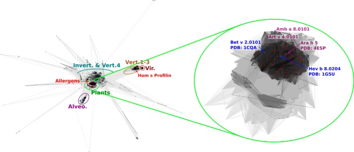
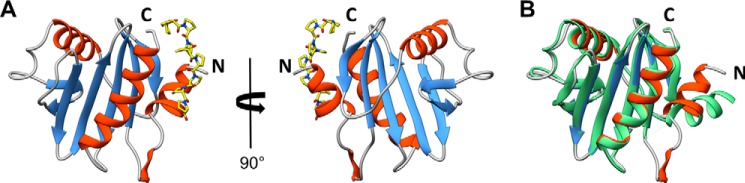
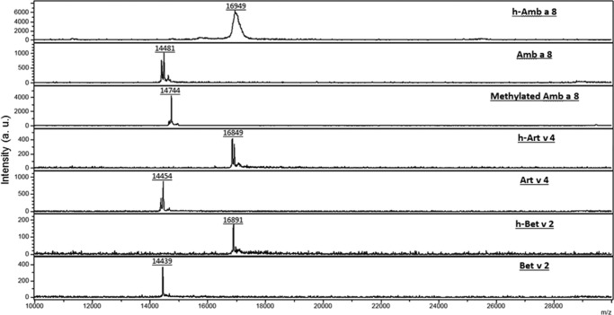
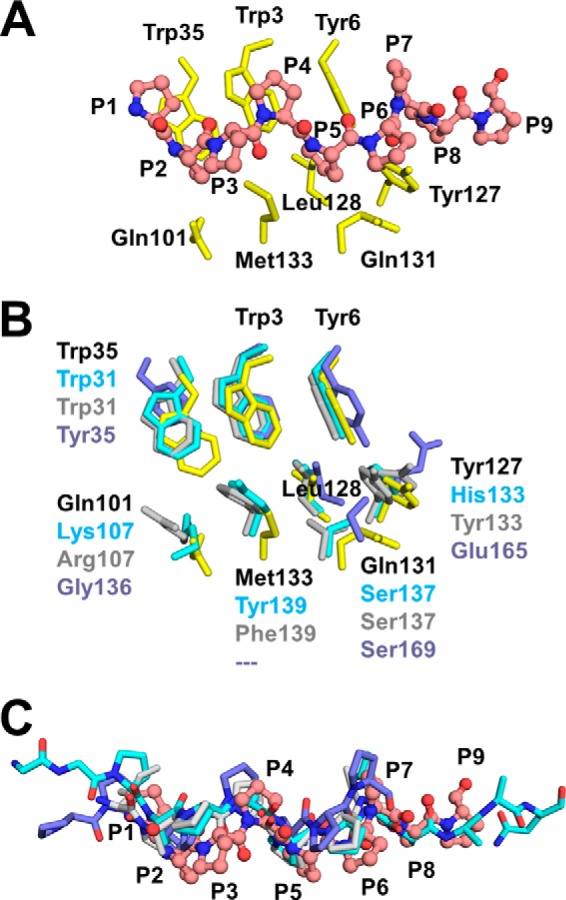
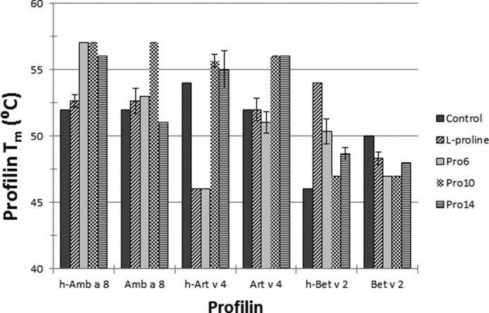
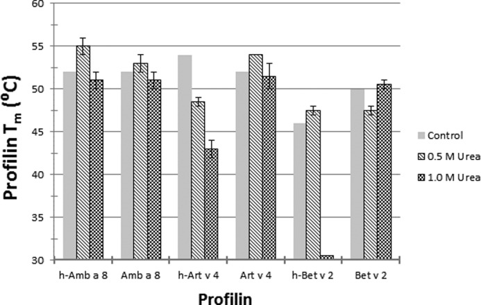
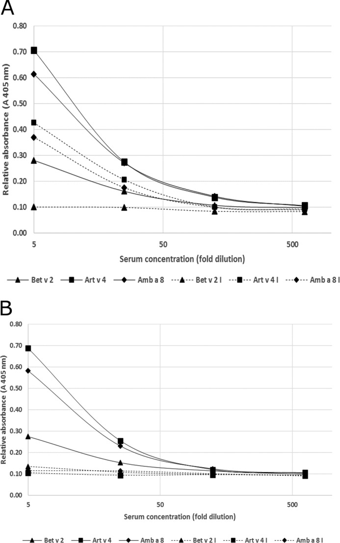
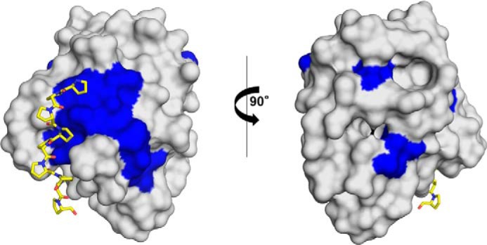
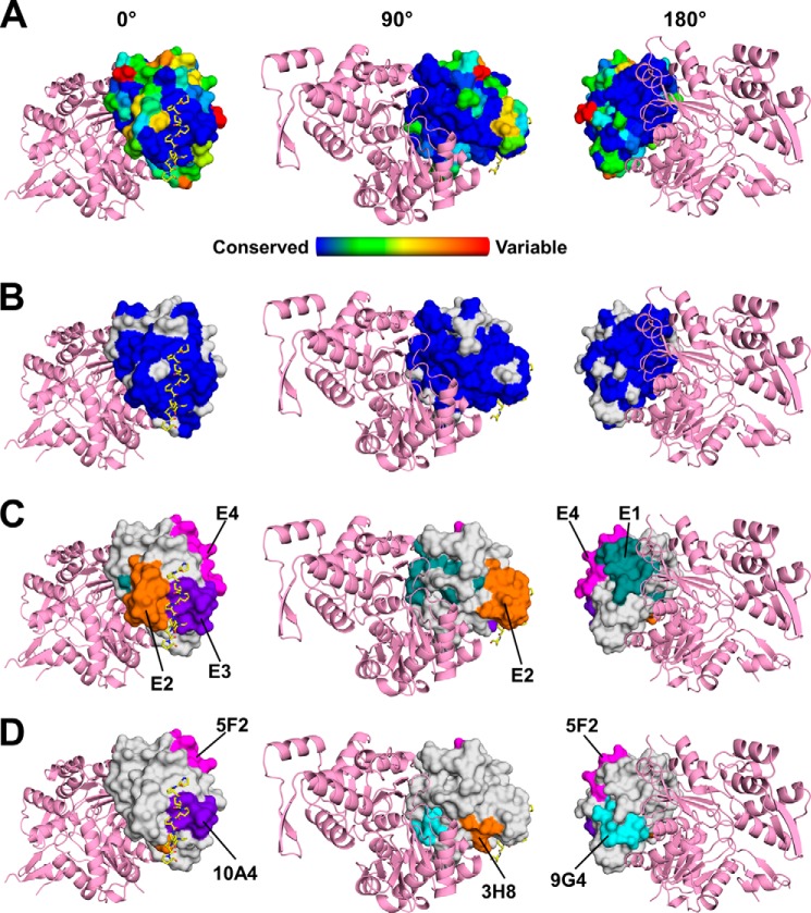
Ragweed allergens affect several million people in the United States and Canada. To date, only two ragweed allergens, Amb t 5 and Amb a 11, have their structures determined and deposited to the Protein Data Bank. Here, we present structures of methylated ragweed allergen Amb a 8, Amb a 8 in the presence of poly(l-proline), and Art v 4 (mugwort allergen). Amb a 8 and Art v 4 are panallergens belonging to the profilin family of proteins. They share significant sequence and structural similarities, which results in cross-recognition by IgE antibodies. Molecular and immunological properties of Amb a 8 and Art v 4 are compared with those of Bet v 2 (birch pollen allergen) as well as with other allergenic profilins. We purified recombinant allergens that are recognized by patient IgE and are highly cross-reactive. It was determined that the analyzed allergens are relatively unstable. Structures of Amb a 8 in complex with poly(l-proline)10 or poly(l-proline)14 are the first structures of the plant profilin in complex with proline-rich peptides. Amb a 8 binds the poly(l-proline) in a mode similar to that observed in human, mouse, and P. falciparum profilin·peptide complexes. However, only some of the residues that form the peptide binding site are conserved.

Keywords: IgE binding; allergen; allergy; crystal structure; epitope mapping; peptide binding; pollen; protein stability.


Figures

FIGURE 1.
FIGURE 1.
Two-dimensional projection of the CLANS clustering results. Proteins are indicated by dots. Lines indicate sequence similarity detectable with BLAST and are colored by a spectrum of shades of gray according to the BLAST p value. Sequences corresponding to structures in the PDB are indicated by blue dots, and sequences of known allergens are indicated by red dots.
FIGURE 2.
FIGURE 2.
A, a structure of Amb a 8·poly(l-Pro)14 complex. Poly(l-Pro) is shown in a stick representation. B, superposition of Amb a 8 and Bet v 2 (PDB entry 1CQA).
FIGURE 3.
FIGURE 3.
Mass spectrometry results for Amb a 8, Art v 4, and Bet v 2. The experiment was performed with a Bruker Ultraflex II MALDI TOF/TOF and processed with flex control software and flexAnalysis version 3.3. Intensity (arbitrary units (a.u.)).
FIGURE 4.
FIGURE 4.
A, interactions between Amb a 8 and poly(l-Pro)14. Amb a 8 residues are shown as yellow sticks, whereas poly(l-Pro) is shown in a ball-and-stick representation and labeled using one-letter codes. B, superposition of profilin complexes with proline-rich peptides. Only side chains of profilin residues involved in binding of proline-rich peptides are shown. The superposition involves Amb a 8 (yellow sticks), human profilin (cyan sticks; PDB entry 3CHW), mouse profilin (gray sticks; PDB entry 2V8F), and profilin from P. falciparum (purple sticks; PDB entry 2JKG). C, superposition of profilin complexes with proline-rich peptides. Only poly(l-Pro) and proline-rich peptide are shown. Poly(l-Pro) from the Amb a 8·poly(l-Pro)14 complex is shown in ball-and-stick representation.
FIGURE 5.
FIGURE 5.
DSF profilin proline screening. Amb a 8, Art v 4, Bet v 2, and their His-tagged counterparts were screened against 10.0 mm l-proline, Pro6, Pro10, or Pro14 to determine change in protein melting temperature (Tm). The control was profilin stability in gel filtration buffer (10 mm Tris, pH 7.4, 150 mm NaCl). Error bars, S.D.
FIGURE 6.
FIGURE 6.
DSF profilin urea screening. Amb a 8, Art v 4, Bet v 2, and their His-tagged counterparts were screened against different concentrations of urea to determine change in protein melting temperature (Tm). The control was profilin stability in gel filtration buffer (10 mm Tris, pH 7.4, 150 mm NaCl). Error bars, S.D.
FIGURE 7.
FIGURE 7.
A, representative inhibition ELISA results with Bet v 2 used as an inhibiting allergen. Bet v 2, Bet v 2 coated on a plate; Art v 4, Art v 4 coated on a plate; Amb a 8, Amb a 8 coated on a plate; I, inhibition by Bet v 2 (30 μg/ml). B, representative inhibition ELISA results using Art v 4 as an inhibiting allergen. Bet v 2, Bet v 2 coated on a plate; Art v 4, Art v 4 coated on a plate; Amb a 8, Amb a 8 coated on a plate; I, inhibition by Art v 4 (30 μg/ml).
FIGURE 8.
FIGURE 8.
Surface representation of Amb a 8. Residues marked in blue are identical in Amb a 8 and human profilins 1. The poly(l-proline) is shown in a stick representation.
FIGURE 9.
FIGURE 9.
A, sequence conservation of profilins calculated with ConSurf is mapped on the surface of Amb a 8. Actin (pink) was modeled using the structure of human profilin 1 in complex with actin (PDB entry 3CHW). Poly(l-Pro) is shown in a stick representation with carbon atoms marked in yellow. The figure shows three different orientations of the molecules corresponding to 0, 90, and 180° rotation along the y axis. B, residues that are identical in Amb a 8, Art v 4, and Bet v 2 are shown in blue. Residues that are not 100% conserved in these allergens are marked in gray. C, epitopes identified in Cuc m 2. The epitopes are labeled using a convention used in the original paper reporting these epitopes (44, 74). D, epitopes identified in Hel a 2.0101. The epitopes are labeled using a convention used in the original paper reporting these epitopes (43).

References

    1. Hauser M., Roulias A., Ferreira F., and Egger M. (2010) Panallergens and their impact on the allergic patient. Allergy Asthma Clin. Immunol. 6, 1. - PMC - PubMed
    1. Mari A. (2001) Multiple pollen sensitization: a molecular approach to the diagnosis. Int. Arch. Allergy Immunol. 125, 57–65 - PubMed
    1. Tinghino R., Twardosz A., Barletta B., Puggioni E. M., Iacovacci P., Butteroni C., Afferni C., Mari A., Hayek B., Di Felice G., Focke M., Westritschnig K., Valenta R., and Pini C. (2002) Molecular, structural, and immunologic relationships between different families of recombinant calcium-binding pollen allergens. J. Allergy Clin. Immunol. 109, 314–320 - PubMed
    1. Wopfner N., Gruber P., Wallner M., Briza P., Ebner C., Mari A., Richter K., Vogel L., and Ferreira F. (2008) Molecular and immunological characterization of novel weed pollen pan-allergens. Allergy 63, 872–881 - PubMed
    1. Ghosh D., Mueller G. A., Schramm G., Edwards L. L., Petersen A., London R. E., Haas H., and Gupta Bhattacharya S. (2014) Primary identification, biochemical characterization, and immunologic properties of the allergenic pollen cyclophilin cat R 1. J. Biol. Chem. 289, 21374–21385 - PMC - PubMed

Publication types

LinkOut - more resources