. 1998 Nov 1;12(21):3343-56.
doi: 10.1101/gad.12.21.3343.

Structure and specificity of nuclear receptor-coactivator interactions

Affiliations

Structure and specificity of nuclear receptor-coactivator interactions

B D Darimont et al. Genes Dev. .

Abstract

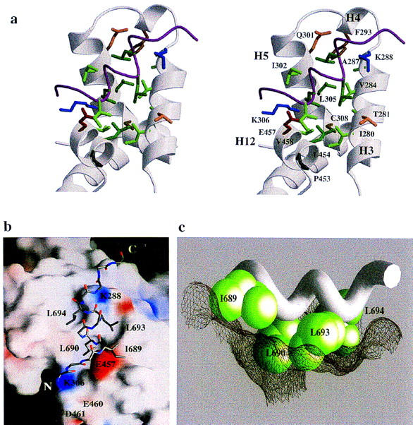
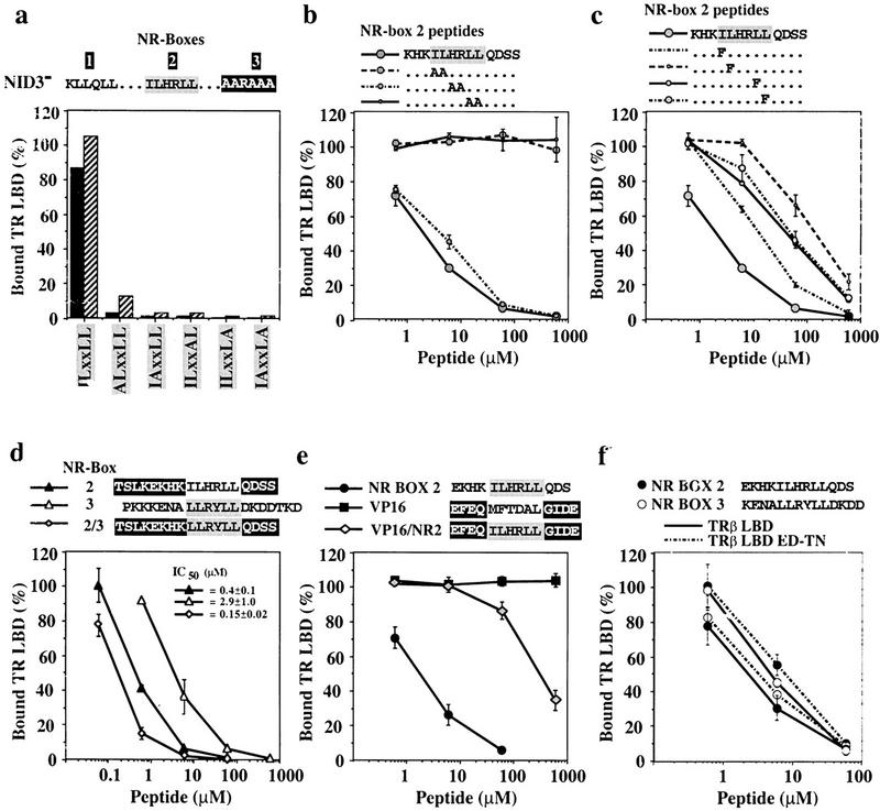
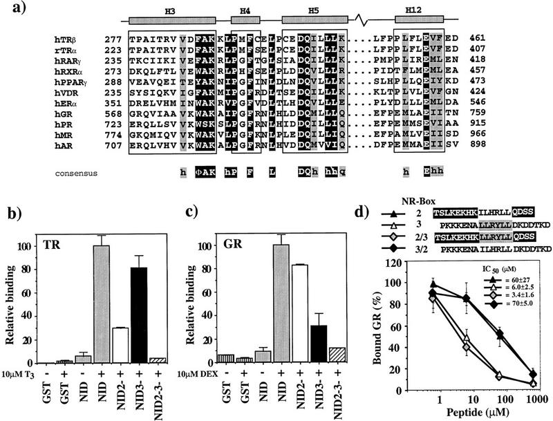
Combinatorial regulation of transcription implies flexible yet precise assembly of multiprotein regulatory complexes in response to signals. Biochemical and crystallographic analyses revealed that hormone binding leads to the formation of a hydrophobic groove within the ligand binding domain (LBD) of the thyroid hormone receptor that interacts with an LxxLL motif-containing alpha-helix from GRIP1, a coactivator. Residues immediately adjacent to the motif modulate the affinity of the interaction; the motif and the adjacent sequences are employed to different extents in binding to different receptors. Such interactions of amphipathic alpha-helices with hydrophobic grooves define protein interfaces in other regulatory complexes as well. We suggest that these common structural elements impart flexibility to combinatorial regulation, whereas side chains at the interface impart specificity.


Figures

Figure 1
Figure 1
(a) Functional domains of p160 family coactivators (Ding et al. 1998; Voegel et al. 1998). (b) The NID of GRIP1 (563–767) contains four predicted α-helices (Rost and Sander 1994); three of them include the conserved LxxLL motifs of NR-boxes 1, 2, and 3. (c) Sequence alignment of LxxLL motifs in members of the p160 coactivator family. Motif leucine residues are green; box-specific conserved residues are red. GRIP1 (Hong et al. 1996, 1997), pCIP (Torchia et al. 1997), and SRC-1 (Oñate et al. 1995) were taken as representatives for the three distinct classes of coactivators in the p160 family. Predicted α-helices are shown for each box.
Figure 2
Figure 2
(a) GRIP1 NR-boxes 1, 2, and 3 interact differentially with TRβ LBD. (GST) An isolated GST domain; (NID) GST fusion of the GRIP1 fragment (563–767)His6 that contains NR-boxes 1, 2, and 3 (shaded bars). In NID2 (open bar) or NID3 (solid bar) the bulky hydrophobic residues of NR-box 2 (ILHRLL) or NR-box 3 (LLRYLL) were replaced by alanine yielding AAHRAA and AARAAA, respectively. NID23 (hatched bar) contains replacement of both NR-boxes 2 and 3. Assays were carried out using 10 nm labeled TRβ LBD and 1.6 μm purified, glutathione–agarose bound GST–NID proteins either in the absence (−) or presence (+) of 10 μm T3. The yield of bound receptor is given as percentage relative to the input. The data show the average of more than three independent experiments together with the standard deviation. (b) NR-box 2 interacts with TRβ LBD with a fourfold higher affinity than NR-box 3. Labeled TRβ LBD (10 nm) was incubated in the presence of 10 μm T3 and various concentrations of purified and glutathione–agarose-bound GST–NID (shaded diamond) and GST–NID2 (open square) or GST–NID3 (solid square) lacking either a functional NR-box 2 or 3, respectively. As in a, the amount of bound receptor is relative to the receptor input. The data represent the average and range of at least two independent experiments. (c) Peptides containing NR boxes compete the interaction of TRβ LBD with the NID. Labeled TRβ LBD (10 nm) was incubated with 1.6 μm glutathione–agarose-bound GST–NID3 in the presence of 10 μm T3 and increasing concentrations of NR-box 2 peptides EKHKILHRLLQDS (solid circle) or TSLKEKHKILHRLLQDSS (solid triangle), or NR-box 3 peptides LLRYLL (open square), KENALLRYLLDKDD (open circle) or PKKKENALLRYLLDKDDTKD (open triangle). Bound receptor is shown relative to the amount retained in the absence of peptide. The data and calculated IC50 values represent the average and standard deviation of three independent experiments.
Figure 3
Figure 3
(a) The contents of the asymmetric unit of the crystallized TRβ LBD:NR-box 2 complex. The repeating unit of the crystal lattice contains a 2:2 complex of TRβ LBD and NR-box 2 peptide. The two monomers of the TRβ LBD are shown as a ribbon drawing, with monomer A in light gray, and monomer B in dark gray. The positions of the two NR-box 2 peptides, depicted as a magenta secondary structure ribbon, are boxed. The interaction of peptide A with monomer A and of peptide B with monomer B observe the same noncrystallographic symmetry relation as the two TRβ LBD monomers. The ligand T3 is shown in a space-filling representation. (b) Electron density for NR-box 2 peptide A and stereo representation of the peptide model. (Left) Initial Fo–Fc electron density map for the NR-box 2 peptide A, using phases calculated from the TRβ LBD model (without peptide), following one cycle of positional refinement against a maximum likelihood target in crystallography NMR systems (CNS) (A.T. Brunger, pers. comm.), with strict noncrystallographic symmetry enforced. The electron density is contoured at 3.0σ (yellow) and 2.0σ (cyan). The peptide appears as a stick representation, using the final refined atomic model. The same map shows strong, corroborating difference density for the central seven residues of peptide B. (Right) Final 2F0–Fc electron density map for the NR-box 2 peptide A (green).
Figure 4
Figure 4
(a) Interface between the NR-box 2 peptide and the TRβ LBD. The side chains of those residues of the TRβ LBD within 4.5 Å of the NR-box 2 peptide are labeled. Acidic residues are red, basic residue are blue, aliphatic residues are green, aromatic residues are brown, and polar residues are orange. The peptide is depicted as a Cα trace, with the side chains of ILxxLL motif shown explicitly. (b) Surface of the TRβ LBD. The side chains of the leucines residues from the NR-box 2 peptide fit within a hydrophobic groove on the surface of the TRβ LBD formed from helices H3, H4, H5, and H12, whereas the side chain of the nonconserved isoleucine residue packs against the outside edge of the groove. The remainder of the peptide is shown as main chain. Areas of positive electrostatic potential are shown in blue; areas of negative electrostatic potential are shown in red. Individual charged residues of the TRβ LBD at the NR-box interface are labeled. Electrostatic and surface calculations used GRASP (Nicholls et al. 1991). (c) The TRβ LBD:LxxLL interface. The side chains of the NR-box 2 ILxxLL motif are shown in a CPK representation, with the main chain of the peptide drawn as a Cα worm. The three leucine residues fit into pockets on the molecular surface of the TRβ LBD, depicted as mesh, whereas the nonconserved isoleucine residue rests on the edge of the surface cleft.
Figure 5
Figure 5
(a) Individual leucine residues of the LxxLL motif are crucial for binding of GRIP1 NID to TRβ LBD. Shown are 1.6 μm (solid bars) or 4.0 μm (hatched bars) of glutathione–agarose-bound GST–NID3 or variants containing alanine substitutions of individual hydrophobic residues of the NR-box 2 ILxxLL motif (ALxxLL: I689A; IAxxLL: L690A; ILxxAL: L693A; ILxxLA: L694A; IAxxLA: L690A + L694A) were incubated with labeled TRβ LBD in the presence of 10 μm T3 (mutations are in boldface type). The amount of bound receptor is relative to the receptor input. These are the results of a representative experiment. (b,c) Pairwise or single conservative substitutions of the ILxxLL leucine residues drastically reduce the affinity of NR-box 2 peptides for TRβ LBD. Interaction of 1.6 μm glutathione–agarose-bound NID3 with 10 nm labeled TRβ LBD + 10 μm T3 was competed with increasing concentrations of variants of the NR-box 2 peptide KHKILHRLLQDSS, containing either pairwise alanine substitutions KHKAAHRLLQDSS, KHKILAALLQDSS, KHKILHRAAQDSS (b), or single phenylalanine substitutions of conserved residues of the hLxxLL motif (KHKFLHRLLQDSS, KHKIFHRLLQDSS, KHKILHRFLQDSS, KHKILHRLFQDSS) (c) (mutations are in boldface type). The amount of bound receptor is relative to the amount of retained receptor in the absence of peptide. The data represent the average and standard deviation of three independent experiments. (d,e) Sequences adjacent to the LxxLL motif affect the affinity of the TRβ LBD:NR-box 2 interaction. Labeled TRβ LBD (10 nm) was incubated with 1.6 μm glutathione–agarose-bound GST–NID3 in the presence of 10 μm T3 and increasing concentrations of peptides containing NR-box 2 (solid triangle), NR-box 3 (open triangle), and the LLRYLL motif of NR-box 3 in the context of flanking sequences from NR-box 2 (shaded diamond) (d), or NR-box 2 (solid circle), VP16 (solid square), and the ILHRLL motif of NR-box 2 in the context of the adjacent sequences from the VP16 peptide (shaded diamond) (e). The amount of bound receptor is relative to the amount of retained receptor in the absence of peptide. The data and IC50 values represent the average and standard deviation of three independent experiments. (f) Labeled TRβ LBD (solid line) or TRβ LBD ED–TN (broken line) (10 nm) was incubated with 1.6 μm glutathione–agarose-bound GST–NID3 in the presence of 10 μm T3 and increasing concentrations of peptides containing NR-box 2 (solid circle), or NR-box 3 (open circle). The amount of bound receptor is relative to the amount of retained receptor in the absence of peptide. The data represent the average and standard deviation of three independent experiments.
Figure 6
Figure 6
(a) Sequence alignment of LBD helices 3, 4, 5, and 12. NR sequences (Wurtz et al. 1996) are shown for LBD regions that interact with GRIP1. Residues representing the NR signature sequence, ΦAKxhPxFxxLxxxDQxxhh, are on dark background. Conserved residues, always hydrophobic (h) or strong polar (q), are shaded. The borders for helices H3, H4, H5, and H12 are those for the TRβ LBD. (Φ) Bulky hydrophobic residue. (b,c) GR and TR interact preferentially with different GRIP1 NR boxes. Labeled TRβ (b) or rat GR (c) (10 nm) was incubated with 1.6 μm glutathione–agarose-bound GST (vertically striped), GST–NID (shaded) or the NID variants NID2 (open), NID3 (solid), and NID23 (hatched) in the absence (−) or presence (+) of 10 μm T3 (TR) or 10 μm dexamethasone (DEX) (GR). The amount of bound receptor is normalized to the fraction bound to GST–NID (100%). The data represent the average and standard deviation of about three independent experiments. (d) Preferential interaction of GR with NR-box 3 is not specified by sequences adjacent to the LLRYLL motif. Labeled GR (10 nm) was incubated with 1.6 μm glutathione–agarose-bound GST–NID3 in the presence of 10 μm DEX and increasing concentrations of NR-box 2 peptide (solid triangle), NR-box 3 peptide (open triangle), and either a chimeric peptide bearing the LLRYLL motif of NR-box 3 with the adjacent sequences from NR-box 2 (shaded diamond), or a chimeric peptide bearing the ILHRLL motif of NR-box 2 with the adjacent sequences from NR-box 3 (solid diamond). The amount of bound receptor is relative to the amount of retained receptor in the absence of peptide. The data and IC50 values represent the average and standard deviation of three independent experiments.

References

    1. Adams PD, Pannu NS, Read RJ, Brunger AT. Cross-validated maximum likelihood enhances crystallographic simulated annealing refinement. Proc Natl Acad Sci. 1997;94:5018–5023. - PMC - PubMed
    1. Anzick SL, Kononen J, Walker RL, Azorsa DO, Tanner MM, Guan XY, Sauter G, Kallioniemi OP, Trent JM, Meltzer PS. AIB1, a steroid receptor coactivator amplified in breast and ovarian cancer. Science. 1997;277:965–958. - PubMed
    1. Apriletti JW, Baxter JD, Lau KH, West B. Expression of the rat α1 thyroid hormone receptor ligand binding domain in Escherichia coli and the use of a ligand-induced conformational change as a method for its purification to homogeneity. Protein Expr Purif. 1995;6:363–370. - PubMed
    1. Barettino D, Vivanco Ruiz MdM, Stunnenberg G. Characterization of the ligand-dependent transactivation domain of thyroid receptor. EMBO J. 1994;13:3039–3049. - PMC - PubMed
    1. Bourguet W, Ruff M, Chambon P, Gronemeyer H, Moras D. Crystal structure of the ligand-binding domain of the human nuclear receptor RXRa. Nature. 1995;375:377–382. - PubMed

Publication types

MeSH terms

Substances

Associated data

LinkOut - more resources