. 2007 Jul;3(7):e121.
doi: 10.1371/journal.pcbi.0030121.

Evolution of function in the "two dinucleotide binding domains" flavoproteins

Affiliations

Evolution of function in the "two dinucleotide binding domains" flavoproteins

Sunil Ojha et al. PLoS Comput Biol. 2007 Jul.

Abstract

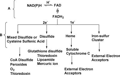
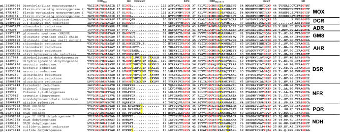
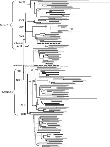
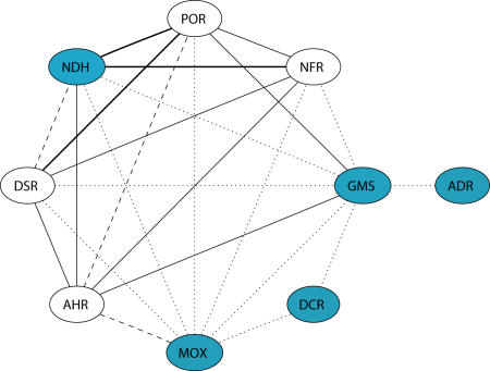
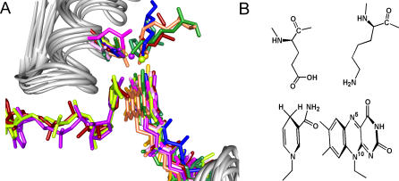
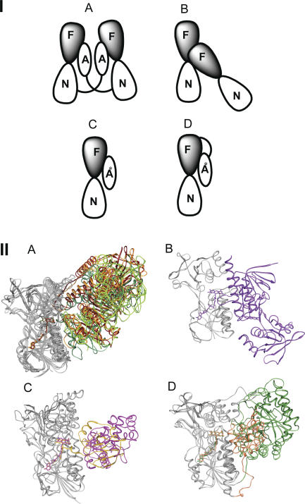
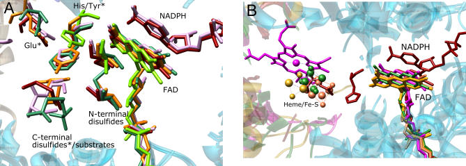
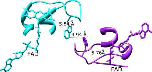
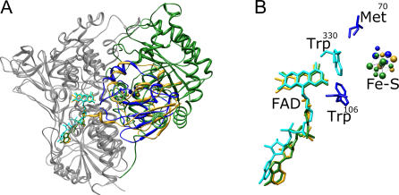
Structural and biochemical constraints force some segments of proteins to evolve more slowly than others, often allowing identification of conserved structural or sequence motifs that can be associated with substrate binding properties, chemical mechanisms, and molecular functions. We have assessed the functional and structural constraints imposed by cofactors on the evolution of new functions in a superfamily of flavoproteins characterized by two-dinucleotide binding domains, the "two dinucleotide binding domains" flavoproteins (tDBDF) superfamily. Although these enzymes catalyze many different types of oxidation/reduction reactions, each is initiated by a stereospecific hydride transfer reaction between two cofactors, a pyridine nucleotide and flavin adenine dinucleotide (FAD). Sequence and structural analysis of more than 1,600 members of the superfamily reveals new members and identifies details of the evolutionary connections among them. Our analysis shows that in all of the highly divergent families within the superfamily, these cofactors adopt a conserved configuration optimal for stereospecific hydride transfer that is stabilized by specific interactions with amino acids from several motifs distributed among both dinucleotide binding domains. The conservation of cofactor configuration in the active site restricts the pyridine nucleotide to interact with FAD from the re-side, limiting the flow of electrons from the re-side to the si-side. This directionality of electron flow constrains interactions with the different partner proteins of different families to occur on the same face of the cofactor binding domains. As a result, superimposing the structures of tDBDFs aligns not only these interacting proteins, but also their constituent electron acceptors, including heme and iron-sulfur clusters. Thus, not only are specific aspects of the cofactor-directed chemical mechanism conserved across the superfamily, the constraints they impose are manifested in the mode of protein-protein interactions. Overlaid on this foundation of conserved interactions, nature has conscripted different protein partners to serve as electron acceptors, thereby generating diversification of function across the superfamily.


Conflict of interest statement

Competing interests. The authors have declared that no competing interests exist.

Figures

Figure 1
Figure 1. Schematic of Reactions Catalyzed by tDBDF
The reductive and oxidative half reactions are shown in brackets with (A) denoting the reductive half-reaction and (B) denoting the oxidative half-reaction. In the oxidative half-reaction, superfamily members can transfer electrons one or two at a time via intermediate acceptors to a variety of different small molecule acceptors or external protein partners.
Figure 2
Figure 2. Sequence Alignment of Representative Sequences of Subgroups Containing at Least One Experimentally Verified Enzyme in the tDBDF Superfamily
The conserved motifs associated with the superfamily are labeled at the top. The numbers of amino acids separating each motif are shown. Functionally important residues including the cysteine residues of the CxxxxC motif of DSR subgroup, the cysteine sulfenic acid of the POR subgroup, and a cysteine residue that binds FAD covalently in cytochrome c sulfide dehydrogenase are colored in blue and highlighted in yellow. An asterisk * designates the lysine and glutamate residues that stabilize the isoalloxazine and nicotinamide ring complex. Not all motifs are conserved in all subgroups, as discussed in the text.
Figure 3
Figure 3. Dendrogram Showing Primary Groups and Subgroups in the tDBDF Superfamily
Nodes defining subgroups that contain at least one characterized member are identified by a circle and named using the abbreviations provided in Table 1. Functionally uncharacterized sequences identified in our searches that do not fall into subgroups and that are not listed in Table 1 are shown using dashed lines. For ease of viewing, only a representative set of sequences is shown.
Figure 4
Figure 4. The Connectivity between Subgroups Based on E-values from Profile-Based HMM Searches
Circles represent subgroups and edges represent connections between two subgroups. The strength of each connection is represented by different types of lines: dotted, E-value > 10−10; dashed, <10−10; solid, <10−20 and bold, <10−30. Blue circles designate new subgroup connections identified in this study using sequence information.
Figure 5
Figure 5. The Active Sites Structure of Members of the tDBDF Superfamily
(A) The superimposed active sites of some divergent members of the tDBDF superfamily. Protein backbones are shown in gray. The cofactors and conserved sidechains important for stabilizing the isoalloxazine and nicotinamide ring complex are shown in color. The stabilizing water residues are shown as balls. Colors, PDB identifiers, and numbers of the displayed residues are as follows. DSR subgroup: dark red, glutathione reductase (1get, Glu181, Lys50); plum, mouse thioredoxin reductase (1zkq, Lys94, Glu232); and sea green, 2-ketopropyl coenzyme M oxidoreductase/carboxylase, (1mo9, Glu228, His90). NDH subgroup: magenta, flavocytochrome c sulfide dehydrogenase (1fcd, Glu167, N/A). NFR subgroup: blue, putidaredoxin reductase (1q1w, Glu163, Lys50). POR subgroup: chartreuse, NADH peroxidase (2npx, Glu163, H2O490). Group2 proteins. DCR subgroup: forest green, 2,4-dienoylCoA reductase (1ps9, Asp508, Lys421). ADR subgroup: goldenrod, adrenodoxin reductase (1e6e, Asp159, H2O16). AHR subgroup: purple, E. Coli thioredoxin reductase (1f6m, Glu167, H257). GMS subgroup: coral, dihydropyrimidine dehydrogenase (1gte, Asp346, Arg235). (Note that the structural information on the MOX subgroup was not available when this analysis was done, thus it was not represented in this figure. However, we have confirmed that it follows the same superfamily theme). (B) Illustration showing an example of common elements of the active site.
Figure 6
Figure 6. Quaternary Structures of Members of the tDBDF Superfamily
(I) Illustration showing the four main types of quaternary structures observed. F, FAD; N, NAD(P)H; A represents a domain that assists in binding small molecules that accept two electrons and/or presents two cysteine residues that can transfer electrons to external electron acceptors; A* represents a domain or protein that accepts one electron at a time using heme or an iron-sulfur cluster. (II) Structural superimposition showing the quaternary structures of some divergent members of the tDBDF superfamily. tDBDF domains are shown in gray. The C-terminal domains of the members of the DSR and NDH subgroups are not shown for ease of viewing. Colors and PDB identifiers are as follows. A) DSR subgroup: dark red, glutathione reductase (1get); sea green, 2-ketopropyl coenzyme M oxidoreductase/carboxylase (1mo9); orange, mercuric reductase. POR subgroup: chartreuse, CoA Disulfide Reductase (1yqz). B) AHR subgroup: purple, E Coli thioredoxin reductase (1f6m). C) NDH subgroup: magenta, cytochrome c sulfide dehydrogenase (1fcd). ADR subgroup: goldenrod, adrenodoxin reductase (1e6e). D) DCR subgroup: forest green, 2,4-dienoylCoA reductase (1ps9). GMS subgroup: coral, dihydropyrimidine dehydrogenase (1gte).
Figure 7
Figure 7. Structural Superposition Showing the Electron Transfer Route for Members of the tDBDF Superfamily
(A) Two-electron transfer route. Residues designated with an asterisk * are from the C-terminal interacting domain of the second subunit of the acceptor proteins. Colors, PDB identifiers, and the numbers of the displayed residues are: dark red, 1grt, glutathione reductase (C42–C47, H439, E444, glutathione disulfide [only the disulfide bond is shown for ease of viewing]); plum, 1zkq, mouse thioredoxin reductase (C86–C91, H143, H497, E502, C522–C523); sea green, 1mo9, 2-ketopropyl coenzyme M oxidoreductase/carboxylase (C82–C87, F501, N503, 2-ketopropyl coenzyme M); orange, mercuric reductase (C207–C212, Y605, E610, C628, C629); chartreuse, 1yqz, CoA disulfide reductase (C43, Y419). (B) One-electron transfer route. One-electron acceptors are forest green, 2,4-dienoylCoA reductase: 4Fe–4S cluster; goldenrod, adrenodoxin reductase: 2Fe–2S cluster; coral, dihydropyrimidine dehydrogenase: 4Fe–4S cluster; magenta, cytochrome c sulfide dehydrogenase: heme. For comparison, H439 and E444 of glutathione reductase from the two-electrons transfer group also are shown.
Figure 8
Figure 8. The Interaction between Subunits of Homodimeric Thioredoxin Reductase, a Member of the AHR Subgroup (E. Coli Thioredoxin Reductase, 1f6m)
The first subunit is shown in cyan and the second is in purple. FAD and tryptophan sidechains are displayed. Distances between atoms are indicated.
Figure 9
Figure 9. Structural Overlap of Putidaredoxin Reductase and Putidaredoxin with Analogous Redox Pairs from Different Subgroups
(A) Structural superposition was performed using coordinates of putidaredoxin reductase (PDB:1q1w), adrenodoxin reductase (1e6e), and 2,4-dienoylCoA reductase (1ps9). Putidaredoxin was superimposed separately on adrenodoxin from the adrenodoxin reductase/adrenodoxin complex (1e6e). The tDBDFs are shown in cyan and the interacting proteins are shown in the following colors: putidaredoxin (PDB:1oqr), blue; adrenodoxin (PDB:1e6e), gold; and N-terminal extension of 2,4-dienoylCoA reductase (1ps9), forest green. (B) Zoomed view of the active site in A, showing one-electron acceptors and the FAD cofactor. Trp106 and Met70 from putidaredoxin are shown in blue and Trp330 from putidaredoxin reductase is shown in cyan.

References

    1. May AC, Johnson MS, Rufino SD, Wako H, Zhu ZY, et al. The recognition of protein structure and function from sequence: Adding value to genome data. Philos Trans R Soc Lond B Biol Sci. 1994;344:373–381. - PubMed
    1. Babbitt PC, Gerlt JA. Understanding enzyme superfamilies. Chemistry as the fundamental determinant in the evolution of new catalytic activities. J Biol Chem. 1997;272:30591–30594. - PubMed
    1. Copley SD, Novak WR, Babbitt PC. Divergence of function in the thioredoxin fold suprafamily: Evidence for evolution of peroxiredoxins from a thioredoxin-like ancestor. Biochemistry. 2004;43:13981–13995. - PubMed
    1. Seibert CM, Raushel FM. Structural and catalytic diversity within the amidohydrolase superfamily. Biochemistry. 2005;44:6383–6391. - PubMed
    1. Burroughs AM, Allen KN, Dunaway-Mariano D, Aravind L. Evolutionary genomics of the HAD superfamily: Understanding the structural adaptations and catalytic diversity in a superfamily of phosphoesterases and allied enzymes. J Mol Biol. 2006;361:1003–1034. - PubMed

Publication types

MeSH terms