. 2000 Jul 18;97(15):8334-9.
doi: 10.1073/pnas.97.15.8334.

NMR structure of the bovine prion protein

Affiliations

NMR structure of the bovine prion protein

F López Garcia et al. Proc Natl Acad Sci U S A. .

Abstract

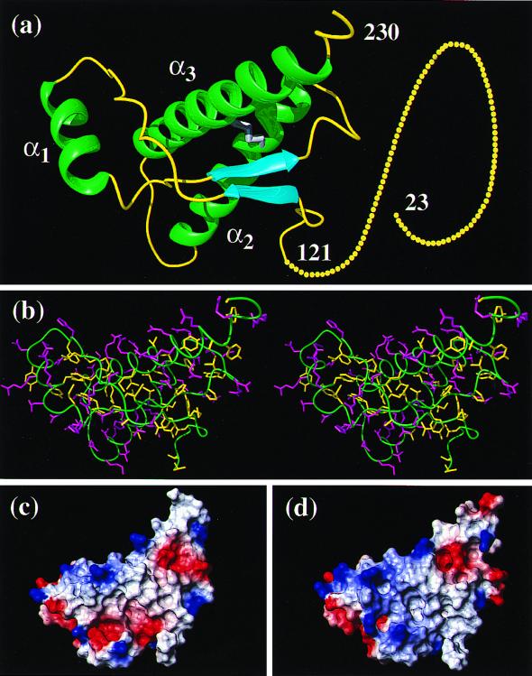
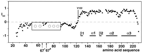
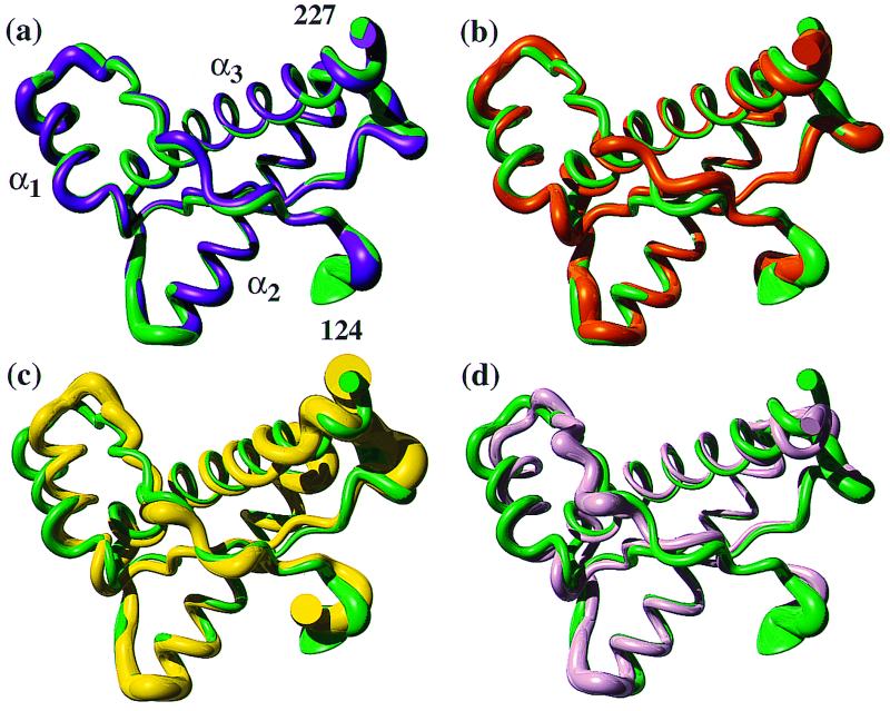
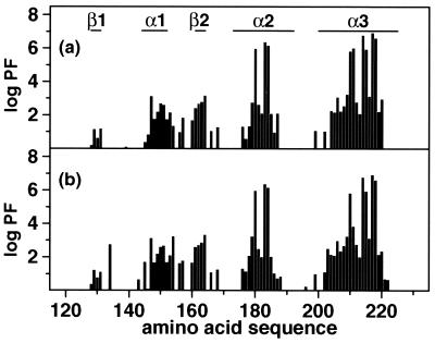
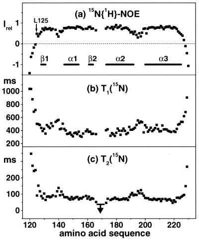
The NMR structures of the recombinant 217-residue polypeptide chain of the mature bovine prion protein, bPrP(23-230), and a C-terminal fragment, bPrP(121-230), include a globular domain extending from residue 125 to residue 227, a short flexible chain end of residues 228-230, and an N-terminal flexibly disordered "tail" comprising 108 residues for the intact protein and 4 residues for bPrP(121-230), respectively. The globular domain contains three alpha-helices comprising the residues 144-154, 173-194, and 200-226, and a short antiparallel beta-sheet comprising the residues 128-131 and 161-164. The best-defined parts of the globular domain are the central portions of the helices 2 and 3, which are linked by the only disulfide bond in bPrP. Significantly increased disorder and mobility is observed for helix 1, the loop 166-172 leading from the beta-strand 2 to helix 2, the end of helix 2 and the following loop, and the last turn of helix 3. Although there are characteristic local differences relative to the conformations of the murine and Syrian hamster prion proteins, the bPrP structure is essentially identical to that of the human prion protein. On the other hand, there are differences between bovine and human PrP in the surface distribution of electrostatic charges, which then appears to be the principal structural feature of the "healthy" PrP form that might affect the stringency of the species barrier for transmission of prion diseases between humans and cattle.


Figures

Figure 1
Figure 1
(a) Cartoon of the three-dimensional structure of the intact bPrP(23–230). Helices are green, β-strands are cyan, segments with nonregular secondary structure within the C-terminal domain are yellow, and the flexibly disordered “tail” of residues 23–121 is represented by 108 yellow dots, each of which represents a residue of the tail (the numeration for hPrP is used, and the insertions and deletions are placed according to the alignment in ref. 23). (b) Stereo-view of an all-heavy atom presentation of the globular domain in bPrP(23–230), with residues 121–230, in the same orientation as in a. The backbone is shown as a green spline function through the Cα positions, hydrophobic side chains are yellow, and polar and charged side chains are violet. (c and d) Surface views of the globular domains of bPrP and hPrP, respectively. The orientation of the molecule is slightly changed relative to a, so that the residue 186 is approximately in the center. The electrostatic surface potential is indicated in red (negative charge), white (neutral), and blue (positive charge). The figures were prepared with the program molmol (42).
Figure 2
Figure 2
Steady-state 15N{1H}-NOEs of the backbone amide groups measured in a 1 mM solution of bPrP(23–230) in 90% H2O/10% D2O containing 10 mM sodium acetate at pH 4.5 and 20°C. In the box enclosing positions 51–91 the pattern of NOE values is indicated for the octapeptide repeat that is inserted between the positions 67 and 68 (hPrP numbering, see ref. 23), where the open rectangle indicates that only one value was measured for two Gly residues. The circles indicate that identical patterns prevail for the other five repeats (see text). The regular secondary structures in the C-terminal globular domain are indicated at the bottom, and the first residue with a positive NOE value, Val-122, is identified.
Figure 3
Figure 3
(a) Superposition of the mean NMR structures of the polypeptide segment 124–227 in bPrP(23–230) (violet) and bPrP(121–230) (green). A spline function was drawn through the Cα positions. The variable radius of the cylindrical rods is proportional to the mean global backbone displacement per residue (43), as evaluated after superposition for best fit of the atoms N, Cα, and C′ of the residues 125–227 in the two bundles of 20 energy-minimized conformers used to represent the solution structures (29). (b–d) Superposition of the segment 125–227 in bPrP(121–230) (green) with the corresponding residues in hPrP(121–230) (b; orange), mPrP(121–231) (c; yellow), and shPrP(90–231) (d; pink), respectively.
Figure 4
Figure 4
Logarithmic plots of amide proton exchange protection factors (PF). (a) bPrP(23–230). (b) bPrP(121–230). Hydrogen–deuterium exchange was measured at 20°C in 99.9% D2O containing 10 mM sodium acetate at pH 4.5. The locations of the regular secondary structure elements are given in a.
Figure 5
Figure 5
Data characterizing the internal mobility of the globular domain of bPrP. (a) Steady-state 15N{1H}-NOEs of bPrP(121–230), where Leu-125 is the first residue with a positive NOE. (b) Longitudinal 15N spin-relaxation times, T1(15N). (c) Transverse 15N spin-relaxation times, T2(15N). The arrow indicates an upper limit for T2(15N) of the residues 166–172. The locations of the regular secondary structure elements are indicated in a.

References

    1. Will R G, Ironside J W, Zeidler M, Cousens S N, Estibeiro K, Alperovitch A, Poser S, Pocchiari M, Hofman A, Smith P G. Lancet. 1996;347:921–925. - PubMed
    1. Collinge J, Palmer M S, Sidle K C L, Hill A F, Gowland I, Meads J, Asante E, Bradley R, Lawrence J D, Lantos P. Nature (London) 1995;378:779–783. - PubMed
    1. Collinge J, Sidle K C L, Meads J, Ironside J, Hill A F. Nature (London) 1996;383:685–690. - PubMed
    1. Bruce M E, Will R G, Ironside J W, McConnell I, Drummond D, Suttie A, McCardle L, Chree A, Hope J, Birkett C, et al. Nature (London) 1997;389:498–501. - PubMed
    1. Hill A F, Desbruslais M, Joiner S, Sidle K C L, Gowland I, Collinge J, Doey L J, Lantos P. Nature (London) 1997;389:448–450. - PubMed

Publication types

LinkOut - more resources