. 2003 Feb 4;100(3):868-73.
doi: 10.1073/pnas.0231020100. Epub 2003 Jan 21.

Structure of the large FK506-binding protein FKBP51, an Hsp90-binding protein and a component of steroid receptor complexes

Affiliations

Structure of the large FK506-binding protein FKBP51, an Hsp90-binding protein and a component of steroid receptor complexes

Cindy R Sinars et al. Proc Natl Acad Sci U S A. .

Abstract

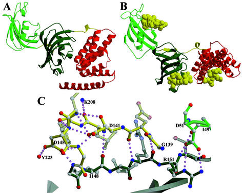
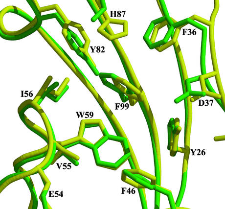
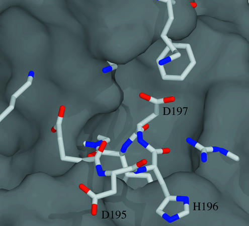
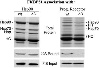
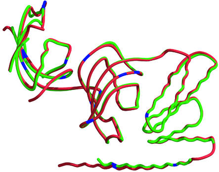
The ability to bind immunosuppressive drugs such as cyclosporin and FK506 defines the immunophilin family of proteins, and the FK506-binding proteins form the FKBP subfamily of immunophilins. Some FKBPs, notably FKBP12 (the 12-kDa FK506-binding protein), have defined roles in regulating ion channels or cell signaling, and well established structures. Other FKBPs, especially the larger ones, participate in important biological processes, but their exact roles and the structural bases for these roles are poorly defined. FKBP51 (the 51-kDa FKBP) associates with heat shock protein 90 (Hsp90) and appears in functionally mature steroid receptor complexes. In New World monkeys, FKBP51 has been implicated in cortisol resistance. We report here the x-ray structures of human FKBP51, to 2.7 A, and squirrel monkey FKBP51, to 2.8 A, by using multiwavelength anomalous dispersion phasing. FKBP51 is composed of three domains: two consecutive FKBP domains and a three-unit repeat of the TPR (tetratricopeptide repeat) domain. This structure of a multi-FKBP domain protein clarifies the arrangement of these domains and their possible interactions with other proteins. The two FKBP domains differ by an insertion in the second that affects the formation of the progesterone receptor complex.


Figures

Figure 1
Figure 1
Crystal structure of FKBP51. (A) The overall structure is as follows: light green is the FK1 domain, dark green is the FK2 domain, red is the TPR domain, and yellow regions are the linkers between domains. (B) In each case, coordinates of a similar domain (FKBP12, FKBP12, and the Hop TPR 2a domain) bound to a ligand [rapamycin (39), rapamycin, and Hsp90 fragment MEEVD (37), respectively] were superposed on FKBP51s domains (FK1, FK2, and TPR). The resulting ligand coordinates are shown in yellow space-filling atoms. (C) Extensive hydrogen bonding of the linker region between FK1 and FK2. The yellow bonds are the linking regions, dark-green bonds are FK2, and light-green bonds are FK1. Figs. 1–5 were rendered with molscript (34) and RASTER3D (35).
Figure 2
Figure 2
Binding pocket of FK1. FKBP12 (yellow with black labels) coordinates were superposed with FK1 of FKBP51 (green with green labels). Only one amino acid differs, at H87 FKBP51, which instead has a serine. The remaining side chains superpose well.
Figure 3
Figure 3
Binding pocket of FK2. The molecular surface of FKBP12, calculated with spock (43), was superposed on FK2 of FKBP51. The binding pocket of FKBP12 is clearly seen in the surface. The FK2 residues, shown as stick models, show how the 3-aa insertion (D195, H196, and D197) and other mutations obstruct the binding pocket.
Figure 4
Figure 4
FK2 involvement in PR association but not Hsp90 binding. Both wild-type human FKBP51 (wt) and a deletion mutant lacking D195, H196, and D197 from the FK2 domain (Δ3) were tested for association with Hsp90 (Left) or PR (Right) complexes. Hsp90 complexes were immunoprecipitated from rabbit reticulocyte lysate that had been supplemented with radiolabeled wt or Δ3, and washed beads were extracted and the proteins were separated by SDS/PAGE. (Top) An image of the Coomassie blue-stained gel with labeled bands from the predominate Hsp90-Hop-Hsp70 and the heavy chain (HC) of antibody used for immunoprecipitating complexes. (Middle and Bottom) Autoradiograms that show the levels of radiolabeled FKBP51 recovered in the gel samples (35S Bound), or representative amounts of the two FKBP forms included in the incubation mixture (35S Input), respectively. Similarly, PR complexes were assembled in reticulocyte lysate mixtures and analyzed for the association of radiolabeled FKBP51. For reference, FKBP51 comigrates with antibody heavy chains.
Figure 5
Figure 5
TPR domains. The known TPR domains were superposed onto the TPR domain of FKBP51 and color-coded according to their proteins: green, FKBP51; red, PP5; lavender, Cyp40; blue, Hop TPR1; and yellow, Hop TPR2a. The backbones of the six helices forming the TPR domain superpose well, whereas α7 continues out from the TPR domains at slightly differing angles.
Figure 6
Figure 6
Structural comparison of human and squirrel monkey FKBP51s. The smFKBP51 (red) was superposed onto huFKBP51 (green). The two proteins have very little difference in their overall structures, with the “80s” loop of the FK1 domains showing the most variability. Blue marks the residues that are different; most of these 15 mutations are conservative.

References

    1. Siekierka J J, Hung S H Y, Poe M, Lin C S, Sigal N H. Nature. 1989;341:755–757. - PubMed
    1. Harding M W, Galat A, Uehling D E, Schreiber S L. Nature. 1989;341:758–760. - PubMed
    1. Van Duyne G D, Standaert R F, Karplus P A, Schreiber S L, Clardy J. Science. 1991;252:839–842. - PubMed
    1. Liu J. Cell. 1991;66:807–815. - PubMed
    1. O'Keefe S J, Tamura J, Kincaid R F, Tocci M J, O'Neill M A. Nature. 1992;357:692–694. - PubMed

Publication types