. 2003 May;12(5):1097-108.
doi: 10.1110/ps.0241803.

Crystal structures of a pantothenate synthetase from M. tuberculosis and its complexes with substrates and a reaction intermediate

Affiliations

Crystal structures of a pantothenate synthetase from M. tuberculosis and its complexes with substrates and a reaction intermediate

Shuishu Wang et al. Protein Sci. 2003 May.

Abstract

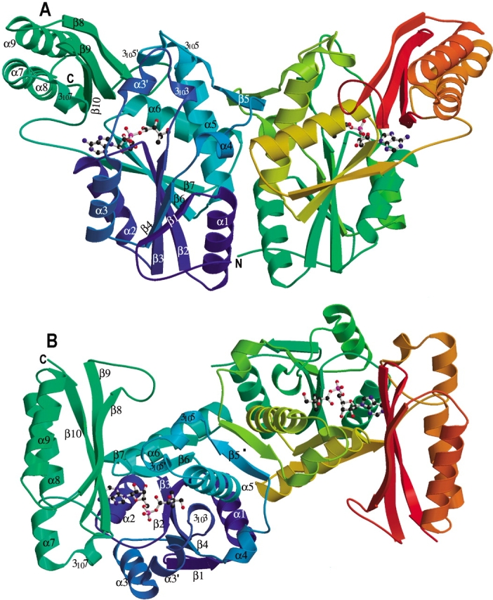
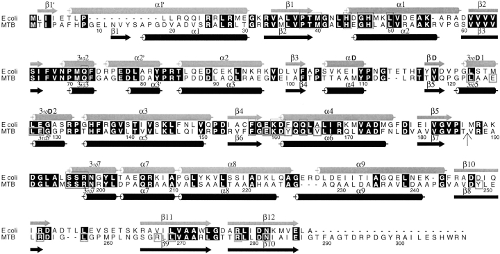
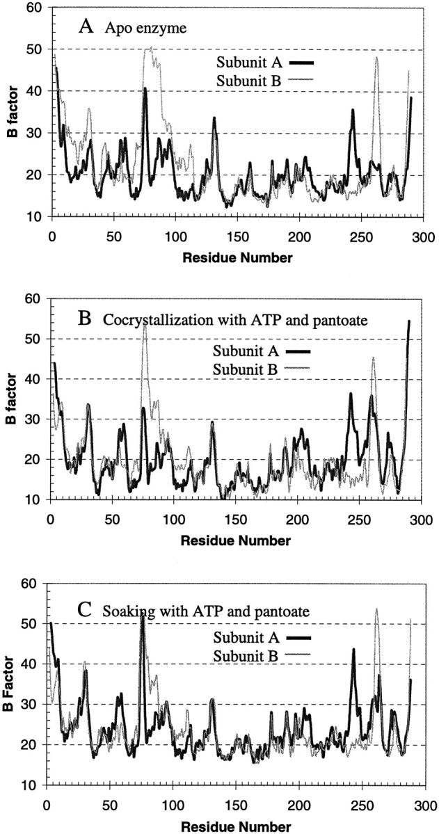
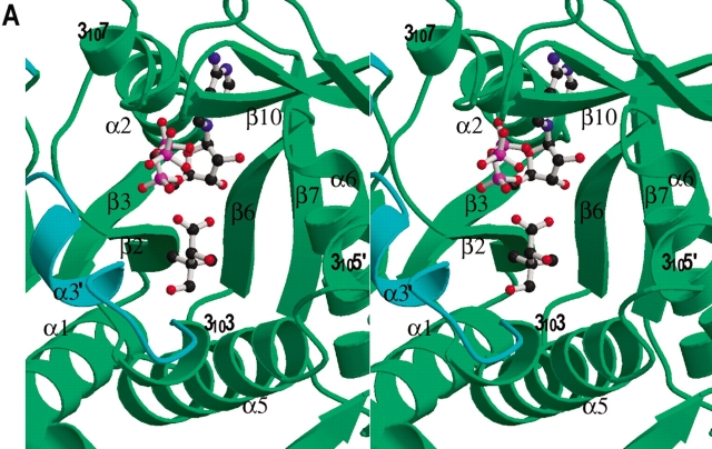
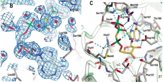
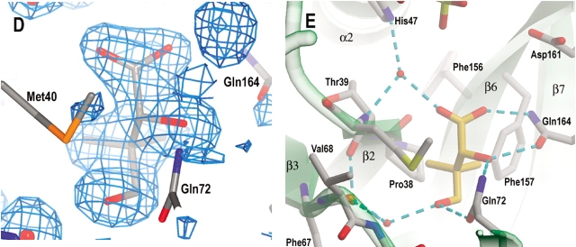
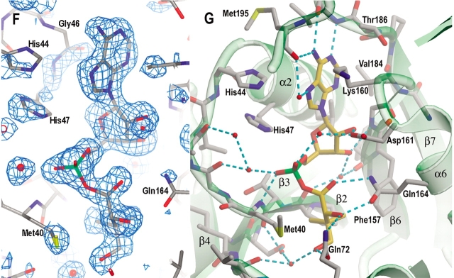
Pantothenate biosynthesis is essential for the virulence of Mycobacterium tuberculosis, and this pathway thus presents potential drug targets against tuberculosis. We determined the crystal structure of pantothenate synthetase (PS) from M. tuberculosis, and its complexes with AMPCPP, pantoate, and a reaction intermediate, pantoyl adenylate, with resolutions from 1.6 to 2 A. PS catalyzes the ATP-dependent condensation of pantoate and beta-alanine to form pantothenate. Its structure reveals a dimer, and each subunit has two domains with tight association between domains. The active-site cavity is on the N-terminal domain, partially covered by the C-terminal domain. One wall of the active site cavity is flexible, which allows the bulky AMPCPP to diffuse into the active site to nearly full occupancy when crystals are soaked in solutions containing AMPCPP. Crystal structures of the complexes with AMPCPP and pantoate indicate that the enzyme binds ATP and pantoate tightly in the active site, and brings the carboxyl oxygen of pantoate near the alpha-phosphorus atom of ATP for an in-line nucleophilic attack. When crystals were soaked with, or grown in the presence of, both ATP and pantoate, a reaction intermediate, pantoyl adenylate, is found in the active site. The flexible wall of the active site cavity becomes ordered when the intermediate is in the active site, thus protecting it from being hydrolyzed. Binding of beta-alanine can occur only after pantoyl adenylate is formed inside the active site cavity. The tight binding of the intermediate pantoyl adenylate suggests that nonreactive analogs of pantoyl adenylate may be inhibitors of the PS enzyme with high affinity and specificity.


Figures

Figure 1.
Figure 1.
Ribbon diagram of the M. tuberculosis pantothenate synthetase dimer. (A) A side view of the dimer structure showing that it resembles the shape of a butterfly. (B) An orthogonal view of (A) from top, with the twofold NCS symmetry axis (labeled with a dot) approximately perpendicular to the paper plane. Secondary structure elements for the subunit A (left) are labeled. Those for subunit B are identical except that the short helix α3′ is not present. The figure was prepared from the coordinates of the intermediate complex (data set 5), with the program Molscript (Kraulis 1991) and Raster3D (Merritt and Murphy 1994). The molecule in the active site of each subunit, shown in ball-and-stick, is the reaction intermediate, pantoyl adenylate.
Figure 2.
Figure 2.
Sequence alignment between the E. coli and M. tuberculosis PS proteins. The sequences were aligned with CLUSTALW (Thompson et al. 1994) and the figure was generated using ALSCRIPT (Barton 1993). Identical residues are highlighted in black. Helices and β-strands of the two structures are marked with cylinders and arrows, respectively. The secondary structure elements and numbering for the E. coli PS are from von Delft et al. (2001). Residues involved in binding of AMPCPP, pantoate, or pantoyl adenylate are boxed in thin lines, while those involved in interactions between N- and C-terminal domains are boxed in thick lines in the M. tuberculosis sequence. A vertical arrow marks the start of the C-terminal domain. All residues involved in binding of substrates and the reaction intermediate are conserved between two sequences, indicating that they have identical reaction mechanism. However, residues involved in domain interactions are not all conserved. See text for more details.
Figure 3.
Figure 3.
Average B factors of main chain atoms plotted against residue number. The dark thick curves are those of subunit A, and the light thin curves are those of subunit B. Subunit B of the apo enzyme (A, thin curve) has high B factors for residues from 75 to 88, with residues 76–78 totally disordered. However, these residues are ordered and have lower B factors in the crystal structure of the reaction intermediate complex from cocrystallization (B). Soaking of the apo enzyme crystals in solutions containing ATP and pantoate (C) also resulted in the disordered residues becoming ordered, and gave a profile of the B factors more similar to that of cocrystallized intermediate complex (B) than to that of the apo enzyme (A).
Figure 4.
Figure 4.
Active site cavity and the binding of AMPCPP, pantoate, and pantoyl adenylate. (A) A stereo view of the active site cavity of subunit A of the complex with both AMPCPP and pantoate. The substrates (both with partial occupancy) are shown as ball-and-stick models. The active site cavity is surrounded by β2-loop-α2, β7-loop, β6-loop-α6, 3105′-loop- α5, and β3-loop-3103–α3′-loop, and covered by 3107 and the β-sheet of C-terminal domain. Residues around helix α3′ (shown in cyan) are disordered in subunit B, which has a fully occupied AMPCPP and a glycerol molecule in the active site. (B) A section of the initial difference electron density map (Fo − Fc) in the active site of subunit B superimposed on the refined model, calculated at 1.7 Å and contoured at the 2σ level. Side chains of Lys160, Ser196, and Arg198 have moved relative to those in the apo enzyme to interact with the phosphate groups, and thus also have positive initial difference electron density. The electron density figures are prepared with PYMOL (DeLano 2002). (C) Detailed binding interactions between AMPCPP (shown with carbon atoms in gold) and protein active site residues of subunit B. The Mg2+ ion is shown as a yellow sphere, and water molecules are shown as red spheres. Hydrogen bonds between AMPCPP and protein atoms, and some water-mediated hydrogen bonds are shown as dashed lines. A glycerol molecule found next to the α-phosphate of AMPCPP, at the pantoate binding site, is also shown. (D) A section of the initial difference electron density (Fo − Fc) around the bound pantoate molecule in the active site of subunit A of the pantoate–β-alanine complex (data set 7) shows that pantoate is very well ordered with full occupancy. The nearby residues did not move relative to those of the apo enzyme, and therefore did not have initial difference density. The electron density was calculated at 1.7 Å and contoured at 2σ. (E) The pantoate molecule (shown in gold for the carbon atoms) is tightly bound and fits snugly in its binding site. Two glutamine side chains form hydrogen bonds to the hydroxyl groups and one carboxyl oxygen of the pantoate. The two methyl groups and the hydrophobic side of pantoate interact with the side chains of Pro38, Met40, and Phe157. (F) A section of the initial difference electron density (Fo − Fc) around the bound pantoyl adenylate molecule in the active site of subunit B of the intermediate complex (data set 6) shows that intermediate is very well ordered with full occupancy. The electron density was calculated at 1.7 Å and contoured at 2σ. (G) The pantoyl adenylate molecule (shown with carbon atoms in gold) is tightly bound and fits snugly in the active site cavity. The adenosine and pantoyl groups are at identical positions as those in the AMPCPP complex and pantoate complex, respectively, and have identical interactions with the active site residues. However, the α-phosphate moved down to have a covalent bond to the pantoate, which allows the phosphate group to have a hydrogen bond to the amide nitrogen of Met40.
Figure 4.
Figure 4.
Active site cavity and the binding of AMPCPP, pantoate, and pantoyl adenylate. (A) A stereo view of the active site cavity of subunit A of the complex with both AMPCPP and pantoate. The substrates (both with partial occupancy) are shown as ball-and-stick models. The active site cavity is surrounded by β2-loop-α2, β7-loop, β6-loop-α6, 3105′-loop- α5, and β3-loop-3103–α3′-loop, and covered by 3107 and the β-sheet of C-terminal domain. Residues around helix α3′ (shown in cyan) are disordered in subunit B, which has a fully occupied AMPCPP and a glycerol molecule in the active site. (B) A section of the initial difference electron density map (Fo − Fc) in the active site of subunit B superimposed on the refined model, calculated at 1.7 Å and contoured at the 2σ level. Side chains of Lys160, Ser196, and Arg198 have moved relative to those in the apo enzyme to interact with the phosphate groups, and thus also have positive initial difference electron density. The electron density figures are prepared with PYMOL (DeLano 2002). (C) Detailed binding interactions between AMPCPP (shown with carbon atoms in gold) and protein active site residues of subunit B. The Mg2+ ion is shown as a yellow sphere, and water molecules are shown as red spheres. Hydrogen bonds between AMPCPP and protein atoms, and some water-mediated hydrogen bonds are shown as dashed lines. A glycerol molecule found next to the α-phosphate of AMPCPP, at the pantoate binding site, is also shown. (D) A section of the initial difference electron density (Fo − Fc) around the bound pantoate molecule in the active site of subunit A of the pantoate–β-alanine complex (data set 7) shows that pantoate is very well ordered with full occupancy. The nearby residues did not move relative to those of the apo enzyme, and therefore did not have initial difference density. The electron density was calculated at 1.7 Å and contoured at 2σ. (E) The pantoate molecule (shown in gold for the carbon atoms) is tightly bound and fits snugly in its binding site. Two glutamine side chains form hydrogen bonds to the hydroxyl groups and one carboxyl oxygen of the pantoate. The two methyl groups and the hydrophobic side of pantoate interact with the side chains of Pro38, Met40, and Phe157. (F) A section of the initial difference electron density (Fo − Fc) around the bound pantoyl adenylate molecule in the active site of subunit B of the intermediate complex (data set 6) shows that intermediate is very well ordered with full occupancy. The electron density was calculated at 1.7 Å and contoured at 2σ. (G) The pantoyl adenylate molecule (shown with carbon atoms in gold) is tightly bound and fits snugly in the active site cavity. The adenosine and pantoyl groups are at identical positions as those in the AMPCPP complex and pantoate complex, respectively, and have identical interactions with the active site residues. However, the α-phosphate moved down to have a covalent bond to the pantoate, which allows the phosphate group to have a hydrogen bond to the amide nitrogen of Met40.

References

    1. Barker, D.G. and Winter, G. 1982. Conserved cysteine and histidine residues in the structures of the tyrosyl and methionyl-tRNA synthetases. FEBS Lett. 145 191–193. - PubMed
    1. Barton, G.J. 1993. ALSCRIPT: A tool to format multiple sequence alignments. Protein Eng. 6 37–40. - PubMed
    1. Brunger, A.T., Adams, P.D., Clore, G.M., DeLano, W.L., Gros, P., Grosse-Kunstleve, R.W., Jiang, J.-S., Kuszewski, J., Nilges, N., Pannu, N.S., et al. 1998. Crystallography & NMR system: A new software suite for macromolecular structure determination. Acta Crystallogr. D54 905–921. - PubMed
    1. CCP4 (Collaborative Computational Project Number 4). 1994. The CCP4 suite: Programs for protein crystallography. Acta Crystallogr. D50 760–763. - PubMed
    1. Cowtan, K. 1994. DM: An automated procedure for phase improvement by density modification. Joint CCP4 ESF-EACBM Newslett. Protein Crystallogr. 31 34–38.

Publication types

LinkOut - more resources