. 2007 Oct;5(10):e257.
doi: 10.1371/journal.pbio.0050257.

Nanosensor detection of an immunoregulatory tryptophan influx/kynurenine efflux cycle

Affiliations

Nanosensor detection of an immunoregulatory tryptophan influx/kynurenine efflux cycle

Thijs Kaper et al. PLoS Biol. 2007 Oct.

Abstract

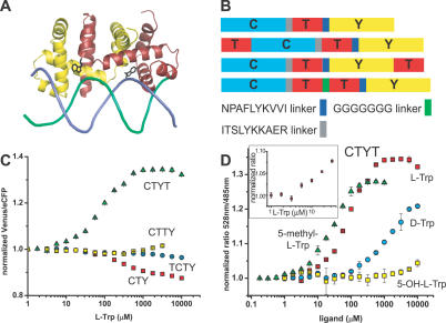
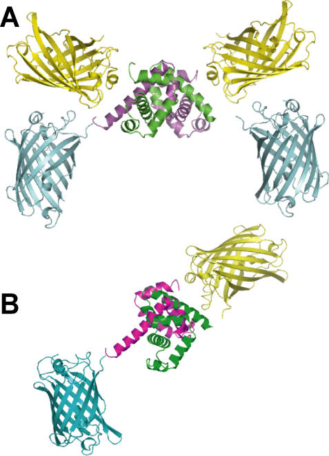
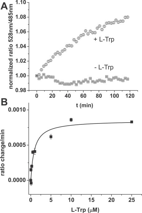
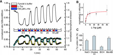
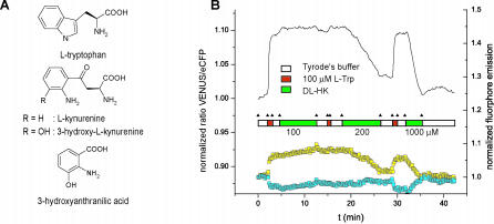
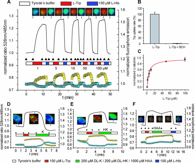
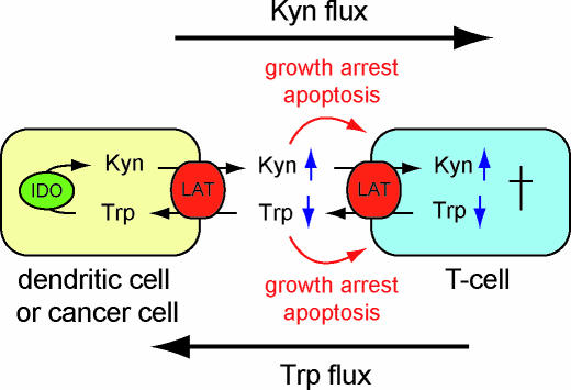
Mammalian cells rely on cellular uptake of the essential amino acid tryptophan. Tryptophan sequestration by up-regulation of the key enzyme for tryptophan degradation, indoleamine 2,3-dioxygenase (IDO), e.g., in cancer and inflammation, is thought to suppress the immune response via T cell starvation. Additionally, the excreted tryptophan catabolites (kynurenines) induce apoptosis of lymphocytes. Whereas tryptophan transport systems have been identified, the molecular nature of kynurenine export remains unknown. To measure cytosolic tryptophan steady-state levels and flux in real time, we developed genetically encoded fluorescence resonance energy transfer nanosensors (FLIPW). The transport properties detected by FLIPW in KB cells, a human oral cancer cell line, and COS-7 cells implicate LAT1, a transporter that is present in proliferative tissues like cancer, in tryptophan uptake. Importantly, we found that this transport system mediates tryptophan/kynurenine exchange. The tryptophan influx/kynurenine efflux cycle couples tryptophan starvation to elevation of kynurenine serum levels, providing a two-pronged induction of apoptosis in neighboring cells. The strict coupling protects cells that overproduce IDO from kynurenine accumulation. Consequently, this mechanism may contribute to immunosuppression involved in autoimmunity and tumor immune escape.


Conflict of interest statement

Competing interests. The authors have declared that no competing interests exist.

Figures

Figure 1
Figure 1. Tryptophan Sensors Based on the E. coli Tryptophan Repressor TrpR
(A) TrpR dimer (yellow, red) in complex with l-tryptophan (black) bound to the trp operator (green, blue) ([28]). (B) FLIPW variants. (C) Normalized FRET ratio change of FLIPW-CTY (red squares), FLIPW-TCTY (cyan circles), FLIPW-CTYT (green triangles), and FLIPW-CTTY (yellow squares) in the presence of l-tryptophan. (D) Normalized FRET ratio change of FLIPW-CTYT in presence of l-tryptophan (red squares), d-tryptophan (cyan circles), 5-hydroxy-l-tryptophan (yellow squares), and 5-methyl-l-tryptophan (green triangles). Inset in (D) highlights the response of FLIPW-CTYT to tryptophan in the low micromolar range. Ratio is defined as fluorescence intensity quotient of emission at 528 nm/485 nm.
Figure 2
Figure 2. Structural Models of FLIPW-CTY and FLIPW-CTYT Tryptophan Sensors
TrpR: green, magenta, eCFP: blue, and Venus: yellow. (A) Dimer of two FLIPW-CTY chains resulting in a TrpR dimer that can bind tryptophan. (B) FLIPW-CTYT monomer. The relative positions of the fluorophore proteins with respect to the TrpR dimer are speculative.
Figure 3
Figure 3. Uptake of Tryptophan by COS-7 Cell Cultures in 96-Well Microplates Monitored with FLIPW-CTYT
(A) FRET ratio change of cell cultures in presence of Tyrode's buffer (squares) and 100 μM l-tryptophan in Tyrode's buffer (circles). Data correspond to means ± SE (n = 12). (B) Velocity of intracellular FLIPW-CTYT response versus external tryptophan concentration fitted with the Michaelis-Menten equation. Cells were incubated with 0.05, 0.1, 0.25, 0.5, 1, 5, 10, and 25 μM l- tryptophan. Data correspond to means ± SE (n = 6). Ratios are defined as fluorescence intensity quotient of emission at 528 nm/485 nm.
Figure 4
Figure 4. Imaging of Intracellular Tryptophan Levels with FLIPW-CTYT in COS-7 Cells
(A) Perfusion of COS-7 cells with various concentrations l-tryptophan (L-Trp) and 100 μM l-histidine (L-His) in Tyrode's buffer. According to the FRET theory, an increase in Venus signal is accompanied by a decrease in eCFP signal. Ratio defined as fluorescence intensity quotient obtained with emission filters 535/40 nm over 480/30 nm. (B) Velocity of intracellular FLIPW-CTYT response versus external tryptophan concentrations used in (A) fitted with the Michaelis-Menten equation. Error bars indicate standard deviation. (C) Effect of Na+-ions and inhibitors on the uptake rate of 100 μM l-tryptophan. Error bars indicate standard deviation.
Figure 5
Figure 5. Imaging of Tryptophan-Kynurenine Exchange with FLIPW-CTYT in COS-7 Cells
(A) Molecular structures of l-tryptophan, 3-hydroxy-l-kynurenine, and 3-hydroxy-anthranilic acid. (B) Perfusion of COS-7 cells with 100 μM l-tryptophan (L-Trp), 100 μM l-histidine (L-His), and 200–1,000 μM 3-hydroxy-dl-kynurenine (DL-HK) in Tyrode's buffer. According to the FRET theory, an increase in Venus signal is accompanied by a decrease in eCFP signal. Ratio defined as fluorescence intensity quotient obtained with emission filters 535/40 nm over 480/30 nm.
Figure 6
Figure 6. Imaging of Tryptophan Uptake and Exchange with FLIPW-CTYT in Human Oral Carcinoma KB Cells
(A) Perfusion of KB cells with various concentrations l-tryptophan (L-Trp) and 100 μM l-histidine (L-His) in Tyrode's buffer. According to the FRET theory, an increase in Venus signal is accompanied by a decrease in eCFP signal. Ratio defined as fluorescence intensity quotient obtained with emission filters 535/40 nm over 480/30 nm. (B) Effect of 5 mM BCH inhibitor on the uptake rate of 100 μM l-tryptophan. Error bars indicate standard deviation. (C) Velocity of intracellular FLIPW-CTYT response versus external tryptophan concentrations used in (A) fitted with the Michaelis-Menten equation. Error bars indicate standard deviation. (D–F) Perfusion of KB cells with 100 μM l-tryptophan (L-Trp), 100 μM l-histidine (L-His), and 200 μM dl-kynurenine (DL-K) (D), 200 μM 3-hydroxy-dl-kynurenine (DL-HK) (E), and 1,000 μM 3-hydroxy-anthranilic acid (HAA) (F) in Tyrode's buffer. Cell images in panels (A) and (D–F) are pseudocolored to demonstrate the ratio change.
Figure 7
Figure 7. Double Trouble for T Cells
Proposed model for the contribution of LAT-mediated tryptophan-kynurenine exchange to inflammation and immune escape. IDO- and LAT-expressing cell types such as dendritic cells or cancer cells replace tryptophan in the local environment with kynurenines. On the one hand, T cells, expressing LAT transporters for the transport of tryptophan [50], are drained of tryptophan; on the other hand, kynurenine levels increase. Both result in T cell growth arrest and apoptosis [39,40,51].

References

    1. Broer S, Cavanaugh JA, Rasko JEJ. Neutral amino acid transport in epithelial cells and its malfunction in Hartnup disorder. Biochem Soc Trans. 2005;33:233–236. - PubMed
    1. Feliubadalo L, Font M, Purroy J, Rousaud F, Estivill X, et al. Non-type I cystinuria caused by mutations in SLC7A9, encoding a subunit (bo,+AT) of rBAT. Nat Genet. 1999;23:52–57. - PubMed
    1. Kim DK, Kanai Y, Matsuo H, Kim JY, Chairoungdua A, et al. The human T-type amino acid transporter-1: Characterization, gene organization, and chromosomal location. Genomics. 2002;79:95–103. - PubMed
    1. Torrents D, Estevez R, Pineda M, Fernandez E, Lloberas J, et al. Identification and characterization of a membrane protein (y+L amino acid transporter-1) that associates with 4F2hc to encode the amino acid transport activity y+L. A candidate gene for lysinuric protein intolerance. J Biol Chem. 1998;273:32437–32445. - PubMed
    1. Pfeiffer R, Rossier G, Spindler B, Meier C, Kuhn L, et al. Amino acid transport of y+L-type by heterodimers of 4F2hc/CD98 and members of the glycoprotein-associated amino acid transporter family. EMBO J. 1999;18:49–57. - PMC - PubMed

Publication types