. 2019 Sep 27;294(39):14454-14466.
doi: 10.1074/jbc.RA119.008231. Epub 2019 Jul 23.

ANKRD9 is a metabolically-controlled regulator of IMPDH2 abundance and macro-assembly

Affiliations

ANKRD9 is a metabolically-controlled regulator of IMPDH2 abundance and macro-assembly

Dawn Hayward et al. J Biol Chem. .

Abstract

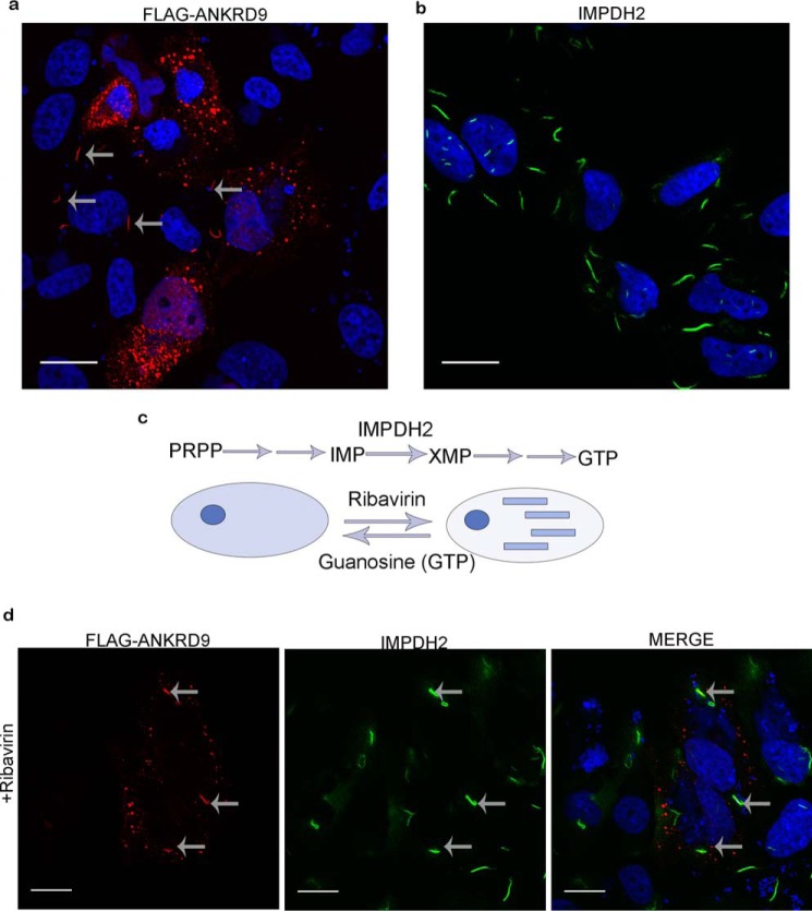
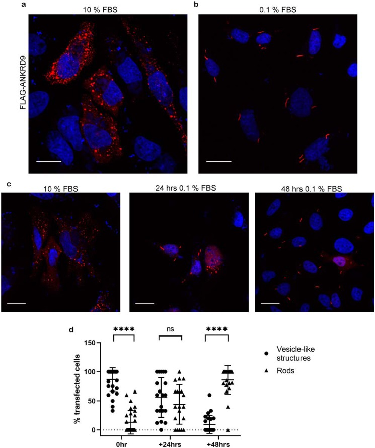
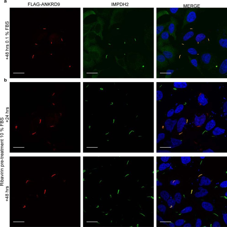
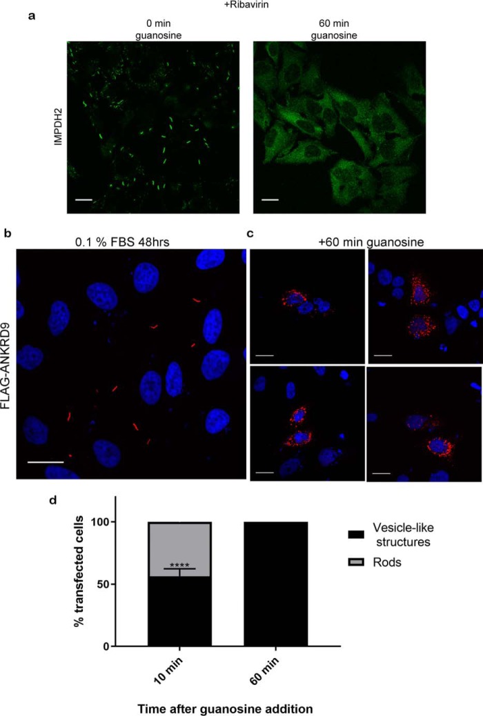
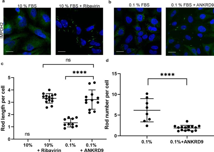
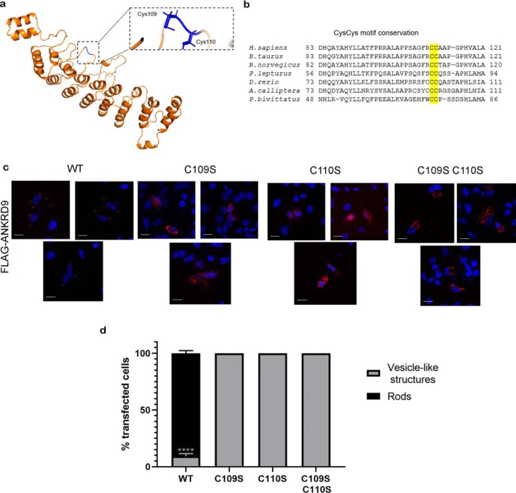
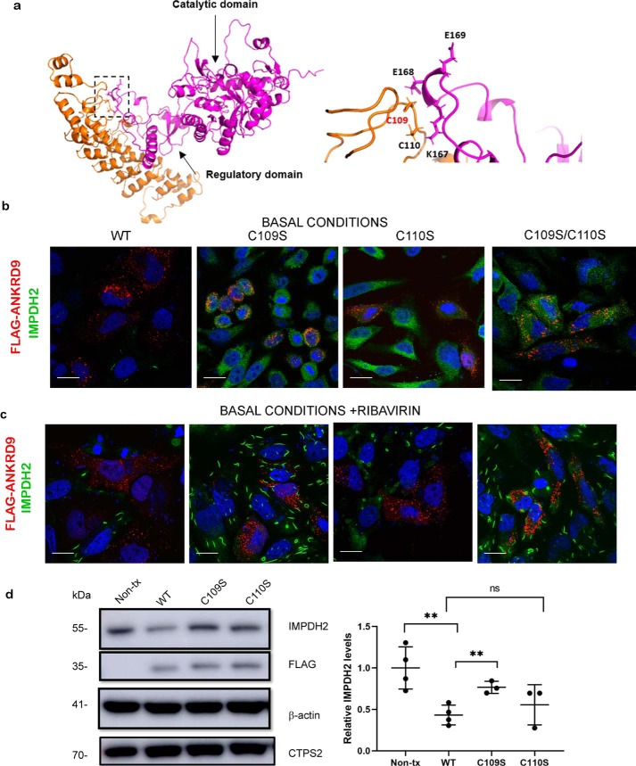
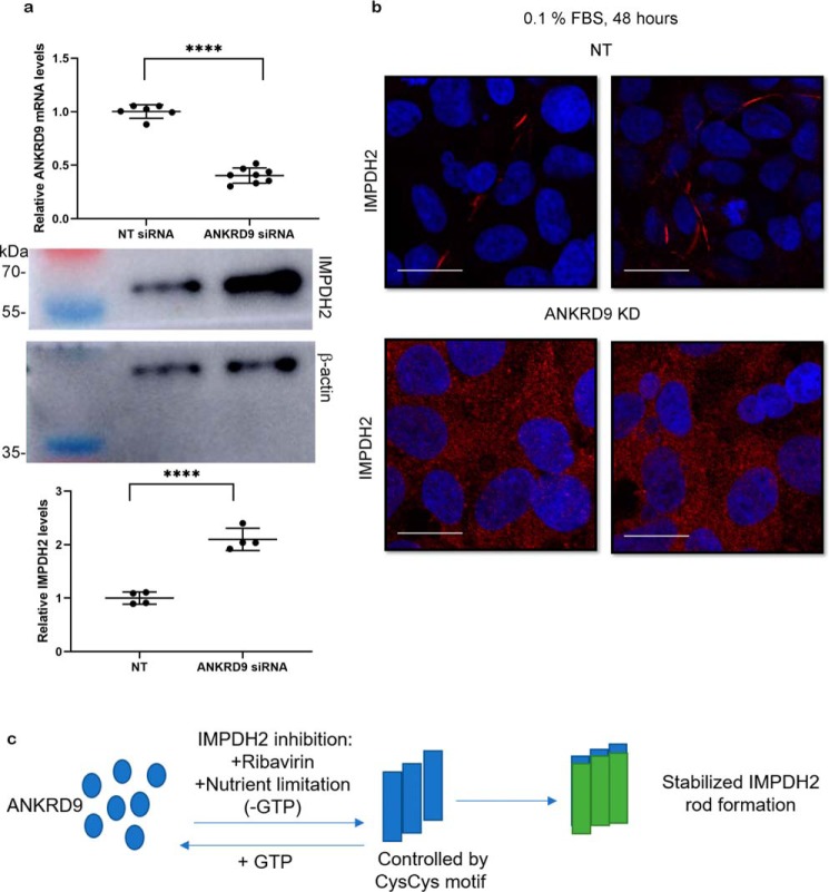
Members of a large family of Ankyrin Repeat Domain (ANKRD) proteins regulate numerous cellular processes by binding to specific protein targets and modulating their activity, stability, and other properties. The same ANKRD protein may interact with different targets and regulate distinct cellular pathways. The mechanisms responsible for switches in the ANKRDs' behavior are often unknown. We show that cells' metabolic state can markedly alter interactions of an ANKRD protein with its target and the functional outcomes of this interaction. ANKRD9 facilitates degradation of inosine monophosphate dehydrogenase 2 (IMPDH2), the rate-limiting enzyme in GTP biosynthesis. Under basal conditions ANKRD9 is largely segregated from the cytosolic IMPDH2 in vesicle-like structures. Upon nutrient limitation, ANKRD9 loses its vesicular pattern and assembles with IMPDH2 into rodlike filaments, in which IMPDH2 is stable. Inhibition of IMPDH2 activity with ribavirin favors ANKRD9 binding to IMPDH2 rods. The formation of ANKRD9/IMPDH2 rods is reversed by guanosine, which restores ANKRD9 associations with the vesicle-like structures. The conserved Cys109Cys110 motif in ANKRD9 is required for the vesicle-to-rods transition as well as binding and regulation of IMPDH2. Oppositely to overexpression, ANKRD9 knockdown increases IMPDH2 levels and prevents formation of IMPDH2 rods upon nutrient limitation. Taken together, the results suggest that a guanosine-dependent metabolic switch determines the mode of ANKRD9 action toward IMPDH2.

Keywords: ANKRD9; GTP; IMPDH2; ankyrin; ankyrin repeat domain; cell metabolism; complex; cytoophidia; nucleotide; protein-protein interaction; rod.


Conflict of interest statement

The authors declare that they have no conflicts of interest with the contents of this article

Figures

Figure 1.
Figure 1.
ANKRD9 adopts two distinct forms. a, HeLa cells were transfected with FLAG-ANKRD9 expressing plasmid and immunostained for the FLAG-tag. FLAG-ANKRD9 (red) shows a vesicle-like and rodlike pattern (gray arrows); cell nuclei are visualized with DAPI (blue); n = 3 of independent experiments. b, IMPDH2 forms rods when inhibited with ribavirin. HeLa cells were treated with 10 mm ribavirin for 1 h then stained for IMPDH2 (green) and DAPI (blue); n = 2. c, IMPDH2 mediates the rate-limiting step (conversion of inosine monophosphate (IMP) to xanthine monophosphate (XMP)) in a pathway beginning with phosphoribosyl pyrophosphate (PRPP) and eventually leading to GTP. IMPDH2 transitions from a diffuse cystolic form (left) to rods (right) when inhibited with ribavirin; this transition is reversed with guanosine. d, HeLa cells were treated with 10 μm ribavirin for 1 h then transfected with FLAG-ANKRD9 and immunostained for FLAG (red) and IMPDH2 (green). Gray arrows point to rods containing both IMPDH2 and ANKRD9. Scale bar, 20 μm.
Figure 2.
Figure 2.
ANKRD9 forms rods upon nutrient depletion. a, HeLa cells were transfected with FLAG-ANKRD9 (red) as in Fig. 1a and incubated in the presence of 10% FBS. n = 3 independent experiments. b, HeLa cells were transfected with FLAG-ANKRD9 and incubated in the presence of 0.1% FBS for 48 h. Rods are indicated by gray arrows; nuclei are stained with DAPI (blue). n = 3 independent experiments. c, representative images of each time point; rods appear in the majority of cells after 48 h in 0.1% FBS media. For 24 h, n = 3. Scale bar, 20 μm. d, percentage of cells containing ANKRD9 in vesicle-like structures (black filled circles) or rods (black filled triangles) after overnight transfection (control), followed by 24 or 48 h in starvation (0.1% FBS) media. The total number of analyzed cells is as follows: n = 137 cells (control, 0 h of 0.1% FBS), n = 149 cells (24 h of 0.1% FBS), n = 131 cells (48 h of 0.1% FBS). Error bars represent S.D. Each individual point represents at least 5 cells. ****, p value < 0.0001., unpaired t test; ns, non-significant.
Figure 3.
Figure 3.
Formation of ANRKD9 rods is caused by depletion of IMPDH2 metabolites. a, HeLa cells were transfected with FLAG-ANKRD9, placed in 0.1% FBS for 48 h and immunostained for FLAG and IMPDH2. n = 3 independent experiments. b, HeLa cells were treated with 10 μm ribavirin for 1 h then transfected with FLAG-ANRKD9 and kept in 10% FBS for 24 and 48 h. Cells were immunostained with FLAG (red) and IMPDH2 (green). Two independent experiments. Merged images show overlap between ANKRD9 and IMPDH2 rods. Scale bar, 20 μm.
Figure 4.
Figure 4.
ANKRD9 transition to rods is reversible with guanosine addition. a, left, IMPDH2 rod formation is reversed with guanosine addition. HeLa cells were treated for 1 h with 10 μm ribavirin to induce IMPDH2 rod formation and immunostained for IMPDH2 (green); right, HeLa cells were treated with 10 μm ribavirin for 1 h to induce rods formation, then 100 μm guanosine was added for 60 min and cells were immunostained for IMPDH2. n = 3 for each condition. Scale bar, 20 μm. b, control, FLAG-ANKRD9 forms rods following incubation of transfected HeLa cells in 0.1% FBS for 48 h. c, the same treatment, followed by incubation with 100 μm guanosine for 1 h. The ANKRD9 rods disappear and ANKRD9 shows a vesicle-like pattern. n = 3. Scale bar, 20 μm. d, percentage of cells with ANKRD9 vesicle-like structures or rods at 10 and 60 min after addition of guanosine. n = 3 for each time point. Error bar represents S.D. ****, p value < 0.0001., unpaired t test. Scale bar, 20 μm.
Figure 5.
Figure 5.
Higher ANKRD9 abundance is associated with longer IMPDH2 rods. a, immunostaining of IMPDH2 in HeLa cells under basal conditions (10% FBS) shows an expected diffuse pattern (left panel). Treatment of these cells with 10 μm ribavirin for 1 h in the absence of recombinant ANKRD9 triggers formation of IMPDH2 rods (right panel). n = 3 for both conditions. Scale bar, 20 μm. b, in the absence of recombinant ANKRD9, nutrient limitation (0.1% FBS for 48 h) causes appearance of short IMPDH2 rods (left panel). Expression of recombinant FLAG-ANKRD9 significantly increases the length of IMPDH2 rods (right panel). n = 3 independent experiments. c, quantification of IMPDH2 rod length in different treatment conditions. The total number of cells analyzed is as follows: n = 150 cells (10% FBS + ribavirin), n = 73 cells (0.1% FBS), and n = 156 cells (0.1% FBS + ANKRD9 expression). Statistical analysis was done using an unpaired t test. ****, p < 0.0001. Error bars represent S.D. Each point is the average rod length of at least 5 cells. d, quantification of the number of rods per cell under nutrient limitation in the absence and presence of recombinant ANKRD9. unpaired t test. ****, p < 0.0001. Error bars represent S.D. Each point is the average rod length of at least 5 cells.
Figure 6.
Figure 6.
A conserved CysCys motif in ANKRD9 is required for transition from vesicle-like structures to rods. a, structural model of ANKRD9 with conserved Cys residues highlighted in blue; the boxed area shows the position of Cys109 and Cys110 in the loop. b, multiple sequence alignment of the Cys109Cys110-containing region illustrates conservation of this motif. c, HeLa cells were transfected with WT ANKRD9 (control) or indicated ANKRD9 mutants and placed in 0. 1% FBS-containing medium for 48 h. Unlike WT, none of the mutants formed rods under these conditions. n = 3 for each condition. Scale bar, 20 μm. d, quantification of ANKRD9 mutant rod formation where vesicle-like structures are in black and rods are in gray. Unlike WT, none of the mutants formed rods under these conditions. n = 3. ****, p < 0.0001 unpaired t test. Error bar represents S.D.
Figure 7.
Figure 7.
The Cys109Cys110 motif contributes to ANKRD9-IMPDH2 interaction. a, left, docking model of ANKRD9 and IMPDH2 (PDB ID 1NF7, see Fig. S7). IMPDH2 catalytic and regulatory domains are indicated by the arrows; the predicted interaction site is indicated by the box. Right, the magnified view of the predicted interaction site between ANKRD9 (orange) and IMPDH2 (purple). The CysCys motif in ANKRD9 interacts with Lys167, Glu168 and Glu169 of IMPDH2. b, Cys mutants do not facilitate IMPDH2 degradation. HeLa cells were transfected with WT ANKRD9 and indicated mutants under basal conditions (10% FBS) and immunostained for FLAG and IMPDH2. Cells expressing WT ANKRD9 showed significantly reduced IMPDH2 staining whereas cells with the Cys mutants did not. n = 3 independent experiments for each condition. Scale bar, 20 μm. c, Cys mutants of ANKRD9 do not form rods with IMPDH2. HeLa cells were transfected with the indicated constructs and subsequently treated with 10 μm ribavirin for 1 h prior to immunostaining for FLAG and IMPDH2. n = 3. Scale bar is 20 μm. d, left, the WT and ANKRD9 mutants were expressed in HeLa cells under basal conditions (10% FBS) and cell lysates, were separated and immunoblotted for FLAG, IMPDH2, CTPS2, and β-actin used as a loading control. WT ANKRD9 but not the C109S mutant significantly decreases IMPDH2 abundance, the C110S mutant is intermediate between the two and similar to WT. Right, densitometry of IMPDH2 intensity after WT ANKRD9 or mutant expression in HeLa cells. n = 3. ****, p value < 0.0001., unpaired t test.
Figure 8.
Figure 8.
ANKRD9 knockdown increases IMPDH2 expression and reduces rod formation under nutrient limiting conditions. a, top, HEK293A cells were transfected with nontargeted (NT) siRNA and ANKRD9 siRNA, and ANKRD9 mRNA levels were quantified. n = 6 for NT siRNA and n = 8 for ANKRD9 siRNA; Bottom: IMPDH2 levels in corresponding cells were analyzed by Western blotting with β-actin used as a loading control (lower panels). n = 3 for each condition. Decrease in ANKRD9 levels is associated with higher IMPDH2 abundance. ****, p < 0.0001 (unpaired t test). Error bar represents S.D. b, ANKRD9 down-regulation diminishes IMPDH2 rod formation under nutrient limitation (0.1% FBS). HEK293A cells were transfected with the indicated siRNAs and placed in 0.1% FBS for 48 h and immunostained for IMPDH2. IMPDH2 rods were apparent in control cells; no IMPDH2 rods are visible in cells treated with ANKRD9 siRNA. n = 3 for each condition. Scale bar, 20 μm. c, cartoon showing that ANKRD9 forms rods with inhibited IMPDH2 when GTP pools are lowered and stabilizes rods.

References

    1. Islam Z., Nagampalli R. S. K., Fatima M. T., and Ashraf G. M. (2018) New paradigm in ankyrin repeats: Beyond protein-protein interaction module. Int. J. Biol. Macromol. 109, 1164–1173 10.1016/j.ijbiomac.2017.11.101 - DOI - PubMed
    1. Li J., Mahajan A., and Tsai M. D. (2006) Ankyrin repeat: A unique motif mediating protein-protein interactions. Biochemistry 45, 15168–15178 10.1021/bi062188q - DOI - PubMed
    1. Sedgwick S. G., and Smerdon S. J. (1999) The ankyrin repeat: A diversity of interactions on a common structural framework. Trends Biochem. Sci. 24, 311–316 10.1016/S0968-0004(99)01426-7 - DOI - PubMed
    1. Mosavi L. K., Minor D. L. Jr., and Peng Z.-Y. (2002) Consensus-derived structural determinants of the ankyrin repeat motif. Proc. Natl. Acad. Sci. U.S.A. 99, 16029–16034 10.1073/pnas.252537899 - DOI - PMC - PubMed
    1. Inada H., Procko E., Sotomayor M., and Gaudet R. (2012) Structural and biochemical consequences of disease-causing mutations in the ankyrin repeat domain of the human TRPV4 channel. Biochemistry 51, 6195–6206 10.1021/bi300279b - DOI - PMC - PubMed

Publication types

MeSH terms

Associated data

LinkOut - more resources