. 2015 Jul 24;290(30):18650-61.
doi: 10.1074/jbc.M114.598227. Epub 2015 May 27.

Histidine 114 Is Critical for ATP Hydrolysis by the Universally Conserved ATPase YchF

Affiliations

Histidine 114 Is Critical for ATP Hydrolysis by the Universally Conserved ATPase YchF

Kirsten S Rosler et al. J Biol Chem. .

Abstract

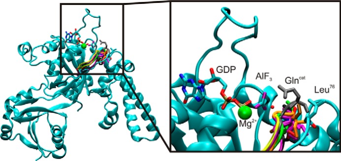
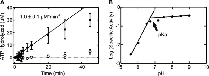
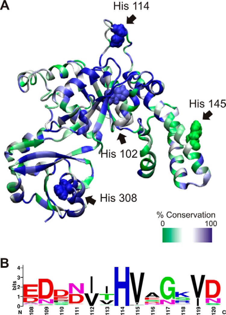
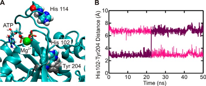
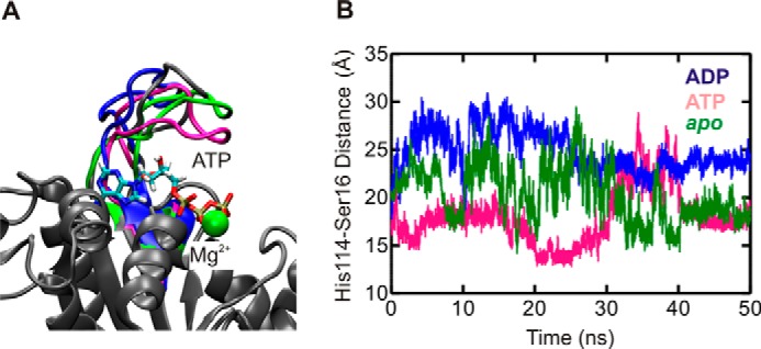
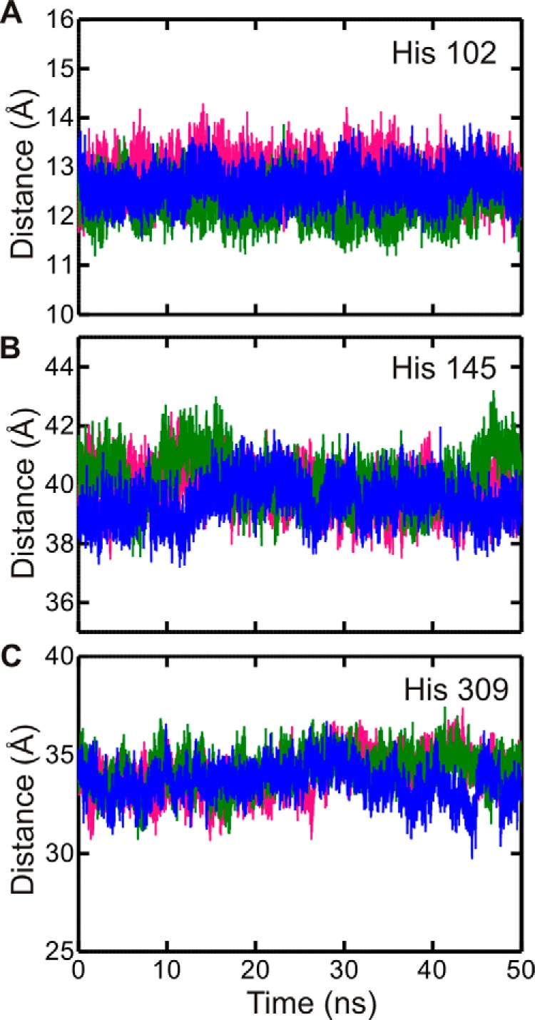
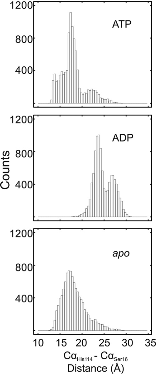
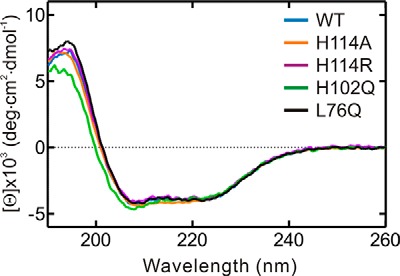
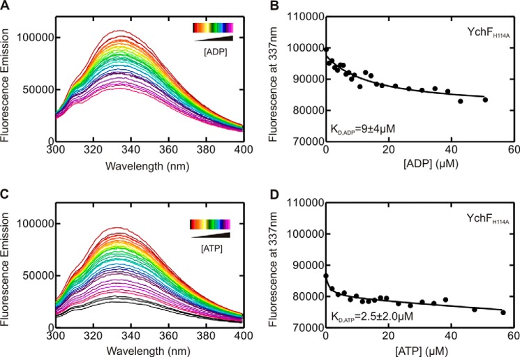
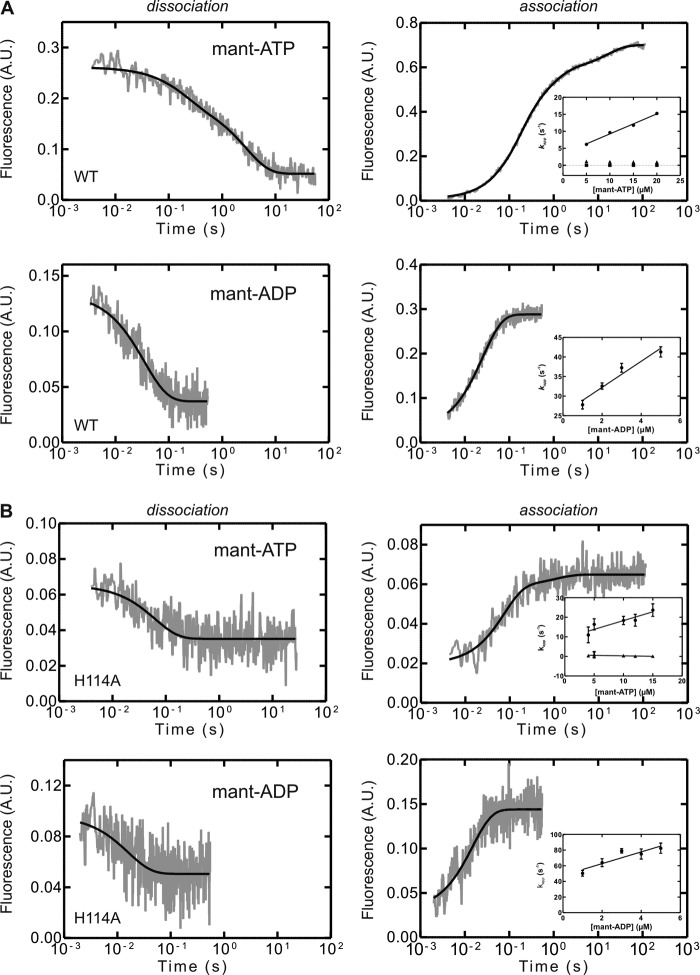
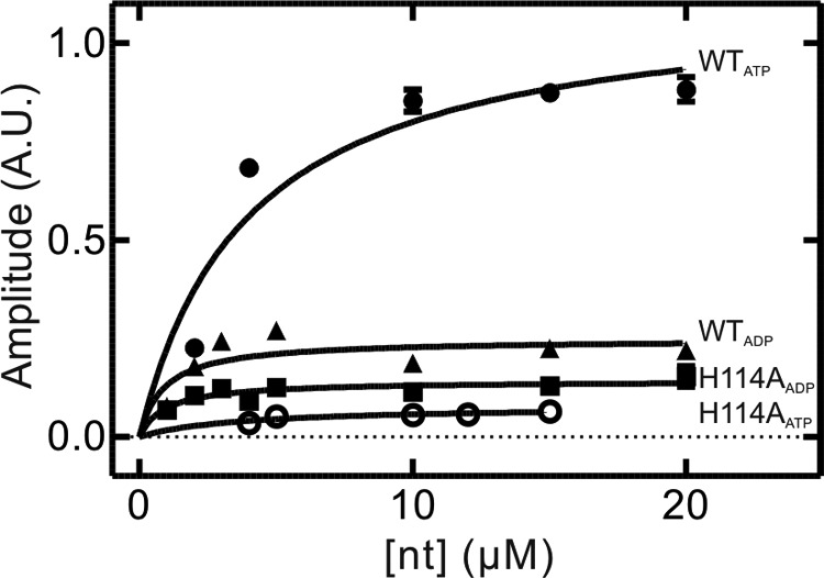
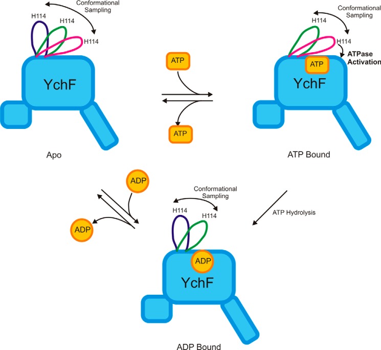
GTPases perform a wide range of functions, ranging from protein synthesis to cell signaling. Of all known GTPases, only eight are conserved across all three domains of life. YchF is one of these eight universally conserved GTPases; however, its cellular function and enzymatic properties are poorly understood. YchF differs from the classical GTPases in that it has a higher affinity for ATP than for GTP and is a functional ATPase. As a hydrophobic amino acid-substituted ATPase, YchF does not possess the canonical catalytic Gln required for nucleotide hydrolysis. To elucidate the catalytic mechanism of ATP hydrolysis by YchF, we have taken a two-pronged approach combining classical biochemical and in silico techniques. The use of molecular dynamics simulations allowed us to complement our biochemical findings with information about the structural dynamics of YchF. We have thereby identified the highly conserved His-114 as critical for the ATPase activity of YchF from Escherichia coli. His-114 is located in a flexible loop of the G-domain, which undergoes nucleotide-dependent conformational changes. The use of a catalytic His is also observed in the hydrophobic amino acid-substituted GTPase RbgA and is an identifier of the translational GTPase family.

Keywords: ATPase; HAS-ATPase; YchF; catalytic mechanism; conformational change; molecular dynamics; molecular switch; nucleotide binding; pre-steady-state kinetics; ribosome.


Figures

FIGURE 1.
FIGURE 1.
YchF is a member of the HAS-GTPase family. Shown is an alignment of the G3 motif of Ras (gray), YqeH (black), MnmE (yellow), HflX (purple), EHD2 (green), FeoB (pink), Era (red), YsxC (brown), and YlqF (orange) with YchF (cyan). Also shown is GDP·AlF3 and the attacking water molecule (red sphere in line with AlF3) from Ras·RasGap (PDB ID 1WQ1). Alignment was based on the structure of the conserved G1 motif (P-loop). Glncat, catalytic Gln.
FIGURE 2.
FIGURE 2.
pH dependence of the intrinsic ATPase activity of YchF. A, time dependence of ATP hydrolysis in the presence (●) or absence (○) of YchF. A linear function was fit to the initial phase of each reaction (first 10 min) to determine the rate of ATP hydrolysis (μm min−1). B, pH dependence of the ATPase activity of YchF (●).
FIGURE 3.
FIGURE 3.
E. coli YchF contains four histidines. A, homology model of E. coli YchF generated with SWISS-MODEL using H. influenzae YchF (PDB ID 1JAL) as template and the amino acid sequence of E. coli YchF (UniProt accession code P0ABU2), represented as a ribbon diagram. The level of conservation of each residue is depicted using a green/white/blue color scale based on the alignment of YchF from 116 bacterial species. The four histidines present in E. coli YchF are indicated and shown as a space-filling model: His-102 (76% conserved), His-114 (98%), His-145 (17%), and His-308 (100%). B, sequence logo of the His-114-containing loop (numbering according to E. coli YchF) based on the multiple-sequence alignment in A.
FIGURE 4.
FIGURE 4.
Structural dynamics of YchF during MD simulations. A, r.m.s.d. of E. coli YchF in complex with ATP (pink) or ADP (blue) or in its apo state (green) with respect to its initial conformation. r.m.s.d. values were calculated using all backbone atoms. B, Cα r.m.s.f. for E. coli YchF in complex with ATP (pink) or ADP (blue) or in its apo state (green). Colored bars at the bottom indicate the different domains of the protein: G-domain (orange), A-domain (teal), and TGS domain (gray).
FIGURE 5.
FIGURE 5.
His-102 forms a stable interaction with Tyr-204. A, YchF is shown in complex with ATP after 50 ns of MD simulations. The locations of His-102 and Tyr-204, involved in a stable interaction, are indicated. B, hydrogen bond distance between Nδ1 of His-102 and Hϵ2 (pink) or Hϵ1 (purple) of Tyr-204.
FIGURE 6.
FIGURE 6.
MD simulations reveal different conformations of the flexible loop. A, final structures of YchF·ATP·Mg2+ (pink), YchF·ADP·Mg2+ (blue), and apo-YchF (green) after 50 ns of simulation aligned with pre-simulation structures (gray). B, distance between the α-carbons of His-114 and Ser-16 over a 50-ns simulation of YchF·ATP·Mg2+ (pink), YchF·ADP·Mg2+ (blue), and apo-YchF (green).
FIGURE 7.
FIGURE 7.
His-102, His-145, and His-308 do not show nucleotide-dependent conformations. Shown is the distance between the α-carbons of His-102 (A), His-145 (B), or His-308 (C) and Ser-16 of the P-loop during a 50-ns MD simulation. YchF·ATP·Mg2+ is shown in pink, YchF·ADP·Mg2+ in blue, and apo-YchF in green.
FIGURE 8.
FIGURE 8.
Nucleotide-dependent conformations of the flexible loop in YchF. Shown are histograms of the Cα–Cα (His-114 to Ser-16) distances (bin size of 0.4 Å) measured during 50-ns MD simulations of the YchF·ATP·Mg2+, YchF·ADP·Mg2+, and apo-YchF models.
FIGURE 9.
FIGURE 9.
CD spectroscopy of WT YchF and variants. The CD spectra of WT YchF (blue), YchF(H114A) (orange), YchF(H114R) (purple), YchF(H102Q) (green), and YchF(L76Q) (black) were recorded in a 1-mm cell. deg, degrees.
FIGURE 10.
FIGURE 10.
YchF(H114A) binds adenine nucleotide di- and triphosphates. Shown are equilibrium fluorescence titrations of 1 μm YchF(H114A) with increasing concentrations of nucleotides. Tyr and Trp in YchF were excited at 280 nm, and fluorescence emission spectra were detected from 295 to 400 nm in the presence of increasing concentrations of ADP (A) or ATP (C). Fluorescence intensities measured at 337 nm are plotted against the concentrations of ADP (B) or ATP (D). KD values were obtained by fitting the data in B and D with a hyperbolic function.
FIGURE 11.
FIGURE 11.
Pre-steady-state kinetics of adenine nucleotide binding and dissociation for WT YchF and YchF(H114A). Representative time courses of the dissociation of a YchF·mant-nucleotide complex (1 μm) in the presence of excess unlabeled nucleotide (100 μm) are shown in the left panels. Representative time courses of mant-nucleotide (5 μm) association with YchF (1 μm) are shown in the right panels, with the concentration dependence of kapp on mant-nucleotide association with YchF shown in the insets. The kapp values were calculated by one-, two-, or three-exponential fitting of the time courses. A, WT YchF; B, YchF(H114A). A.U., arbitrary units.
FIGURE 12.
FIGURE 12.
KD determination using amplitude plots. Shown are amplitudes of the overall signal change observed in stopped-flow time courses plotted as a function of nucleotide concentration. Lines represent hyperbolic fits to obtain equilibrium binding constants (KD) for mant-ATP binding to WT YchF (●) and YchF(H114A) (○) and for mant-ADP binding to WT YchF (▴) and YchF(H114A) (■). A.U., arbitrary units.
FIGURE 13.
FIGURE 13.
Nucleotide binding induces conformational changes in WT YchF. Binding of ADP results in the open conformation of the flexible loop containing the conserved His-114 essential for catalysis, whereas interaction with ATP causes a conformational change in the flexible loop leading to a closed conformation and ultimately to the catalytically competent state (ATPase activation).

References

    1. Bourne H. R., Sanders D. A., McCormick F. (1990) The GTPase superfamily: a conserved switch for diverse cell functions. Nature 348, 125–132 - PubMed
    1. Verstraeten N., Fauvart M., Versées W., Michiels J. (2011) The universally conserved prokaryotic GTPases. Microbiol. Mol. Biol. Rev. 75, 507–542 - PMC - PubMed
    1. Caldon C. E., March P. E. (2003) Function of the universally conserved bacterial GTPases. Curr. Opin. Microbiol 6, 135–139 - PubMed
    1. Bourne H. R., Sanders D. A., McCormick F. (1991) The GTPase superfamily: conserved structure and molecular mechanism. Nature 349, 117–127 - PubMed
    1. Sprang S. R. (1997) G protein mechanisms: insights from structural analysis. Annu. Rev. Biochem. 66, 639–678 - PubMed

Publication types

MeSH terms

LinkOut - more resources