. 2020 Feb 21;295(8):2186-2202.
doi: 10.1074/jbc.RA119.010486. Epub 2019 Nov 26.

Moonlighting matrix metalloproteinase substrates: Enhancement of proinflammatory functions of extracellular tyrosyl-tRNA synthetase upon cleavage

Affiliations

Moonlighting matrix metalloproteinase substrates: Enhancement of proinflammatory functions of extracellular tyrosyl-tRNA synthetase upon cleavage

Parker G Jobin et al. J Biol Chem. .

Abstract

Tyrosyl-tRNA synthetase ligates tyrosine to its cognate tRNA in the cytoplasm, but it can also be secreted through a noncanonical pathway. We found that extracellular tyrosyl-tRNA synthetase (YRS) exhibited proinflammatory activities. In addition to acting as a monocyte/macrophage chemoattractant, YRS initiated signaling through Toll-like receptor 2 (TLR2) resulting in NF-κB activation and release of tumor necrosis factor α (TNFα) and multiple chemokines, including MIP-1α/β, CXCL8 (IL8), and CXCL1 (KC) from THP1 monocyte and peripheral blood mononuclear cell-derived macrophages. Furthermore, YRS up-regulated matrix metalloproteinase (MMP) activity in a TNFα-dependent manner in M0 macrophages. Because MMPs process a variety of intracellular proteins that also exhibit extracellular moonlighting functions, we profiled 10 MMPs for YRS cleavage and identified 55 cleavage sites by amino-terminal oriented mass spectrometry of substrates (ATOMS) positional proteomics and Edman degradation. Stable proteoforms resulted from cleavages near the start of the YRS C-terminal EMAPII domain. All of the MMPs tested cleaved at ADS386387LYV and VSG405406LVQ, generating 43- and 45-kDa fragments. The highest catalytic efficiency for YRS was demonstrated by MMP7, which is highly expressed by monocytes and macrophages, and by neutrophil-specific MMP8. MMP-cleaved YRS enhanced TLR2 signaling, increased TNFα secretion from macrophages, and amplified monocyte/macrophage chemotaxis compared with unprocessed YRS. The cleavage of YRS by MMP8, but not MMP7, was inhibited by tyrosine, a substrate of the YRS aminoacylation reaction. Overall, the proinflammatory activity of YRS is enhanced by MMP cleavage, which we suggest forms a feed-forward mechanism to promote inflammation.

Keywords: aminoacyl tRNA synthetase; inflammation; innate immunity; macrophage; matrix metalloproteinase (MMP); moonlighting proteins; multifunctional protein; proteolysis; toll-like receptor (TLR).


Conflict of interest statement

The authors declare that they have no conflicts of interest with the contents of this article

Figures
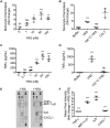
Figure 1.
Figure 1.
Increased THP1 monocyte chemotaxis, TNFα secretion, and chemokine secretion induced by YRS. Transwell chemotaxis assay of THP1 monocytes toward increasing concentrations of recombinant human YRS (A) and 50 nm YRS, 50 nm heat-denatured YRS (100 °C YRS) or buffer (B). CCL7 (50 nm) was used as a positive control. After 90 min, cells in the lower chamber were counted. Data were plotted as fold change compared with buffer (mean ± S.D., n = 3) of N = 2 independent experiments for both A and B. ELISA of TNFα protein levels in THP1 M0 macrophage-conditioned media after treatment for 3 h with increasing concentrations of YRS (C) and 50 nm YRS, heat-denatured YRS (100 °C YRS) (D), or buffer control (mean ± S.D., n = 4) of N = 2 independent experiments for each of C and D. E, cytokine protein levels in the conditioned media of human peripheral blood mononuclear-derived macrophages treated for 3 h ± 50 nm YRS detected by a human cytokine array. The cytokines and chemokines with significant changes in expression ± YRS are boxed. See Fig. S2 for identities of cytokine and chemokine spots. F, mean pixel intensities from E were measured by densitometric analysis and plotted as fold changes + YRS compared with −YRS (mean ± S.D., n = 4) of N = 2 independent experiments. Statistical significance was determined: against buffer for A–D using a one-way ANOVA with Dunnett's multiple comparison post-tests and between ± YRS conditions for F using an unpaired two tailed Student's t test. ***, p < 0.001; ns, not significant. Error bars represent S.D.
Figure 2.
Figure 2.
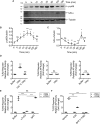
YRS stimulates NF-κB signaling through TLR2. A, representative immunoblots of phosphorylated (p)-p65 NF-κB and inhibitor of NF-κB (IκB-α) following treatment of PMA-differentiated THP1-derived human macrophages with 50 nm recombinant human YRS for the times shown. An immunoblot of α-tubulin is shown as the loading control. Uncropped immunoblots can be found in Fig. S4C. Quantification of relative band densities of p-p65 NF-κB (B) and IκB-α (C) plotted as mean ± S.D. of N = 3 independent experiments. D, HEK293 cells expressing TLR2, -4, or -9 with a NF-κB alkaline phosphatase (AP) reporter system were treated for 18 h with 50 nm recombinant human YRS, heat-denatured YRS (100 °C YRS), or buffer. E, TLR2 reporter cells were pre-treated for 1 h with 5 μg/ml TLR2-blocking antibody (αTLR2), isotype control IgA2, or buffer prior to treatment ± 50 nm YRS for 18 h. F, TLR2 reporter cells were pre-treated for 1 h with 100 μm TLR2 inhibitor C29, 10 μm NF-κB activation inhibitor BAY11-7082 that targets IκB kinase, or vehicle (1% (v/v) DMSO) prior to treatment ± 50 nm YRS for 18 h. The relative activity of alkaline phosphatase was plotted as follows: D, fold changes compared with buffer (means ± S.D., n = 4) of N = 2 independent experiments; TLR9, N = 1; E and F, fold change compared with the buffer −YRS control (means ± S.D., n = 4) of N = 2 independent experiments. Statistical significance was determined as follows: B and C, against 0 h using a one-way ANOVA with Dunnett's multiple comparison post-tests; D, against buffer using a one-way ANOVA with Dunnett's multiple comparison post-tests for TLR2 and an unpaired two-tailed Student's t test for TLR4/9; E and F, against buffer (or vehicle) and antibody (or inhibitor) treatment in the presence of YRS (+YRS) using a two-tailed unpaired Student's t test. *, p < 0.05; **, p < 0.01; ***, p < 0.001; ns, not significant. Error bars represent S.D.
Figure 3.
Figure 3.
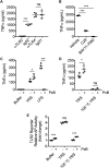
Inhibition of TLR2 signaling reduces YRS-mediated TNFα release from THP1 macrophages. TNFα released to the conditioned medium of PMA-differentiated THP1-derived macrophages in response to treatment with 50 nm recombinant human YRS for 3 h after pre-treatment for 1 h with 5 μg/ml TLR2- or TLR4-blocking antibodies (αTLR2, αTLR4) or isotype controls (IgA2, for αTLR2; IgG1, for αTLR4) (mean ± S.D., n = 4) of N = 3 independent experiments (A), or TLR2 inhibitor C29 (100 μm), IκB kinase inhibitor BAY11-7082 (10 μm), or vehicle (1% (v/v) DMSO) (mean ± S.D., n = 4) of N = 2 independent experiments (B). TNFα measured by ELISA in the conditioned media of PMA-differentiated THP1-derived macrophages treated for 3 h ± 10 μg/ml polymyxin B (PxB) with 50 nm YRS, 100 ng/ml LPS, or buffer (C) or 50 nm heat-denatured YRS (100 °C YRS) (D). Data were plotted as means ± S.D. (n = 4) of N = 3 independent experiments. E, HEK293 cells expressing TLR2 with an NF-κB alkaline phosphatase (AP) reporter system were treated for 18 h with 50 nm recombinant human YRS, heat-denatured YRS (100 °C YRS) or buffer ± 10 μg/ml polymyxin B. The relative activity of alkaline phosphatase was plotted as fold changes compared with buffer, polymyxin B (means ± S.D., n = 4) of N = 2 independent experiments. Statistical significance was determined as follows. A, between each isotype control and antibody using a two-tailed unpaired Student's t test; B, against vehicle using a one-way ANOVA with Dunnett's multiple comparison post-tests; C–E, between the −PxB and +PxB conditions using a two-tailed unpaired Student's t test. *, p < 0.05; ***, p < 0.001; ns, not significant. Error bars represent S.D.
Figure 4.
Figure 4.
YRS increases macrophage-secreted MMP activity in a TNFα-dependent manner. A, PMA-differentiated THP1-derived macrophages were treated for 24 h with 50 nm recombinant human YRS, or 40 ng/ml TNFα, or buffer, with or without a mAb inhibitor of TNFα (100 ng/ml inflixamab, IN). MMPs in the concentrated (20×) conditioned media were activated with 1 mm APMA, and cleavage of a quenched fluorescence peptide substrate Mca-Pro-Leu-Gly-Leu-Dpa-Ala-Arg-NH2 was measured (mean ± S.D., n = 3) in N = 3 independent experiments. Statistical significance was determined against “buffer + inflixamab” using a one-way ANOVA with Dunnett's multiple comparison post-test. **, p < 0.01; ns, not significant. Error bars represent S.D. B, to induce MMP expression, PMA-differentiated THP1-derived macrophages were treated for 24 h with 40 ng/ml TNFα. MMPs in the concentrated (20×) conditioned media were activated with 1 mm APMA for 20 min at 22 °C prior to addition of YRS (∼62 kDa) ± MMP inhibitor (10 μm marimastat), at a protein ratio of 5:1 medium protein/YRS, for 18 h at 37 °C. YRS-containing conditioned medium samples were resolved by 10% SDS-PAGE and stained with Coomassie Brilliant Blue G-250. The ∼66-kDa band is BSA in the concentrated conditioned medium. A representative gel of N = 3 independent experiments is shown. The uncropped gel is shown in Fig. S5A.
Figure 5.
Figure 5.
Coomassie Brilliant Blue-stained SDS-PAGE analysis of YRS cleavage by recombinant MMPs in vitro. A, recombinant human YRS (∼62 kDa) was incubated for 18 h at 37 °C with recombinant MMPs, neutrophil elastase, or plasmin (1:10 molar ratio protease/YRS), N = 2 independent experiments. B, YRS was incubated for 18 h at 37 °C at the protease/YRS molar ratios shown. Uncropped gels are shown in Fig. S5, B and C. Controls (+) are the highest concentration of protease without YRS. Cleavage products were analyzed by 12% (A) or 10% (B) SDS-PAGE with Coomassie Brilliant Blue G-250 staining. Cleavage products are indicated by arrows. C, kcat/Km values for cleavage of YRS.
Figure 6.
Figure 6.
MMP-cleaved YRS protein bands sequenced by LC-MS/MS and Edman degradation. A, LC-MS/MS was used to identify the most N-terminal peptide of MMP-cleaved YRS bands. After MMP digestion of 2 μm recombinant human YRS for 18 h at 37 °C (MMP/YRS molar ratios used were guided by results shown in Fig. 5B), cleavage products or the full-length ∼62 kDa YRS (top left) were resolved by 10% SDS-PAGE and stained with Coomassie Brilliant Blue G-250. Bands were excised, digested with trypsin, and analyzed by LC-MS/MS. N-terminal peptides identified from YRS cleavage products after trypsin digestion are shown. Molecular masses shown in italics were estimated from Rf values. The YRS negative control lane was duplicated here between MMP2, MMP3, MMP7, MMP8, and MMP9 and again between MMP10, MMP12, MMP13, and MMP14 according to the grouping of digests on the same gels for analysis as shown on the uncropped full gels presented in Fig. S5D. B, YRS was incubated for 18 h at 37 °C ± MMP7, MMP8, or MMP12 at 1:10 MMP/YRS molar ratios. Cleavage products were resolved by 16.5% SDS-PAGE, transferred to PVDF membrane, stained with Coomassie Brilliant Blue G-250, and subjected to Edman degradation. N-terminal sequences are shown and the original stained blots, from which bands were excised for Edman degradation, are shown in full in Fig. S6.
Figure 7.
Figure 7.
Identification of MMP cleavage sites in YRS. Major and minor MMP cleavage sites in recombinant human YRS were determined by ATOMS N-terminal positional proteomics or by Edman degradation of proteins blotted to PVDF membranes as shown in Fig. 6B. Cleavage sites identified by ATOMS (↓), Edman degradation (*, underlined), or by both (**) are shown. Note, ATOMS is a highly-sensitive technique that identifies cleavage sites, but does not determine relative cleavage rates. Thus, several ATOMS sites were not observed by SDS-PAGE due to the low abundance of their cleavage fragments. Yellow, Rossmann fold catalytic domain; green, anticodon recognition domain; blue, endothelial monocyte-activating polypeptide II-like (EMAPII) domain; pink, E91LR93 tripeptide motif; red, R371VGKIIT377 heptapeptide motif.
Figure 8.
Figure 8.
Major stable YRS proteoforms generated by MMP cleavage of YRS. A, MMP cleavage sites in YRS were determined by N-terminal sequencing by Edman degradation of proteins shown in Fig. 6B or by ATOMS N-terminal positional proteomics shown in Fig. 7. Schematic diagrams of YRS with the MMP cleavage sites leading to predicted N-terminal fragments of YRS (ΔYRS) are shown. Fragments predicted from cleavage sites are aligned by molecular mass with fragments observed on SDS-polyacrylamide gels in Fig. 6A. Molecular masses of predicted fragments were determined using the ExPASy tool (https://web.expasy.org/compute_pi/), and the molecular mass of YRS fragments observed by SDS-PAGE were determined from relative migration distances compared with molecular mass standards (Rf). MMPs generating each fragment are indicated. Yellow, Rossmann fold catalytic domain; green, anticodon recognition domain; blue, endothelial monocyte-activating polypeptide II-like (EMAPII) domain; pink, E91LR93 tripeptide motif; red, R371VGKIIT377 heptapeptide motif. B, table of cleavage sites common to all MMPs identified by ATOMS and Edman sequencing. Predicted mass, the calculated molecular mass of the ΔYRS(1–383) and ΔYRS(1–405) cleavage products. Rf, apparent molecular masses of the major cleavage fragments resolved by electrophoresis.
Figure 9.
Figure 9.
MMP7 and MMP8 cleavage of YRS increases monocyte chemotaxis, TLR2 signaling, NF-κB activation, and TNFα release by macrophages. A, cleavage of recombinant human YRS (∼62 kDa) by MMP7 and MMP8 (1:10 protease/YRS molar ratio) for 18 h at 37 °C visualized by Coomassie Brilliant Blue G-250-stained 10% SDS-PAGE. ΔYRS-cleavage products. The uncropped gel is presented in Fig. S5D. B, transwell chemotaxis assay (90 min) of THP1 monocytes migrating toward 50 nm YRS, YRS cleaved by MMP7 or MMP8, MMPs alone or buffer. Data are presented as fold change compared with buffer alone (mean ± S.D., n = 3) of N = 3 independent experiments. HEK293 cells co-expressing TLR2 (C) or TLR4 (D) and a NF-κB alkaline phosphatase (AP) reporter system were treated for 18 h with 50 nm YRS, YRS cleaved by MMP7 or MMP8, MMPs alone or buffer. The relative activity of alkaline phosphatase is plotted as fold change for TLR compared with buffer alone (mean ± S.D., n = 4) of N = 2 independent experiments for both C and D. E, ELISA measurement of TNFα released to the conditioned media of PMA-differentiated THP1-derived macrophages treated for 3 h with 50 nm intact YRS, MMP7, or MMP8-cleaved YRS or MMPs alone or buffer (plotted as mean ± S.D., n = 4) of N = 3 independent experiments. Statistical significance was determined between YRS treated with buffer and YRS cleaved by MMP7 and MMP8 using a two-tailed unpaired Student's t test. *, p < 0.05; **, p < 0.01; ***, p < 0.001; ns, not significant. Error bars represent S.D.
Figure 10.
Figure 10.
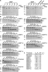
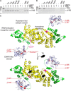
MMP8 cleavage of YRS is inhibited by Tyr in vitro. Recombinant human YRS (∼62 kDa) was pre-incubated with 0.5 mm Tyr, Trp, Met, Ap4A, Ap5A, AMP, ATP, or buffer as shown for 30 min at room temperature. Recombinant human MMP7 (A) or MMP8 (B) was added (1:100 MMP/YRS molar ratio) and incubated for 18 h at 37 °C. Cleavage products (ΔYRS) were analyzed by 10% SDS-PAGE, N = 2 independent experiments. Cleavage products were visualized by Coomassie Brilliant Blue G-250 staining and are indicated by arrows. C, structural model of apo-YRS (upper) and with bound Tyr (lower) showing major sites of MMP cleavage. The 3D structures of N-terminal YRS (residues 1–342) homodimers without (above, PDB 1N3L) and with (below, PDB 4QBT) Tyr bound. The Rossmann fold catalytic domains (yellow) and the tRNA-anticodon recognition domains (green) essential for aminoacylation, were modeled with the 3D structure of the C-terminal EMAPII-like domain (blue) of YRS (PDB 1NTG) to represent a full-length YRS molecule. The sequences of E91LR93 and R371VGKIIT377, important for the cytokine activity of YRS, are shown in pink and red, respectively. MMP8 cleavage sites L387 and L406 are in red.
Figure 11.
Figure 11.
Feed-forward model of the YRS-MMP temporal relationship in the induction and potentiation of proinflammatory pathways. 1) Extracellular YRS activation of TLR2 signaling; 2) triggering TNFα and chemokine release; 3) chemokines recruit neutrophils and TNFα and NF-κB activation up-regulate MMP expression; 4) macrophage MMP7 and neutrophil MMP8 processing of YRS generates truncated proteoforms (ΔYRS). 5) ΔYRS drives inflammation by enhanced activation of TLR2 signaling in a feed-forward mechanism.

References

    1. Parks W. C., Wilson C. L., and López-Boado Y. S. (2004) Matrix metalloproteinases as modulators of inflammation and innate immunity. Nat. Rev. Immunol. 4, 617–629 10.1038/nri1418 - DOI - PubMed
    1. Rodríguez D., Morrison C. J., and Overall C. M. (2010) Matrix metalloproteinases: what do they not do? New substrates and biological roles identified by murine models and proteomics. Biochim. Biophys. Acta 1803, 39–54 10.1016/j.bbamcr.2009.09.015 - DOI - PubMed
    1. McQuibban G. A., Gong J. H., Tam E. M., McCulloch C. A., Clark-Lewis I., and Overall C. M. (2000) Inflammation dampened by gelatinase A cleavage of monocyte chemoattractant protein-3. Science 289, 1202–1206 10.1126/science.289.5482.1202 - DOI - PubMed
    1. Butler G. S., and Overall C. M. (2009) Updated biological roles for matrix metalloproteinases and new “Intracellular” substrates revealed by degradomics. Biochemistry 48, 10830–10845 10.1021/bi901656f - DOI - PubMed
    1. Jobin P. G., Butler G. S., and Overall C. M. (2017) New intracellular activities of matrix metalloproteinases shine in the moonlight. Biochim. Biophys. Acta Mol. Cell Res. 1864, 2043–2055 10.1016/j.bbamcr.2017.05.013 - DOI - PubMed

Publication types