. 2003 Nov 11;100(23):13264-9.
doi: 10.1073/pnas.2232255100. Epub 2003 Oct 28.

Identification of the protein kinase A regulatory RIalpha-catalytic subunit interface by amide H/2H exchange and protein docking

Affiliations

Identification of the protein kinase A regulatory RIalpha-catalytic subunit interface by amide H/2H exchange and protein docking

Ganesh S Anand et al. Proc Natl Acad Sci U S A. .

Abstract

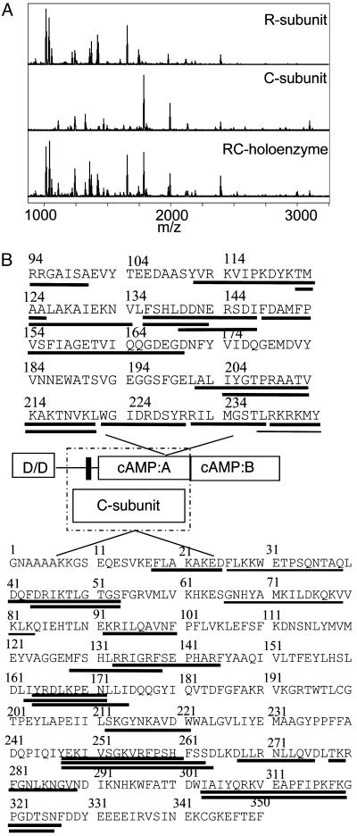
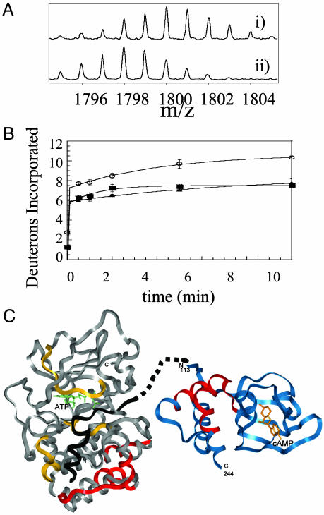
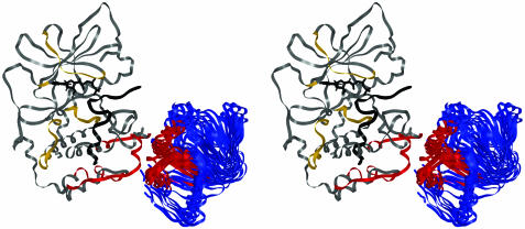
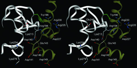
An important goal after structural genomics is to build up the structures of higher-order protein-protein complexes from structures of the individual subunits. Often structures of higher order complexes are difficult to obtain by crystallography. We have used an alternative approach in which the structures of the individual catalytic (C) subunit and RIalpha regulatory (R) subunit of PKA were first subjected to computational docking, and the top 100,000 solutions were subsequently filtered based on amide hydrogen/deuterium (H/2H) exchange interface protection data. The resulting set of filtered solutions forms an ensemble of structures in which, besides the inhibitor peptide binding site, a flat interface between the C-terminal lobe of the C-subunit and the A- and B-helices of RIalpha is uniquely identified. This holoenzyme structure satisfies all previous experimental data on the complex and allows prediction of new contacts between the two subunits.


Figures

Fig. 1.
Fig. 1.
(A) MALDI-TOF mass spectrum of the peptic digest from RIα (Top), from the C-subunit (Middle), and from the RIα2–C2 holoenzyme (Bottom). (B) Sequence of the RIα(94–244) and C-subunit showing the fragments that were observed in the mass spectrum of the peptic digest. Lines underneath the sequence are fragments for which quantifiable data were obtained. A schematic is provided showing the domains of each protein, and the dashed box encloses those domains known to participate in the R–C interaction. In the regulatory subunit, D/D refers to the dimerization/docking domain, and the black bar shows the pseudosubstrate/inhibitor sequence.
Fig. 2.
Fig. 2.
(A) Expansion of the MALDI-TOF mass spectra to show one of the 17 fragments (m/z = 1,793.97, residues 247–261) from the analysis of the C-subunit that experienced slowed exchange in the RIα2–C2 holoenzyme complex. (Ai) The isotopic envelope for the fragment from the free C-subunit bound to MgATP after 10 min of deuteration. (Aii) The isotopic envelope for the same fragment from the RIα2–C2 holoenzyme after 10 min of deuteration. (B) Plot of deuterium incorporation into the amide positions of the region of C-subunit for the peptide fragment; m/z = 1,793.97 from C-subunit +MgATP (○), RIα2–C2 holoenzyme (▪), and RIα(94–244)-C holoenzyme (▴). (C) The structure of the C-subunit is shown in gray; the residues protected by the R-subunit are shown in red; the inhibitor peptide PKI(5–24), which mimics the pseudosubstrate, is shown in black; and the residues protected by it are shown in yellow (8). The structure of RIα(113–244) is shown in blue with the residues protected by the C-subunit in red. The pseudosubstrate/inhibitor sequence is shown connected by dots to the N terminus of the structured part of RIα.
Fig. 3.
Fig. 3.
Stereo image of the top cluster of eight solutions that represent the final docked structure. The structure of the C-subunit is colored as in Fig. 2. The regions of RIα(113–244) that showed decreased solvent accessibility exclusively on binding the C-subunit are colored red.
Fig. 4.
Fig. 4.
Stereo image of a close-up of the R–C subunit interface from the lowest energy filtered dot solution. The C-subunit is shown in gray, and RIα is in gold. Glu-143 (R) is in contact with Lys-213 (C). The side chain of Trp-196 (C) is in green and is predicted to contact Arg-230/Arg-231 (R). Arg-194 (C) is predicted to contact Asp-227 (R). Lys-254 (C) and Glu-248 (C) are also in the interface.

References

    1. Gibson, R. M., Ji-Buechler, Y. & Taylor, S. S. (1997) J. Biol. Chem. 272, 16343-16350. - PubMed
    1. Huang, L. J. & Taylor, S. S. (1998) J. Biol. Chem. 273, 26739-26746. - PubMed
    1. Zheng, J., Knighton, D. R., Ten Eyck, L. F., Karlsson, R., Xuong, N., Taylor, S. S. & Sowadski, J. M. (1993) Biochemistry 32, 2154-2161. - PubMed
    1. Su, Y., Dostmann, W. R. G., Herberg, F. W., Durick, K., Xuong, N.-h., Ten Eyck, L. F., Taylor, S. S. & Varughese, K. I. (1995) Science 269, 807-819. - PubMed
    1. Ten Eyck, L. F., Mandell, J. G., Roberts, V. A. & Pique, M. E. (1995) in Proceedings of the 1995 ACM/IEEE Supercomputing Conference (IEEE Comput. Soc. Press, Los Alamitos, CA).

Publication types

Substances

Associated data

LinkOut - more resources