. 2004 May 11;101(19):7293-8.
doi: 10.1073/pnas.0401595101. Epub 2004 May 3.

The C-terminal domain of DNA gyrase A adopts a DNA-bending beta-pinwheel fold

Affiliations

The C-terminal domain of DNA gyrase A adopts a DNA-bending beta-pinwheel fold

Kevin D Corbett et al. Proc Natl Acad Sci U S A. .

Abstract

DNA gyrase is unique among enzymes for its ability to actively introduce negative supercoils into DNA. This function is mediated in part by the C-terminal domain of its A subunit (GyrA CTD). Here, we report the crystal structure of this approximately 35-kDa domain determined to 1.75-A resolution. The GyrA CTD unexpectedly adopts an unusual fold, which we term a beta-pinwheel, that is globally reminiscent of a beta-propeller but is built of blades with a previously unobserved topology. A large, conserved basic patch on the outer edge of this domain suggests a likely site for binding and bending DNA; fluorescence resonance energy transfer-based assays show that the GyrA CTD is capable of bending DNA by > or =180 degrees over a 40-bp region. Surprisingly, we find that the CTD of the topoisomerase IV A subunit, which shares limited sequence homology with the GyrA CTD, also bends DNA. Together, these data provide a physical explanation for the ability of DNA gyrase to constrain a positive superhelical DNA wrap, and also suggest that the particular substrate preferences of topoisomerase IV might be dictated in part by the function of this domain.


Figures

Fig. 1.
Fig. 1.
(A) Overall structure of BbGac. Blades 1-6 are purple, blue, green, yellow, orange, and red, respectively. Strands A-D of blade 6 are labeled, as well as strand C of blade 1 (C′). (B) Comparison of the strand connectivity within the blades of BbGac versus a canonical β-propeller. (C) Comparison of the overall strand topology between the BbGac β-pinwheel and a canonical β-propeller. One repeat unit of each is highlighted in red. (D) Packing of blades 1 and 6 of BbGac. Strands A-D of blade 6 are labeled. The C strand of blade 1 (C′) packs against the B strand of blade 6.
Fig. 2.
Fig. 2.
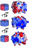
Electrostatic surface representations of BbGac. The large basic patch (blue) encompasses four blades (1, 4, 5, and 6) and stretches around approximately two-thirds of the outer edge of the domain. The protein orientation and distribution of basic (blue) and acidic (red) regions are shown in schematic diagrams next to each image.
Fig. 3.
Fig. 3.
FRET measurements of DNA bending by GyrA and ParC CTDs. (A) Representative fluorescence spectra of donor-only-labeled 40-bp duplex DNA (yellow), donor plus acceptor-labeled (blue), and donor plus acceptor-labeled plus 10 μM BbGac (green), with maximum donor fluorescence normalized to 1. (B) FRET enhancement by BbGac (green) and E. coli ParC CTD (blue), of the 40-bp DNA substrate. (C) Schematic view of the GyrA CTD (blue and red) bending a 40-bp DNA substrate labeled with donor (D) and acceptor (A) fluorophores, along with approximate distances involved.
Fig. 4.
Fig. 4.
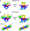
A model for supercoiling by DNA gyrase. Schematic views of strand passage by a type IIA topo catalytic core (no CTD) (A) and DNA gyrase (B). The DNA-binding/cleavage cores are shown in blue and red, the ATPase domains in yellow, the GyrA CTD in green, and the bound G and T segments in magenta and cyan, respectively. The handedness of the DNA crossover is noted by (+) or (-). We note that a flexible linkage between the N- and C-terminal regions of the A subunit (shown as a black line) might allow the CTD to move with respect to the N-terminal region and thereby help “shuttle” a T segment from one side of the G segment to the other and out of the protein.

References

    1. Champoux, J. J. (2001) Annu. Rev. Biochem. 70, 369-413. - PubMed
    1. Wang, J. C. (2002) Nat. Rev. Mol. Cell Biol. 3, 430-440. - PubMed
    1. Hickson, I. D. (2003) Nat. Rev. Cancer 3, 169-178. - PubMed
    1. Brown, P. O. & Cozzarelli, N. R. (1979) Science 206, 1081-1083. - PubMed
    1. Liu, L. F., Liu, C. C. & Alberts, B. M. (1980) Cell 19, 697-707. - PubMed

Publication types

Associated data

LinkOut - more resources