Comparative Study
. 2004 Oct 5;43(39):12539-48.
doi: 10.1021/bi048737e.

Structures of the Escherichia coli PutA proline dehydrogenase domain in complex with competitive inhibitors

Affiliations
Comparative Study

Structures of the Escherichia coli PutA proline dehydrogenase domain in complex with competitive inhibitors

Min Zhang et al. Biochemistry. .

Abstract

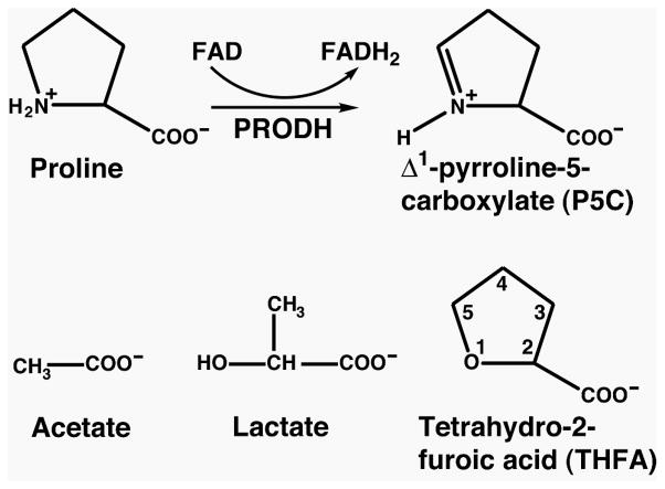
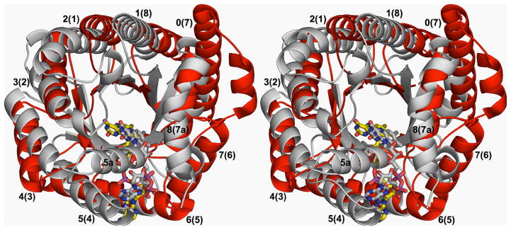
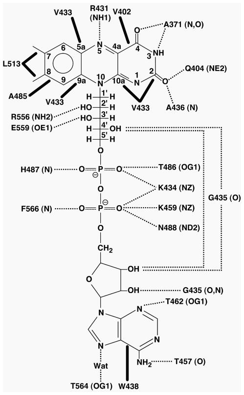
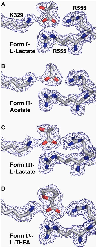
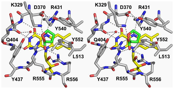
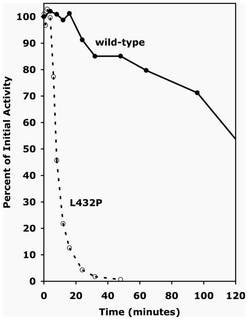
Proline dehydrogenase (PRODH) catalyzes the first step of proline catabolism, the flavin-dependent oxidation of proline to Delta(1)-pyrroline-5-carboxylate. Here we present a structure-based study of the PRODH active site of the multifunctional Escherichia coli proline utilization A (PutA) protein using X-ray crystallography, enzyme kinetic measurements, and site-directed mutagenesis. Structures of the PutA PRODH domain complexed with competitive inhibitors acetate (K(i) = 30 mM), L-lactate (K(i) = 1 mM), and L-tetrahydro-2-furoic acid (L-THFA, K(i) = 0.2 mM) have been determined to high-resolution limits of 2.1-2.0 A. The discovery of acetate as a competitive inhibitor suggests that the carboxyl is the minimum functional group recognized by the active site, and the structures show how the enzyme exploits hydrogen-bonding and nonpolar interactions to optimize affinity for the substrate. The PRODH/L-THFA complex is the first structure of PRODH with a five-membered ring proline analogue bound in the active site and thus provides new insights into substrate recognition and the catalytic mechanism. The ring of L-THFA is nearly parallel to the middle ring of the FAD isoalloxazine, with the inhibitor C5 atom 3.3 A from the FAD N5. This geometry suggests direct hydride transfer as a plausible mechanism. Mutation of conserved active site residue Leu432 to Pro caused a 5-fold decrease in k(cat) and a severe loss in thermostability. These changes are consistent with the location of Leu432 in the hydrophobic core near residues that directly contact FAD. Our results suggest that the molecular basis for increased plasma proline levels in schizophrenic subjects carrying the missense mutation L441P is due to decreased stability of human PRODH2.


Figures

Figure 1
Figure 1
The reaction catalyzed by PutA proline dehydrogenase domain (top), and chemical structures of the three inhibitors used in this study (bottom).
Figure 2
Figure 2
Ribbon drawing of PutA86-669 with bound L-lactate. The β/α barrel substructure is shown in green/red (residues 263–561). Helices of the barrel are labeled α0-α8, and selected residue numbers are indicated. Yellow cylinders indicate the helical arm that wraps around the barrel. Blue cylinders denote helices of a poorly resolved structural domain consisting of residues 141–262. The dashed curve indicates disordered residues. The purple cylinders denote helices positioned after the barrel. FAD and L-lactate are drawn as ball-and-stick models in yellow and white, respectively. This and other figures were prepared with PyMol (38).
Figure 3
Figure 3
Stereoscopic view depicting the superposition of the β/α barrels of PutA86-669 (white) and methylenetetrahydrofolate reductase (red, PDB entry 1B5T) (43). The FAD cofactor of PutA86-669 and methylenetetrahydrofolate reductase are shown in white and yellow, respectively. The helices of PutA are numbered 0–8, with the corresponding helices in MTHFR appearing in parentheses. This alignment was calculated using the CE server (50). The RMSD between the barrels of PutA and 1B5T is 3.4 Å for 188 aligned residues.
Figure 4
Figure 4
Schematic diagram of selected protein-FAD interactions. The dotted lines indicate hydrogen bonds and ion pairs, with the interacting atoms listed in parentheses. The thick solid lines indicate non-polar interactions. Note there is also an intramolecular hydrogen bond indicated, between the FAD ribose and ribityl groups.
Figure 5
Figure 5
Views of the active sites of the four structures showing bound inhibitors interacting with Arg555, Arg556, and Lys329. Panels A–D correspond to crystal forms I–IV, respectively (see Table 1). The electron density maps are simulated annealing Fo-Fc omit maps contoured at 2.5 σ.
Figure 6
Figure 6
Stereoscopic view depicting the active site of the PutA86-669/L-THFA complex (crystal form IV). The dashed lines indicate hydrogen bonds and ion pairs. The dashed line connecting C5 of THFA and N5 of FAD indicates a distance of 3.3 Å. The protein is shown in white, FAD in yellow, and L-THFA in green.
Figure 7
Figure 7
Structural context of the L432P mutation of PutA86-669. Residues 431–433 are part of β5, while residues 437–438 are part of α5a. The protein is shown in white, FAD in yellow, and L-THFA in green.
Figure 8
Figure 8
Thermostability analysis for wild-type PutA86-669 and L432P at 45 °C. The filled circles and solid curve correspond to data for the wild-type enzyme. The open circles and dashed curve represent data for L432P.

References

    1. Phang JM. The Regulatory Functions of Proline and Pyrroline-5-Carboxylic Acid. Current Topics in Cell Regulation. 1985;25:92–132. - PubMed
    1. Adams E, Frank L. Metabolism of proline and the hydroxyprolines. Annu Rev Biochem. 1980;49:1005–61. - PubMed
    1. Donald SP, Sun XY, Hu CA, Yu J, Mei JM, Valle D, Phang JM. Proline oxidase, encoded by p53-induced gene-6, catalyzes the generation of proline-dependent reactive oxygen species. Cancer Res. 2001;61:1810–5. - PubMed
    1. Polyak K, Xia Y, Zweier JL, Kinzler KW, Vogelstein B. A model for p53-induced apoptosis. Nature. 1997;389:300–5. - PubMed
    1. Maxwell SA, Davis GE. Differential gene expression in p53-mediated apoptosis-resistant vs. apoptosis-sensitive tumor cell lines. Proc Natl Acad Sci USA. 2000;97:13009–14. - PMC - PubMed

Publication types

MeSH terms

LinkOut - more resources