. 2006 Feb 8;25(3):605-14.
doi: 10.1038/sj.emboj.7600950. Epub 2006 Jan 19.

A divergent external loop confers antagonistic activity on floral regulators FT and TFL1

Affiliations

A divergent external loop confers antagonistic activity on floral regulators FT and TFL1

Ji Hoon Ahn et al. EMBO J. .

Abstract

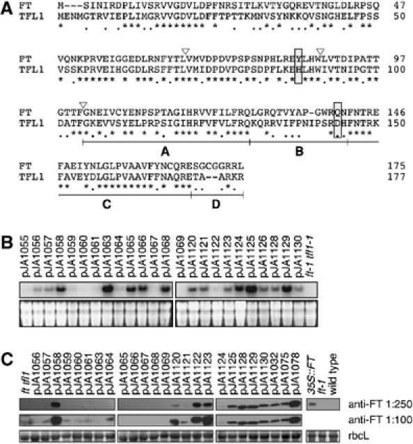
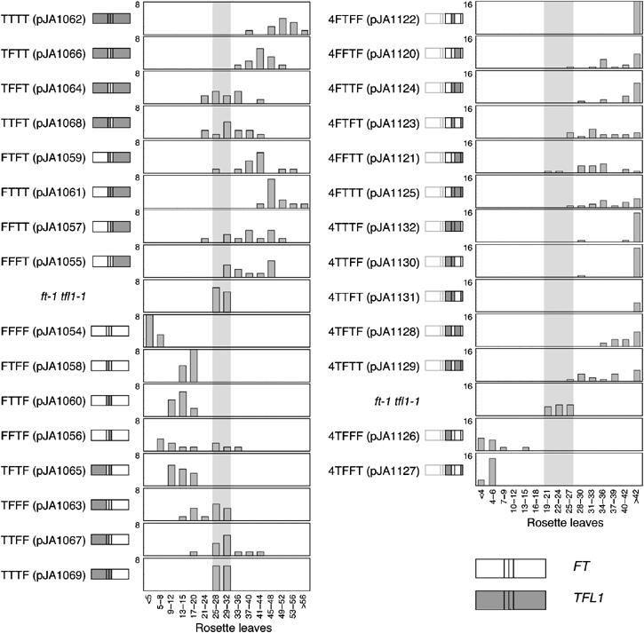
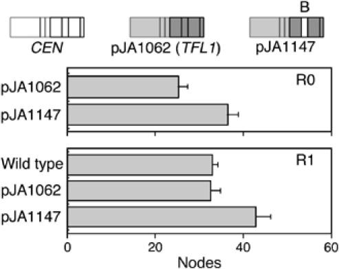
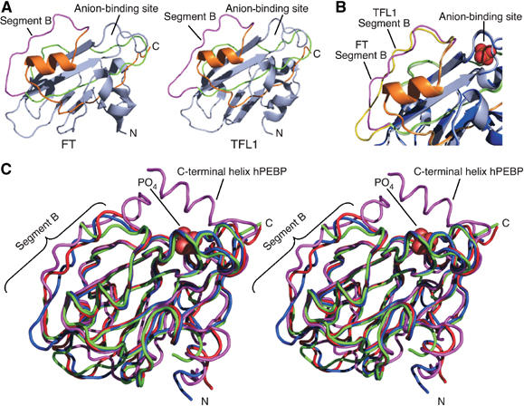
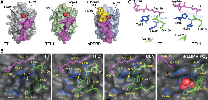
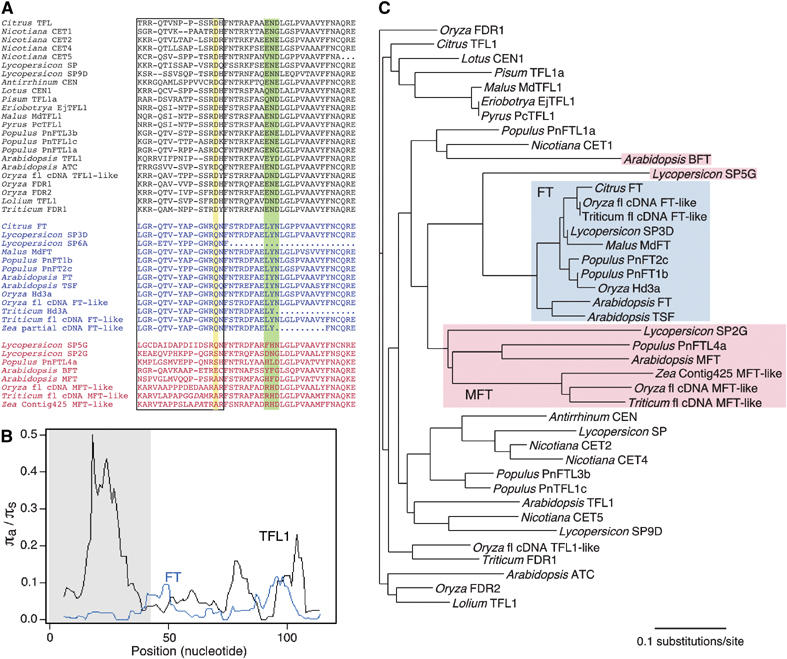
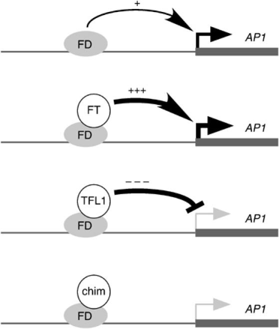
The Arabidopsis genes FT and TERMINAL FLOWER1 (TFL1) encode related proteins with similarity to human Raf kinase inhibitor protein. FT, and likely also TFL1, is recruited to the promoters of floral genes through interaction with FD, a bZIP transcription factor. FT, however, induces flowering, while TFL1 represses flowering. Residues responsible for the opposite activities of FT and TFL1 were mapped by examining plants that overexpress chimeric proteins. A region important in vivo localizes to a 14-amino-acid segment that evolves very rapidly in TFL1 orthologs, but is almost invariant in FT orthologs. Crystal structures show that this segment forms an external loop of variable conformation. The only residue unambiguously distinguishing the FT and TFL1 loops makes a hydrogen bond with a residue near the entrance of a potential ligand-binding pocket in TFL1, but not in FT. This pocket is contacted by a C-terminal peptide, which also contributes to the opposite FT and TFL1 activities. In combination, these results identify a molecular surface likely to be recognized by FT- and/or TFL1-specific interactors.


Figures

Figure 1
Figure 1
Sequence and expression of chimeras. (A) Amino-acid sequences of the parental FT and TFL1 proteins. Asterisks indicate identical residues, dots residues with similar biochemical properties, and dashes gaps introduced to optimize the sequence alignment. Triangles show the exon boundaries of FT and TFL1. The four segments used to generate the segmental chimeras within the fourth exon are shown as A, B, C and D. The Tyr85/His88 and Gln140/Asp144 residues, which form a hydrogen bond in TFL1, but not FT, and which are likely the most critical residues for distinguishing FT and TFL1 activity, are boxed. (B) RNA accumulation in transgenic plants. Blots were probed with a mixture of FT and TFL1 probes. The bottom panels show ethidium bromide-stained gels as loading controls. (C) Protein blot analysis using anti-FT antibody. The bottom panels show Ponceau S-stained blots (major band is large subunit of Rubisco).
Figure 2
Figure 2
Flowering times of Arabidopsis plants expressing FT/TFL1 chimeras. The structure of each chimeric gene is shown next to the name of each construct. Sequences of FT and TFL1 are shown as open and gray boxes, respectively. Left, exon chimeras. Most transgenes that contain the fourth exon of TFL1 flower (top) cause later flowering than what is seen in the nontransgenic ft tfl1 control. Chimeras that contain the fourth exon of FT cause mostly late flowering (bottom). Right, swaps of segments in the fourth exon, with exons one to three from FT. Chimeras that cause later flowering are shown above the nontransgenic ft tfl1 control. In this experiment, the control plants flowered earlier than in the exon-swapping experiment (22.9±1.1 versus 28.6±0.8 rosette leaves), due to changed growth conditions.
Figure 3
Figure 3
Flowering times of tobacco plants with CEN/TFL1 segment B swap. Primary transformants (R0) and progeny (R1) are shown. Only plants that overexpress a chimeric gene with segment B of the fourth exon of CEN in the TFL1 background are late flowering.
Figure 4
Figure 4
Crystal structures of FT and TFL1. (A) Cartoon diagrams of FT and TFL1. For residues encoded by the fourth exon, segment A is colored green, segment B magenta, and segment C orange. Segment D forms the C-terminus of each protein, but is disordered in both crystal structures. Amino (N) and carboxy (C) termini are labeled. (B) Close-up showing an overlay of segment B and surrounding regions of FT and TFL1. A phosphate ion, from the hPEBP structure, is modeled as orange and red spheres in the anion-binding site. (C) Stereo view of an overlay of FT (red), TFL1 (blue), CEN (green), and hPEBP (magenta) structures as ribbon traces. A phosphate ion bound within the anion ligand-binding site of hPEBP is shown as red spheres. The C-terminal helix in hPEBP, not present in the FT and TFL1 structures, is indicated.
Figure 5
Figure 5
Details of the ligand-binding sites and external loop. (A) Surface representation of FT, TFL1 and hPEBP in similar orientation. Phosphate ligands (red and orange spheres, from hPEBP structure) are shown bound within the anion-binding site of each structure. The surfaces formed by segment B are colored magenta. The surface formed by the C-terminal helix in hPEBP (yellow) makes the ligand-binding site less accessible than in FT and TFL1. The two key conserved residues that bind the ligand (histidine and aspartic acid) are labeled. (B) Details of the amino acids lining the ligand-binding site. The pocket of hPEBP includes a bound phosphate ion (red and magenta spheres). Amino acids are colored by segment: residues in segment A (or equivalent) are colored green, residues in segment B magenta, and residues that precede segment A blue. (C) Interactions of segment B residues with amino acids at the entrance to the binding pocket. Hydrogen bonds are shown as light dashed lines (distances marked in Å), with amino acids colored by segment as in (B). Residues His87 and Val120 in FT and His90 and Phe123 in TFL1 are shown for orientation (see panel B). Only TFL1 shows an interaction between Asp144 and His88 (red line), while the corresponding residues in FT, Gln140 and Tyr85 do not interact.
Figure 6
Figure 6
Sequence comparison of FT/TFL1 family members from flowering plants. (A) Alignments of segments B and C encoded by the fourth exons of TFL1-like genes (top, black), FT-like genes (middle, blue) and other members of the family (bottom, red). Dashes denote gaps, dots missing data. Additional indels in the two lower most sequences are indicated by italics. Segment B is boxed; the Asp144/Gln140 residue distinguishing all FT and TFL1 members is highlighted in yellow. The triad in segment C, which is also more conserved in FT than TFL1 proteins, is highlighted in green. (B) Average pairwise ratio of nonsynonymous (πa) to synonymous substitutions (πs) (sliding window, window size 12, step 1) (see also Supplementary Table S3). TFL1 genes show an excess of nonsynonymous substitutions in segment B. (C) Phylogenetic neighbor-joining tree. FT orthologs form a well-resolved clade, with the somewhat more divergent MFT-like proteins as sister clade. TFL1 sequences do not appear monophyletic because of their rapid divergence.
Figure 7
Figure 7
Model for interaction of FD with FT, TFL1 and certain chimeras. FD, which binds to the AP1 promoter (Wigge et al, 2005), is proposed to be a weak activator on its own. FT converts FD into a strong activator, while TFL1 converts FD into a strong repressor, explaining why TFL1 overexpressers are not merely late-flowering, like ft mutants, but also show floral defects (Ratcliffe et al, 1998). Certain chimeras (chim), such 4FFTF, might not have true TFL1 function, but still cause moderate late flowering, because they interfere with the weak activator function of FD by itself.

References

    1. Abe M, Kobayashi Y, Yamamoto S, Daimon Y, Yamaguchi A, Ikeda Y, Ichinoki H, Notaguchi M, Goto K, Araki T (2005) FD, a bZIP protein mediating signals from the floral pathway integrator FT at the shoot apex. Science 309: 1052–1056 - PubMed
    1. Alvarez J, Guli CL, Yu X-H, Smyth DR (1992) Terminal flower: a gene affecting inflorescence development in Arabidopsis thaliana. Plant J 2: 103–116
    1. Amaya I, Ratcliffe OJ, Bradley DJ (1999) Expression of CENTRORADIALIS (CEN) and CEN-like genes in tobacco reveals a conserved mechanism controlling phase change in diverse species. Plant Cell 11: 1405–1418 - PMC - PubMed
    1. Ausubel FM, Brent R, Kingston RZ, Moore DD, Seideman JG, Smith JA, Struhl K (1991) Current Protocols in Molecular Biology. New York, NY: Wiley
    1. Banfield MJ, Barker JJ, Perry AC, Brady RL (1998) Function from structure? The crystal structure of human phosphatidylethanolamine-binding protein suggests a role in membrane signal transduction. Structure 6: 1245–1254 - PubMed

Publication types

MeSH terms