. 2005 May 27;349(1):11-26.
doi: 10.1016/j.jmb.2005.03.059. Epub 2005 Apr 7.

Structural analysis and solution studies of the activated regulatory domain of the response regulator ArcA: a symmetric dimer mediated by the alpha4-beta5-alpha5 face

Affiliations

Structural analysis and solution studies of the activated regulatory domain of the response regulator ArcA: a symmetric dimer mediated by the alpha4-beta5-alpha5 face

Alejandro Toro-Roman et al. J Mol Biol. .

Abstract

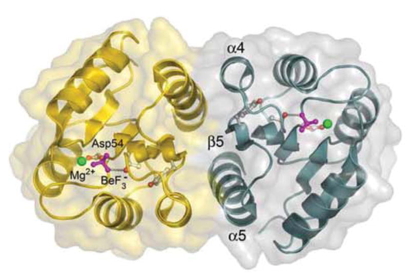
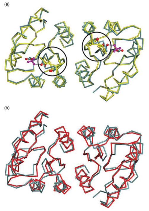
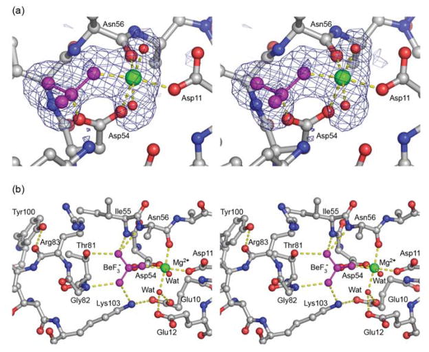
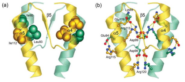
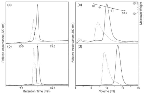
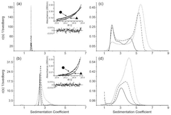
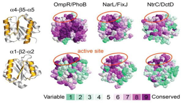
Escherichia coli react to changes from aerobic to anaerobic conditions of growth using the ArcA-ArcB two-component signal transduction system. This system, in conjunction with other proteins, regulates the respiratory metabolic pathways in the organism. ArcA is a member of the OmpR/PhoB subfamily of response regulator transcription factors that are known to regulate transcription by binding in tandem to target DNA direct repeats. It is still unclear in this subfamily how activation by phosphorylation of the regulatory domain of response regulators stimulates DNA binding by the effector domain and how dimerization and domain orientation, as well as intra- and intermolecular interactions, affect this process. In order to address these questions we have solved the crystal structures of the regulatory domain of ArcA in the presence and absence of the phosphoryl analog, BeF3-. In the crystal structures, the regulatory domain of ArcA forms a symmetric dimer mediated by the alpha4-beta5-alpha5 face of the protein and involving a number of residues that are highly conserved in the OmpR/PhoB subfamily. It is hypothesized that members of this subfamily use a common mechanism of regulation by dimerization. Additional biophysical studies were employed to probe the oligomerization state of ArcA, as well as its individual domains, in solution. The solution studies show the propensity of the individual domains to associate into oligomers larger than the dimer observed for the intact protein, and suggest that the C-terminal DNA-binding domain also plays a role in oligomerization.


Figures

Figure 1
Figure 1
Ribbon diagram and surface representation of the BeF3-activated regulatory domain of ArcA. The protomers that make up the dimer are shown in gold and teal. The two-fold rotationally symmetric dimer is mediated by the α4-β5-α5 face, which buries 850 Å2 of surface area at the interface. Side chains of Asp54 and Tyr100 (gray and red), BeF3 (magenta) and the catalytic Mg2+ ion (green) are shown as ball-and-stick models. Figures were created using Pymol (http://www.pymol.org/).
Figure 2
Figure 2
(a) Superposition of the Cα backbone of ArcAN-BeF3 (teal) versus that of ArcAN (yellow). Circles identify the β4-α4 loops, which are found to be in different conformations in the two dimers. (b) Superposition of ArcAN-BeF3 versus MicAN, an OmpR/PhoB subfamily member from S. pneumoniae (red, PDB accession code 1NXW).
Figure 3
Figure 3
Stereo views of the active site of ArcAN-BeF3. (a) Close up view of the active site showing the Fo-Fc electron density for the BeF3 (magenta), Mg2+ (green) and two water molecules (red spheres). The Mg2+ ion is octahedrally coordinated to Asp54, Asn56, Asp11, BeF3 and the two water molecules. The difference density map was calculated with the occupancies of the BeF3, Mg2+ and two water molecules set to zero and contoured at 3 σ. (b) Extended view of the active site. The BeF3 moiety, in addition to serving as a ligand for Mg2+, makes contacts with the side chain oxygen of Thr81, the Nζ of Lys103 and the backbone nitrogens of Gly82, Ile55 and Asn56. Tyr100 stabilizes the β4-α4 loop by forming a hydrogen bond with the carbonyl oxygen of Arg83. Three side chain atoms of Asn56 have been removed for clarity. Carbon, oxygen and nitrogen atoms from the protein chain are shown in gray, red and blue, respectively.
Figure 4
Figure 4
Intermolecular interactions at the α4-β5-α5 dimer interface. (a) The core of a hydrophobic patch (spheres) that brings helices α4 and α5 together is formed between Ile90 (α4), Leu93 (α4) and Ile112 (α5). (b) The interface is further stabilized by an extensive network of salt bridges (ball-and-stick models, yellow dotted lines) formed between Lys89 (α4)-Glu109 (α5), Asp99 (β5)-Arg113 (α5), Glu94 (α4)-Arg115 (α5) and Asp98 (α4-β5 loop)-Arg120 (α5). Protomers are distinguished by colors gold and teal.
Figure 5
Figure 5
Sequence alignment of the α4-β5-α5 face of members from the OmpR/PhoB subfamily from E. coli and other organisms for which structures of the regulatory domain are available. Sequence numbering is for ArcA. Abbreviations are as follow: E.c., E. coli; T.m., T. maritima; S.p., S. pneumoniae; B.s., Bacillus subtilis. Residues mediating the dimer interface of ArcAN-BeF3 are highly conserved in this subfamily. Residues involved in formation of the hydrophobic patch and the intermolecular salt bridges are highlighted in blue and gold, respectively. A red highlight represents residue pairs that are not conserved but could complement each other at the interface. The alignment was performed using sequence data corresponding to the complete regulatory domains. Members used for the alignment are E. coli ArcA (sp: P03026), TorR (sp: P38684), KdpE (sp: P21866), PhoB (sp: P08402), PhoP (sp: P23836), OmpR (sp: P03025), CpxR (sp: P16244), RstA (gb: AAC74680), BasR (sp: P30843), QseB (gb: AAC76061), CusR (gb: AAC73672), PcoR (sp: Q47456), YedW (gb: AAC75035), BaeR (sp: P30846), and CreB (sp: P08368); T. maritima DrrD (gb: AAD35484) and DrrB (gb: AAD35220); S. pneumoniae MicA (emb: CAB54568); B. subtilis PhoP (sp: P13792). Accession numbers are from GenBank (gb), Swiss-Prot (sp), NCBI Reference Sequence (ref) and EMBL Data Library (emb). Members for which a structure is available are E. coli PhoB (PDB accession code 1B00), T. maritima DrrD and DrrB (1KGS and 1P2F, respectively),, S. pneumoniae MicA (1NXW), and B. subtilis PhoP (1MVO).
Figure 6
Figure 6
Analysis of ArcA phosphorylation and oligomerization. Unphosphorylated and phosphorylated proteins are represented by solid and dotted lines, respectively. Phosphoramidate was used as a small molecule phosphodonor. (a and b) Reverse-phase HPLC analysis of the phosphorylation of ArcA (a) and ArcAN (b) at loading concentrations of 128.7 μM and 3.7 μM, respectively. The ability of ArcA and ArcAN to completely phosphorylate in vitro was evident from the observed shifts in migration in the presence of phosphodonor. (c and d) Analytical size-exclusion chromatography of unphosphorylated and phosphorylated ArcA (c) and ArcAN (d). Earlier retention times of the phosphorylated species are consistent with either a monomer to dimer or dimer to tetramer transition. The following molecular weight standards were used for calibration (c) of the size-exclusion column as described in the Materials and Methods: albumin (66 KDa), ovalbumin (44 KDa), trypsin inhibitor (21 KDa), and RNAse A (13.7 KDa). Loading concentrations of 2.2 μM and 128.7 μM were used for ArcA and ArcAN, respectively.
Figure 7
Figure 7
Continuous sedimentation coefficient (c(s)) distributions and sedimentation equilibrium (SE) profiles of ArcAN, ArcAC, and ArcA. (a) c(s) distributions of ArcAN run at loading concentrations of 36 μM (solid), 71 μM (dashed), 143 μM (dotted) collected using an interference optical system. The c(s) distributions were generated as described in the Methods section. The inset shows the SE distributions, collected using absorbance optics (280 nm), for the 36 μM concentration spun at 27 K (circles) and 33 K (triangles) rpm and the residual plots of the fits to a single species model. (b) c(s) distributions for ArcA at 18 μM (solid), 37 μM (dashed), and 73 μM (dotted) collected using interference optics. The inset shows the SE profiles for the 9 μM concentration spun at 27 K (circles) and 33 K (triangles) rpm and the residual plot of the fits to a monomer-dimer model. (c) ArcAC c(s) distributions run at loading concentrations of 20 μM (solid), 41 μM (dashed), and 81 μM (dotted) collected using absorbance optics. (d) ArcAN-P c(s) distributions for ArcAN-P at 36 μM (solid), 71 μM (dashed), and 143 μM (dotted) collected using absorbance optics. The buffer for the SV experiments was 25 mM Tris-Cl, 100 mM NaCl, 1 mM DTT and 1 mM EDTA, pH 7.5 while for the SE experiments it was 25 mM Tris-Cl, 100 mM NaCl and 1 mM DTT, pH 7.5. All sedimentation coefficients have been corrected to the density and viscosity of water at 20 °C.
Figure 8
Figure 8
Consurf (http://consurf.tau.ac.il) analysis of the conservation of surface residues of the regulatory domain for members of the three major subfamilies of RR transcription factors (OmpR/PhoB, NarL/FixJ, and NtrC/DctD). Results from ClustalW multiple sequence alignments were used to map the conservation of residues, using the Consurf maximum likelihood method for calculating conservation scores, onto the structures of ArcAN-BeF3, Sinorhizobium meliloti FixJN-P (PDB accession code 1D5W) and S. enterica NtrCN-P (1DC7) as model members of the OmpR/PhoB, NarL/FixJ and NtrC/DctD subfamilies, respectively. Ribbon diagrams of ArcAN at the far left represent the face of the protein that is shown (gold) for each model. The coloring scheme for conservation ranges from cyan (variable) to dark red (conserved). The results show a highly conserved α4-β5-α5 face for members of the OmpR/PhoB subfamily, in contrast to the other two subfamilies, consistent with a common mechanism of dimerization upon activation. Aside from the active site, marked by orange ellipses, the α1-β2-α2 face is not conserved in any of the three subfamilies. Members of the OmpR/PhoB subfamily used for the sequence alignment are E. coli ArcA (sp: P03026), PhoP (sp: P23836), PhoB (sp: P08402), TorR (sp: P38684), KdpE (sp: P21866), CpxR (sp: P16244), OmpR (sp: P03025), BaeR (sp: P30846), RstA (gb: AAC74680), CusR (gb: AAC73672), BasR (sp: P30843), CreB (sp: P08368), YedW (gb: AAC75035) and QseB (gb: AAC76061); T. maritima DrrB (gb: AAD35220) and DrrD (gb: AAD35484); S. pneumoniae MicA (emb: CAB54568); Shewanella oneidensis YgiX (ref: NP_717707); Staphylococcus epidermidis SrrA (ref: NP_764731); B. subtilis PhoP (sp: P13792). Members of the NarL/FixJ subfamily are E. coli NarL (emb: CAA33023), FimZ (emb: CAA35973), UvrY (gb: AAC74981), RcsB (gb: AAC75277), EvgA (ref: NP_416870), NarP (gb: AAA16411) and UhpA (gb: AAC76692); S. meliloti FixJ (ref: NP_435915); Agrobacterium tumefaciens TraR (ref: NP_059701); Bacillus licheniformis ComA (gb: AAU24816); Listeria monocytogenes DegU (gb: AAT74538); Bordetella bronchiseptica NodW (ref: NP_887648). Members of the NtrC/DctD subfamily are E. coli AtoC (gb: AAC75280), YfhA (gb: AAC75607) and HydG (gb: AAC76978); S. enterica NtrC (gb: AAV79614); Rhodopirellula baltica SasR (ref: NP_869833); Brucella suis NtrC (gb: AAN30037); Xanthomonas axonopodis NtrC (ref: NP_644040); Helicobacter hepaticus FlgR (ref: NP_861166); Burkholderia pseudomallei DctD (emb: CAH37507); A. tumefaciens NtrC (ref: NP_532136). Accession numbers are from GenBank (gb), Swiss-Prot (sp), NCBI Reference Sequence (ref) and EMBL Data Library (emb).

References

    1. Stock AM, Robinson VL, Goudreau PN. Two-component signal transduction. Annu Rev Biochem. 2000;69:183–215. - PubMed
    1. West AH, Stock AM. Histidine kinases and response regulator proteins in two-component signaling systems. Trends Biochem Sci. 2001;26:369–376. - PubMed
    1. Barrett JF, Hoch JA. Two-component signal transduction as a target for microbial anti-infective therapy. Antimicrob Agents Chemother. 1998;42:1529–1536. - PMC - PubMed
    1. Macielag MJ, Goldschmidt R. Inhibitors of bacterial two-component signalling systems. Expert Opin Investig Drugs. 2000;9:2351–2369. - PubMed
    1. Stephenson K, Hoch JA. Developing inhibitors to selectively target two-component and phosphorelay signal transduction systems of pathogenic microorganisms. Curr Med Chem. 2004;11:765–773. - PubMed

Publication types

MeSH terms

LinkOut - more resources