. 2004 Dec 14;101(50):17371-6.
doi: 10.1073/pnas.0407965101. Epub 2004 Dec 1.

Structure of the streptococcal endopeptidase IdeS, a cysteine proteinase with strict specificity for IgG

Affiliations

Structure of the streptococcal endopeptidase IdeS, a cysteine proteinase with strict specificity for IgG

Katja Wenig et al. Proc Natl Acad Sci U S A. .

Abstract

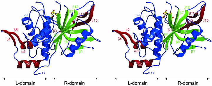
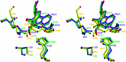
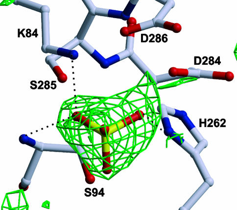
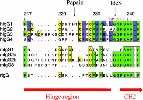
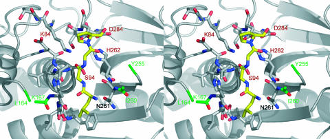
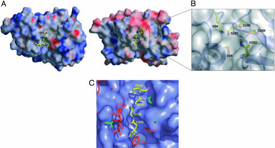
Pathogenic bacteria have developed complex and diverse virulence mechanisms that weaken or disable the host immune defense system. IdeS (IgG-degrading enzyme of Streptococcus pyogenes) is a secreted cysteine endopeptidase from the human pathogen S. pyogenes with an extraordinarily high degree of substrate specificity, catalyzing a single proteolytic cleavage at the lower hinge of human IgG. This proteolytic degradation promotes inhibition of opsonophagocytosis and interferes with the killing of group A Streptococcus. We have determined the crystal structure of the catalytically inactive mutant IdeS-C94S by x-ray crystallography at 1.9-A resolution. Despite negligible sequence homology to known proteinases, the core of the structure resembles the canonical papain fold although with major insertions and a distinct substrate-binding site. Therefore IdeS belongs to a unique family within the CA clan of cysteine proteinases. Based on analogy with inhibitor complexes of papain-like proteinases, we propose a model for substrate binding by IdeS.


Figures

Fig. 1.
Fig. 1.
Stereo ribbon diagram of IdeS-C94S. Compared to papain, additional structural elements are drawn in red, analogue α-helices are shown in blue, and β-strands are shown in green. The catalytic triade residues at the enzyme active site are drawn as in ball-and-stick form.
Fig. 2.
Fig. 2.
Comparison of IdeS-C94S (yellow), papain (green) (Protein Data Bank ID code 1POP), and cathepsin B (purple) (Protein Data Bank ID code 1CSB) active sites. The figure was prepared by superposition of papain and cathepsin B on IdeS and fitting the active-site cysteines on residue 94 of IdeS-C94S.
Fig. 3.
Fig. 3.
Ball-and-stick model of the catalytic triad of IdeS-C94S with the initial FobsFcalc electron density map. This difference in electron density, contoured at 2.5 σ, presumably accounts for a sulfate ion from the crystallization liquid.
Fig. 4.
Fig. 4.
Sequence comparison of the IgG-Fc hinge region and the cleavage site of human, mouse, and rabbit IgG subclasses.
Fig. 5.
Fig. 5.
Stereo top view of the active-site cleft of IdeS-C94S, shown together with a tetrapeptide (yellow, carbon; blue, nitrogen; red, oxygen) derived from the natural cleavage sequence in the hinge peptide of IgG. The first and last visible amino acids of the flexible loop regions of IdeS (Leu-164, Lys-167, Ile-260, and Tyr-255) are shown in green. The orientation of the molecule is that of Fig. 6B.
Fig. 6.
Fig. 6.
Solid surface comparison of the active site of papain and IdeS. (A and B) The molecular surface colored by electrostatic potential of papain in complex with its inhibitor E64 (A, shown as ball-and-stick form, Protein Data Bank ID code 1PE6) in comparison with IdeS-C94S with the modeled tetrapeptide Pro-Gly-Gly-Leu (B, ball-and-stick form) and a clipping of the active site. To facilitate a comparison both structures are oriented equally and show the top view of the active-site cleft (rotated 90° about the x axis toward the viewer relative to Fig. 1). (C) Overlay of papain (Protein Data Bank ID code 1POP) shown as blue surface and IdeS-C94S shown as ball-and-stick in red with the start and end point of the flexible loops shown in green. The modeled tetrapeptide (ball-and-stick form) is shown in front of the molecular surface of the catalytic cleft (yellow, carbon; blue, nitrogen; red, oxygen). Especially the nonprimed site of the active-site cleft is much narrower in IdeS than in papain.

References

    1. Cunningham, M. W. (2000) Clin. Microbiol. Rev. 13, 470–511. - PMC - PubMed
    1. Travis, J. & Potempa, J. (2000) Biochim. Biophys. Acta 1477, 35–50. - PubMed
    1. Travis, J., Potempa, J. & Maeda, H. (1995) Trends Microbiol. 3, 405–407. - PubMed
    1. von Pawel-Rammingen, U., Johansson, B. P. & Björck, L. (2002) EMBO J. 21, 1607–1615. - PMC - PubMed
    1. von Pawel-Rammingen, U., Johansson, B. P., Tapper, H. & Björck, L. (2002) Nat. Med. 8, 1044–1045. - PubMed

Publication types

MeSH terms

Associated data

LinkOut - more resources