. 2005 Jan 18;102(3):622-7.
doi: 10.1073/pnas.0407792102. Epub 2005 Jan 6.

Solution NMR-derived global fold of a monomeric 82-kDa enzyme

Affiliations

Solution NMR-derived global fold of a monomeric 82-kDa enzyme

Vitali Tugarinov et al. Proc Natl Acad Sci U S A. .

Abstract

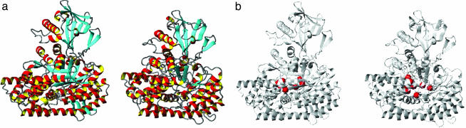
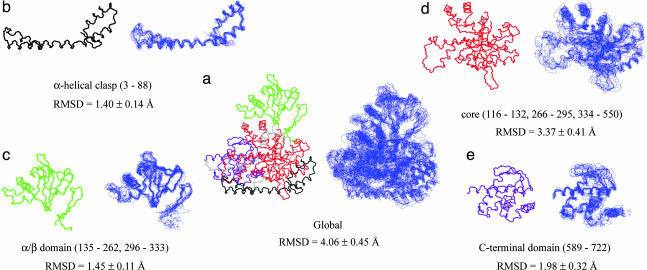
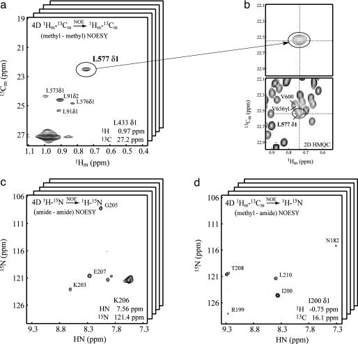
The size of proteins that can be studied by solution NMR spectroscopy has increased significantly because of recent developments in methodology. Important experiments include those that make use of approaches that increase the lifetimes of NMR signals or that define the orientation of internuclear bond vectors with respect to a common molecular frame. The advances in NMR techniques are strongly coupled to isotope labeling methods that increase sensitivity and reduce the complexity of NMR spectra. We show that these developments can be exploited in structural studies of high-molecular-weight, single-polypeptide proteins, and we present the solution global fold of the monomeric 723-residue (82-kDa) enzyme malate synthase G from Escherichia coli, which has been extensively characterized by NMR in the past several years.


Figures

Fig. 2.
Fig. 2.
Comparison of x-ray and NMR-derived structures of MSG. (a) Ribbon diagrams of the x-ray structure of MSG (Left, PDB ID code 1D8C; ref. 17) and the lowest energy NMR structure (Right) calculated on the basis of 1,531 NOE, 1,101 dihedral angle, 415 residual dipolar couplings, and 300 carbonyl-shift restraints. (b) Ribbon representations of MSG (Left shows x-ray structure, and Right shows the lowest-energy NMR structure) are shown with the Cα carbons of residues that either contact or are proximal to glyoxylate (D270, E272, R338, E427, F453, L454, D455, and D631) in the active site of the protein, indicated with red spheres. The image was prepared by using molmol (39).
Fig. 3.
Fig. 3.
Comparison of x-ray and NMR structures on a per-domain basis. (a) The x-ray structure (PDB ID code 1D8C; ref. 17) and the 10 lowest-energy NMR structures of MSG calculated on the basis of experimental restraints. Backbone traces of the x-ray structure (Left) and NMR structures (Right) are displayed and superimposed by aligning residues in elements of regular secondary structure. The α-clasp, α/β, core, and C-terminal domains are shown in black, green, red, and purple, respectively, in the x-ray structure, with the linkers shown in gray. Individual domains [α-clasp (b), α/β (c), core (d), and C-terminal (e)] are shown and superimposed by fitting over residues in regular secondary structure. The rmsd of the NMR ensemble (10 structures) and the x-ray are indicated for heavy backbone atoms of regular secondary structure elements for the entire molecule and individual domains.
Fig. 1.
Fig. 1.
Representative planes from 4D NOE data sets. (a)F1(1H)—F2(13C) plane from the 4D CH3—CH3 NOESY spectrum showing correlations to L433δ1. (b) The correlation involving L577δ1 can be assigned despite the fact that this residue is in a very crowded region of the 2D 1H—13C correlation map. (c)F3(15N)—F4(1HN) plane from the HN—HN 4D data set showing correlations to Lys 206 HN. (d)F3(15N)—F4(1HN) plane from the methyl—HN 4D matrix, showing NOEs between I200δ1 and proximal amide protons.

References

    1. Wüthrich, K. (1986) NMR of Proteins and Nucleic Acids (Wiley, New York).
    1. Lian, L. Y., Barsukov, I. L., Sutcliffe, M. J., Sze, K. H. & Roberts, G. C. (1994) Methods Enzymol. 239, 657-700. - PubMed
    1. Pervushin, K., Riek, R., Wider, G. & Wüthrich, K. (1997) Proc. Natl. Acad. Sci. USA 94, 12366-12371. - PMC - PubMed
    1. Pervushin, K., Riek, R., Wider, G. & Wüthrich, K. (1998) J. Am. Chem. Soc. 120, 6394-6400.
    1. Tugarinov, V., Hwang, P. M., Ollerenshaw, J. E. & Kay, L. E. (2003) J. Am. Chem. Soc. 125, 10420-10428. - PubMed

Publication types

MeSH terms

Associated data