. 2016 Sep 23;291(39):20372-86.
doi: 10.1074/jbc.M116.737577. Epub 2016 Jul 26.

Family-specific Kinesin Structures Reveal Neck-linker Length Based on Initiation of the Coiled-coil

Affiliations

Family-specific Kinesin Structures Reveal Neck-linker Length Based on Initiation of the Coiled-coil

Rebecca K Phillips et al. J Biol Chem. .

Abstract

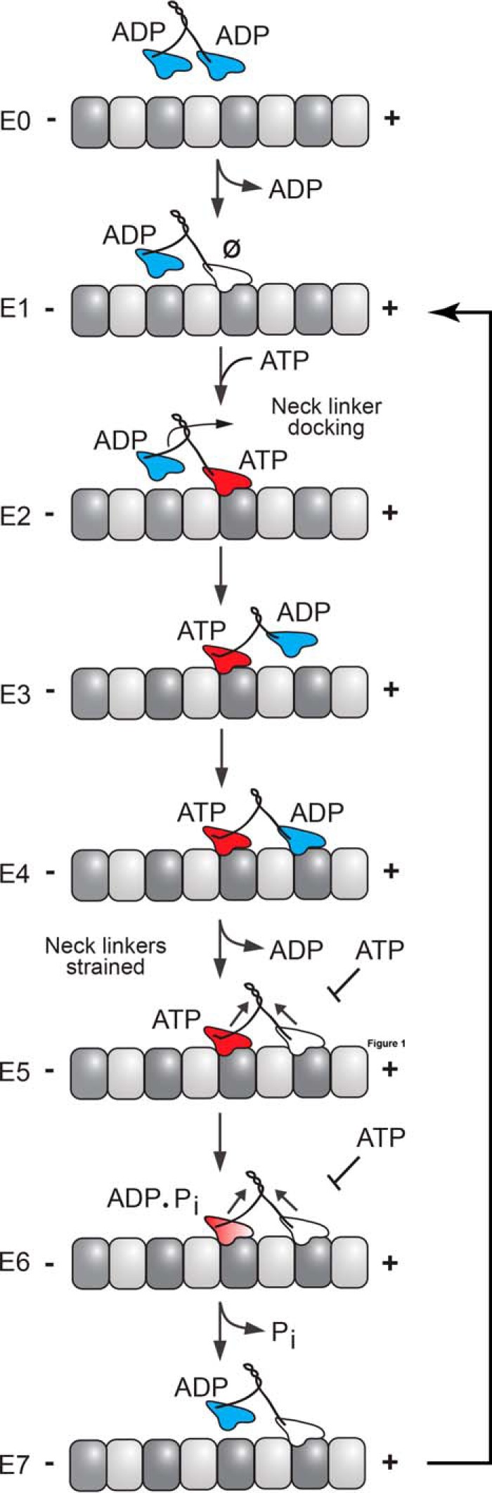
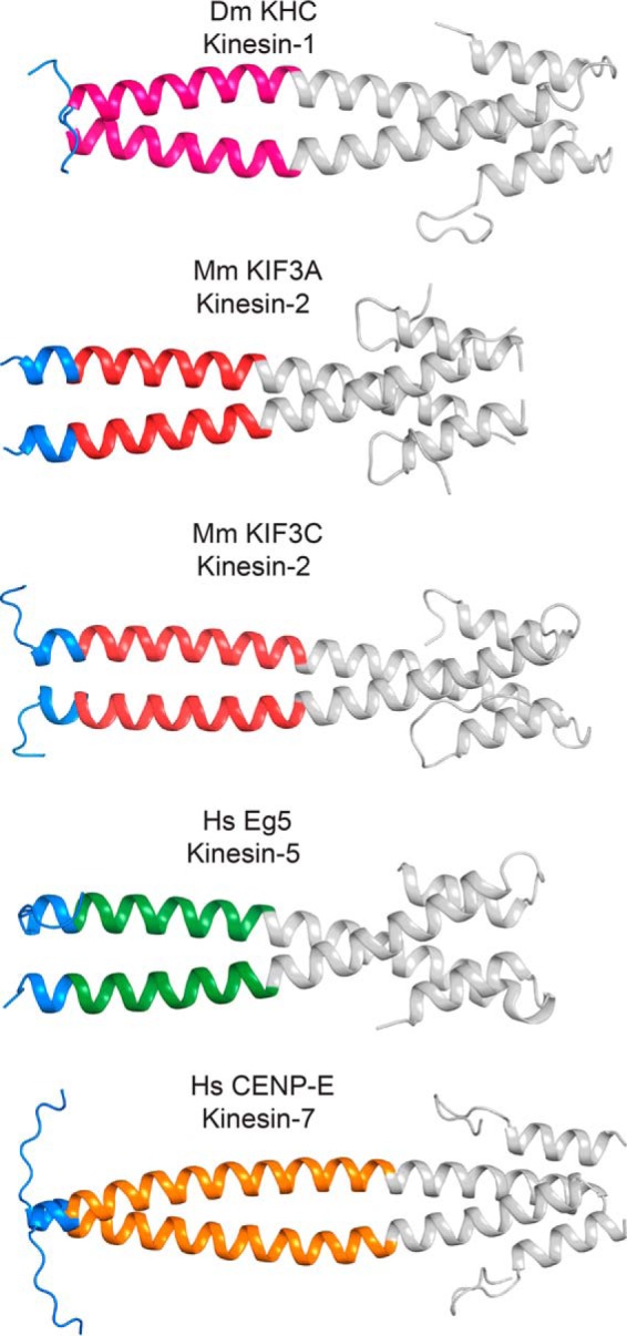
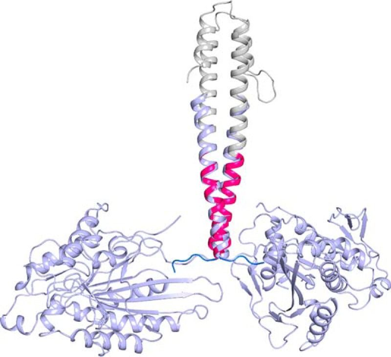
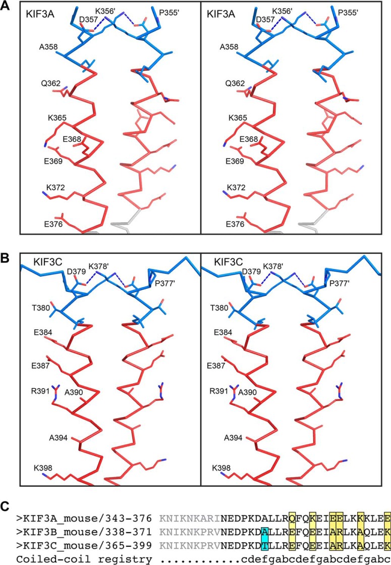
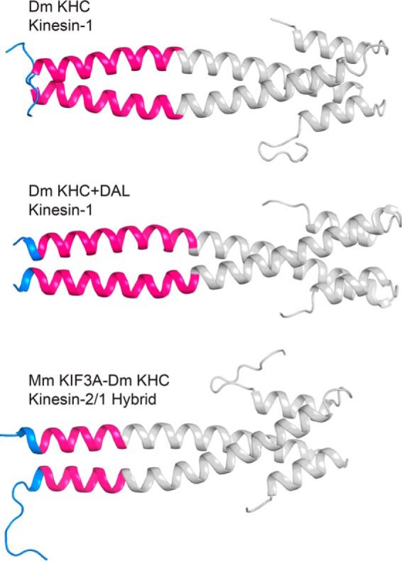
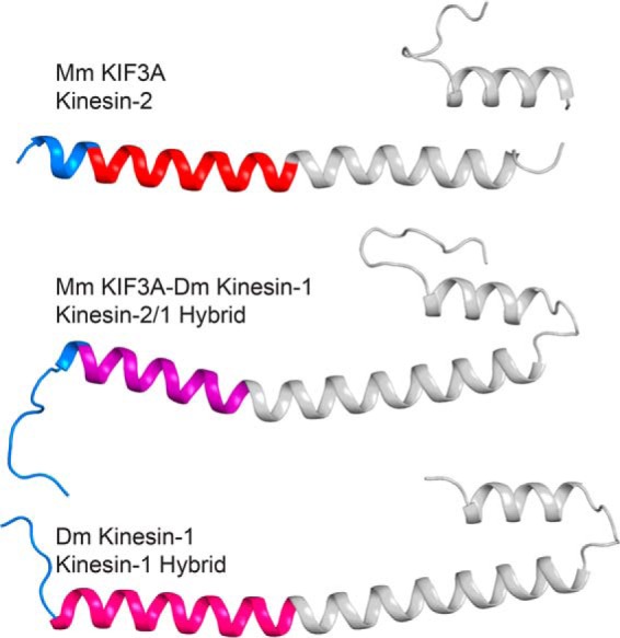
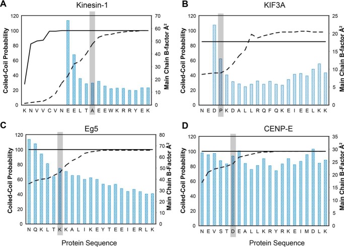
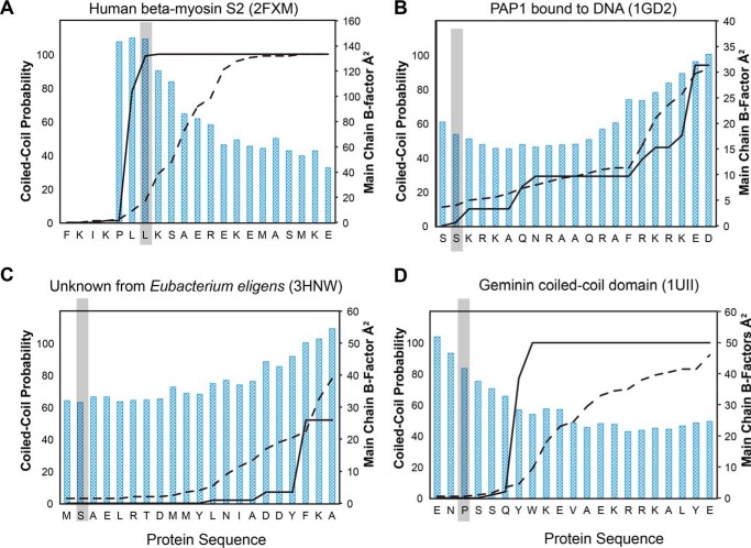
Kinesin-1, -2, -5, and -7 generate processive hand-over-hand 8-nm steps to transport intracellular cargoes toward the microtubule plus end. This processive motility requires gating mechanisms to coordinate the mechanochemical cycles of the two motor heads to sustain the processive run. A key structural element believed to regulate the degree of processivity is the neck-linker, a short peptide of 12-18 residues, which connects the motor domain to its coiled-coil stalk. Although a shorter neck-linker has been correlated with longer run lengths, the structural data to support this hypothesis have been lacking. To test this hypothesis, seven kinesin structures were determined by x-ray crystallography. Each included the neck-linker motif, followed by helix α7 that constitutes the start of the coiled-coil stalk. In the majority of the structures, the neck-linker length differed from predictions because helix α7, which initiates the coiled-coil, started earlier in the sequence than predicted. A further examination of structures in the Protein Data Bank reveals that there is a great disparity between the predicted and observed starting residues. This suggests that an accurate prediction of the start of a coiled-coil is currently difficult to achieve. These results are significant because they now exclude simple comparisons between members of the kinesin superfamily and add a further layer of complexity when interpreting the results of mutagenesis or protein fusion. They also re-emphasize the need to consider factors beyond the kinesin neck-linker motif when attempting to understand how inter-head communication is tuned to achieve the degree of processivity required for cellular function.

Keywords: coiled-coil; kinesin; kinesin neck-linker; microtubule; molecular motor; protein structure; x-ray crystallography.


Figures

FIGURE 1.
FIGURE 1.
Schematic representation of the kinesin chemomechanical cycle that illustrates key transitions that influence processivity. A processive run is started by binding of either head followed by rapid ADP release to form the E1 intermediate where the leading head that forms the initial contact is microtubule-bound but nucleotide-free (Ø), whereas the trailing head is detached with ADP tightly bound. ATP binding to the leading head induces a series of structural transitions, including neck-linker docking that allows the trailing ADP-head to move 16 nm ahead to its new microtubule-binding site (E2–E4). ADP release from the new leading head (E4 and E5) results in the E5 two-head bound state, thereby generating intermolecular strain, which inhibits ATP binding at the now leading head. ATP hydrolysis at the trailing head followed by phosphate (Pi) release generates a microtubule weakly bound ADP state (E6 and E7). Detachment of the trailing head relieves the intermolecular strain (E7), and initiates the next motor cycle.
FIGURE 2.
FIGURE 2.
Crystal structures of kinesin-1, -2, -5, and -7 reveal unique class-specific neck-linker α7 neck-coil domains. The neck-linker motif predicted to occur based on the earlier studies of kinesin-1 is colored blue. Helix α7, as observed in kinesin-1, is colored according to the kinesin family to which it belongs. EB1, a coiled-coil fusion protein, is colored gray. There are varied amounts of ordered neck-linker motifs. These structures show that the true start of helix α7 is variable across the kinesin superfamily. Table 4 provides the protein sequence of each fusion protein and their coiled-coil registry. Figs. 2–6 were prepared in part with PyMOL.
FIGURE 3.
FIGURE 3.
Overlay of the kinesin-1 neck-linker structure and dimeric kinesin-1 crystal structure (PDB code 3KIN) (14). The kinesin-1 neck-linker is colored as in Fig. 2. Dimeric kinesin-1 is shown in light blue.
FIGURE 4.
FIGURE 4.
Selected side-chain interactions in and the sequence differences between KIF3A and KIF3C neck-linkers shown in stereo. KIF3A and KIF3C are colored as in Fig. 2, with side chains shown as sticks and colored by element. In both KIF3A (A) and KIF3C (B), there is a hydrogen bonding interaction between a lysine and an aspartate that stabilizes part of the coiled-coil. The sequences of the neck-linker and α7 are also shown (C) where the residues depicted in gray were not included in the constructs but represent the full-length linker. Interestingly, none of the differences in sequence between KIF3A and KIF3C are predicted to influence formation of a heterodimer.
FIGURE 5.
FIGURE 5.
Crystal structures of native D. melanogaster kinesin-1 neck-linker protein, kinesin-1 + DAL, and the hybrid of the M. musculus KIF3A neck-linker with the kinesin-1 helix α7 coil. All are fused to EB1 dimerization domain (gray). Each helix α7 domain, as predicted based on the earlier studies of kinesin-1, is colored hot pink with the neck-linker peptide colored blue.
FIGURE 6.
FIGURE 6.
Comparison of KIF3A, the KIF3A-kinesin-1 hybrid protein, and kinesin-1 structures. Predicted neck-linkers are shown in blue and the EB1 domain in gray. KIF3A is at the top with its helix α7 colored red, KIF3A-kinesin-1 hybrid protein in the middle with its helix α7 in purple, and kinesin-1 at the bottom with its helix α7 in hot pink. Note the variability in neck-linker length based on the start of helix α7.
FIGURE 7.
FIGURE 7.
Predicted coiled-coil probability and temperature factors versus sequence for the class-specific kinesins. A, kinesin-1; B, KIF3A; C, Eg5; and D, CENP-E. The COILS-28 probabilities are shown as a solid line with MARCOIL probabilities shown with a dashed line. The start residue of the helix α7 coil as determined in the x-ray crystal structure is highlighted in gray. The predicted starting residues of the coiled-coil for KIF3A, Eg5, and CENP-E do not agree with the observed coiled-coil in the x-ray structures.
FIGURE 8.
FIGURE 8.
Coiled-coil probability and temperature factors versus sequence for selected PDB files. COILS-28 probabilities shown as a solid line and MARCOIL probabilities shown as a dashed line. The start of the coiled-coil is highlighted in gray. All sequences begin on a g residue. PDB codes included are (A) 2FXM (63), (B) 1GD2 (64), (C) 3HNW, and (D) 1UII (79).

References

    1. Crick F. H. (1953) The packing of α-helices: simple coiled-coils. Acta Crystallogr. 6, 689–697
    1. Rackham O. J., Madera M., Armstrong C. T., Vincent T. L., Woolfson D. N., and Gough J. (2010) The evolution and structure prediction of coiled coils across all genomes. J. Mol. Biol. 403, 480–493 - PubMed
    1. Delorenzi M., and Speed T. (2002) An HMM model for coiled-coil domains and a comparison with PSSM-based predictions. Bioinformatics 18, 617–625 - PubMed
    1. Lupas A., Van Dyke M., and Stock J. (1991) Predicting coiled coils from protein sequences. Science 252, 1162–1164 - PubMed
    1. Parry D. A., Fraser R. D., and Squire J. M. (2008) Fifty years of coiled-coils and α-helical bundles: a close relationship between sequence and structure. J. Struct. Biol. 163, 258–269 - PubMed

LinkOut - more resources