. 2005 Apr 26;102(17):5981-6.
doi: 10.1073/pnas.0502101102. Epub 2005 Apr 18.

Crystal structure of prostate-specific membrane antigen, a tumor marker and peptidase

Affiliations

Crystal structure of prostate-specific membrane antigen, a tumor marker and peptidase

Mindy I Davis et al. Proc Natl Acad Sci U S A. .

Abstract

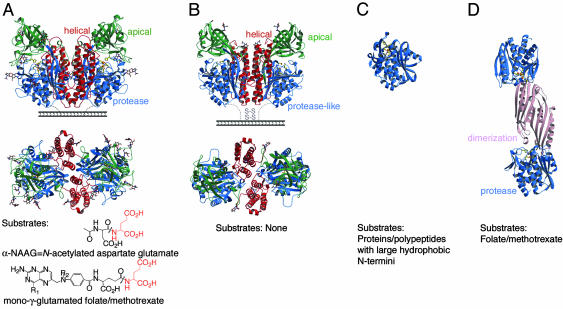
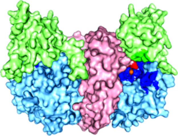
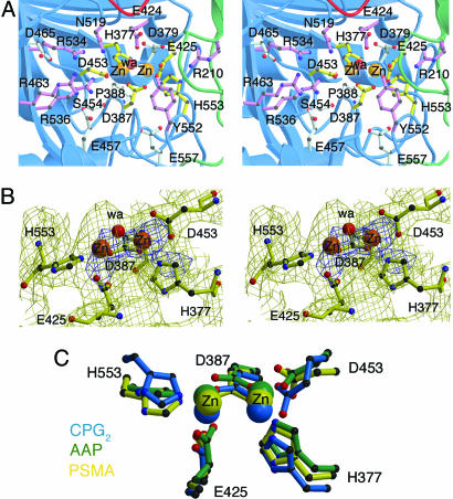
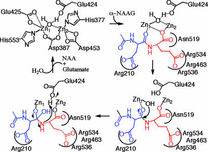
Prostate-specific membrane antigen (PSMA) is highly expressed in prostate cancer cells and nonprostatic solid tumor neovasculature and is a target for anticancer imaging and therapeutic agents. PSMA acts as a glutamate carboxypeptidase (GCPII) on small molecule substrates, including folate, the anticancer drug methotrexate, and the neuropeptide N-acetyl-l-aspartyl-l-glutamate. Here we present the 3.5-A crystal structure of the PSMA ectodomain, which reveals a homodimer with structural similarity to transferrin receptor, a receptor for iron-loaded transferrin that lacks protease activity. Unlike transferrin receptor, the protease domain of PSMA contains a binuclear zinc site, catalytic residues, and a proposed substrate-binding arginine patch. Elucidation of the PSMA structure combined with docking studies and a proposed catalytic mechanism provides insight into the recognition of inhibitors and the natural substrate N-acetyl-l-aspartyl-l-glutamate. The PSMA structure will facilitate development of chemotherapeutics, cancer-imaging agents, and agents for treatment of neurological disorders.


Figures

Fig. 1.
Fig. 1.
Structural comparison of PSMA and related proteins. The common fold of the protease or protease-like domain consists of a central seven- to eight-stranded mixed β-sheet and 6-11 flanking α-helices. Ribbon diagrams of side and top view of PSMA (A), side and top view of TfR1 (PDB ID code 1CX8) (B), AAP (PDB ID code 1RTQ) (C), CPG2 (PDB ID code 1CG2) (D). The protease (or protease-like) domains are shown in blue, the apical domains of PSMA and TfR1 in green, the helical domains in red, and the CPG2 dimerization domain in pink. Water molecules are red, zinc ions are orange, zinc-binding residues are yellow sticks, and carbohydrate residues are shown in a color-coded ball-and-stick representation. Representative substrates are shown below the appropriate molecule. R1 = OH or NH2, and R2 = H or CH3 for folate and methotrexate, respectively. Note: A mono-γ-glutamated folate is shown for clarity; in poly-γ-glutamated folate/methotrexate, the additional glutamates (up to seven) are attached to the glutamate shown via a γ-peptide linkage.
Fig. 2.
Fig. 2.
A surface rendering in which the helical domain is light red, the apical domain is light green, the protease domain is light blue, and zinc ions are orange. The residues lining the substrate-binding cavity are highlighted in a darker version of the color corresponding to the domain from which the residue originates.
Fig. 3.
Fig. 3.
The PSMA active site. (A) Stereoview of the PSMA active site. Zinc ions are orange spheres, and a water molecule is shown as a red sphere. Zinc-binding residues are yellow sticks, water- or substrate-binding ligands are purple sticks, and residues with structural roles are light blue sticks. (B) Stereoview of the PSMA model in the region of the PSMA active site superimposed on a 3.5-Å 2Fobs-Fcalc annealed omit electron density map contoured at 1σ (yellow) and 4σ (blue). The zinc ions and zinc-binding residues were omitted from the model before generation of the map. Some of the extra density corresponds to nonzinc-binding residues that were omitted from the figure for clarity. (C) Overlay of the active sites of PSMA (yellow), aminopeptidase AAP (green) (PDB ID code 1RTQ), and carboxypeptidase CPG2 (blue) (PDB ID code 1CG2) with the zinc ions as spheres and the zinc-binding residues in ball-and-stick representation.
Fig. 4.
Fig. 4.
A mechanistic scheme for α-NAAG (glutamate is red, and NAA is blue) binding and cleavage by PSMA (black). The zinc-binding PSMA residues are assumed to remain bound during the reaction mechanism and are omitted in the substrate-binding and cleavage steps for clarity. The C-terminal carboxyl group binds the zinc ions in a bridging fashion, and there are electrostatic interactions between the glutamate side chain and the arginine patch and the aspartate side chain and Arg-210. The zinc-bound water molecule is poised to attack the peptide bond of the bound substrate, leading to a tetrahedral intermediate, bond cleavage, and product dissociation. An analogous mechanism would be expected for the cleavage of α-NAAG by NLD2 (2).

References

    1. Ghosh, A. & Heston, W. D. W. (2004) J. Cell. Biochem. 91, 528-539. - PubMed
    1. Pangalos, M. N., Neefs, J. M., Somers, M., Verhasselt, P., Bekkers, M., van der Helm, L., Fraiponts, E., Ashton, D. & Gordon, R. D. (1999) J. Biol. Chem. 274, 8470-8483. - PubMed
    1. Berger, U. V., Luthi-Carter, R., Passani, L. A., Elkabes, S., Black, I., Konradi, C. & Coyle, J. T. (1999) J. Comp. Neurol. 415, 52-64. - PubMed
    1. Gong, M. C., Chang, S. S., Sadelain, M., Bander, N. H. & Heston, W. D. W. (1999) Cancer Metastasis Rev. 18, 483-490. - PubMed
    1. Elgamal, A. A., Holmes, E. H., Su, S. L., Tino, W. T., Simmons, S. J., Peterson, M., Greene, T. G., Boynton, A. L. & Murphy, G. P. (2000) Semin. Surg. Oncol. 18, 10-16. - PubMed

Publication types

Associated data