. 2005 Oct 18;102(42):14994-9.
doi: 10.1073/pnas.0506254102. Epub 2005 Oct 7.

Crystal structure of the archaeal ammonium transporter Amt-1 from Archaeoglobus fulgidus

Affiliations

Crystal structure of the archaeal ammonium transporter Amt-1 from Archaeoglobus fulgidus

Susana L A Andrade et al. Proc Natl Acad Sci U S A. .

Abstract

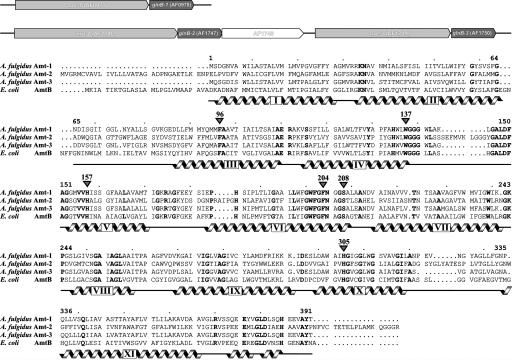
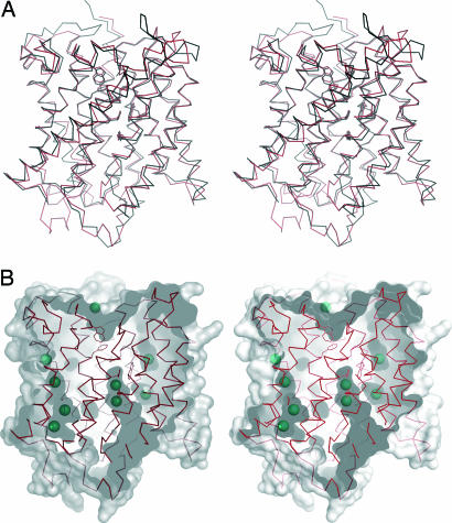
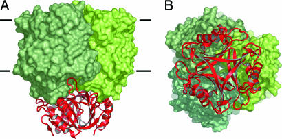
Ammonium transporters (Amts) are integral membrane proteins found in all kingdoms of life that fulfill an essential function in the uptake of reduced nitrogen for biosynthetic purposes. Amt-1 is one of three Amts encoded in the genome of the hyperthermophilic archaeon Archaeoglobus fulgidus. The crystal structure of Amt-1 shows a compact trimer with 11 transmembrane helices per monomer and a central channel for substrate conduction in each monomer, similar to the known crystal structure of AmtB from Escherichia coli. Xenon derivatization has been used to identify apolar regions of Amt-1, emphasizing not only the hydrophobicity of the substrate channel but also the unexpected presence of extensive internal cavities that should be detrimental for protein stability. The substrates ammonium and methylammonium have been used for cocrystallization experiments with Amt-1, but the identification of binding sites that are distinct from water positions is not unambiguous. The well ordered cytoplasmic C terminus of the protein in the Amt-1 structure has allowed for the construction of a docking model between Amt-1 and a homology model for its physiological interaction partner, the P(II) protein GlnB-1. In this model, GlnB-1 binds tightly to the cytoplasmic face of the transporter, effectively blocking conduction through the three individual substrate channels.


Figures

Fig. 1.
Fig. 1.
Amts of A. fulgidus. (Upper) The genome of the archaeon contains three copies of paired genes for an Amt and a protein of the PII family. (Lower) Sequence alignment of A. fulgidus Amt-1–3 and E. coli AmtB. Helices, as derived from the crystal structures, are marked in gray, the numbering corresponds to Amt-1.
Fig. 2.
Fig. 2.
Structure of A. fulgidus Amt-1. (A) Stereo representation of the Amt-1 monomer. The membrane is indicated in gray, with the extracellular side above and the cytoplasmic side below. The protein chain is colored from blue at the N terminus to red at the C terminus, the 11 transmembrane helices numbered in accordance with Fig. 1. (B) The Amt-1 trimer seen from the extracellular side. (C) B factor representation of Amt-1. The region with the highest B factors is in the loop between helices V and VI, connecting the pseudosymmetry-related halves of the protein.
Fig. 3.
Fig. 3.
Structural details of Amt-1. (A) Structural comparison of Amt-1 (red) with E. coli AmtB (black). (B) Xenon binding to internal cavities in Amt-1. The surface representation shows the substrate channel and several internal cavities as well as the positions of 9 of a total of 15 Xe atoms located in the structure. The residues depicted as sticks are the same as in Fig. 4.
Fig. 4.
Fig. 4.
Ammonium recruitment site and substrate channel. formula image presumably forms a hydrogen bond to the side chain of S208 on the extracellular side of Amt-1 (at the top), stabilized by a π interaction with the side chain of W137. The hydrophobic channel leading to the cytoplasmic side is blocked by F96 and F204, with the latter having significantly elevated B factors, an indication of structural flexibility. The Nδ atoms of two conserved histidine residues, H157 and H305, are in hydrogen-bonding distance and their imidazole rings are almost perfectly coplanar.
Fig. 5.
Fig. 5.
Docking model for the interaction of Amt-1 with GlnB-1. (A) Side view. GlnB-1 binds tightly to the transporter, with the protruding T loops inserting deeply into the substrate channels. (B) View from the cytoplasmic side. Both GlnB-1 and Amt-1 are stable trimers, and in the docked state, their threefold axes coincide.

References

    1. Kleiner, D. (1985) FEMS Microbiol. Rev. 32, 87–100.
    1. Lide, D. R. (2001) Handbook of Chemistry and Physics (Chemical Rubber Publishing, Cleveland).
    1. Marini, A. M., Vissers, S., Urrestarazu, A. & Andre, B. (1994) EMBO J. 13, 3456–3463. - PMC - PubMed
    1. Ludewig, U., von Wirén, N., Rentsch, D. & Frommer, W. B. (2001) Genome Biol. 2, 1010.1–1010.5. - PMC - PubMed
    1. Blakey, D., Leech, A., Thomas, G. H., Coutts, G., Findlay, K. & Merrick, M. (2002) Biochem. J. 364, 527–535. - PMC - PubMed

Publication types

MeSH terms

LinkOut - more resources