Comparative Study
. 2006 Dec 8;24(5):653-663.
doi: 10.1016/j.molcel.2006.10.024.

Recognition of a functional peroxisome type 1 target by the dynamic import receptor pex5p

Affiliations
Comparative Study

Recognition of a functional peroxisome type 1 target by the dynamic import receptor pex5p

Will A Stanley et al. Mol Cell. .

Abstract

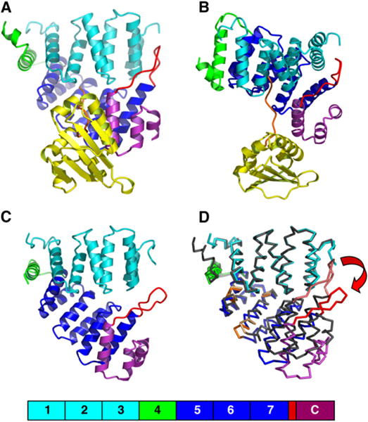
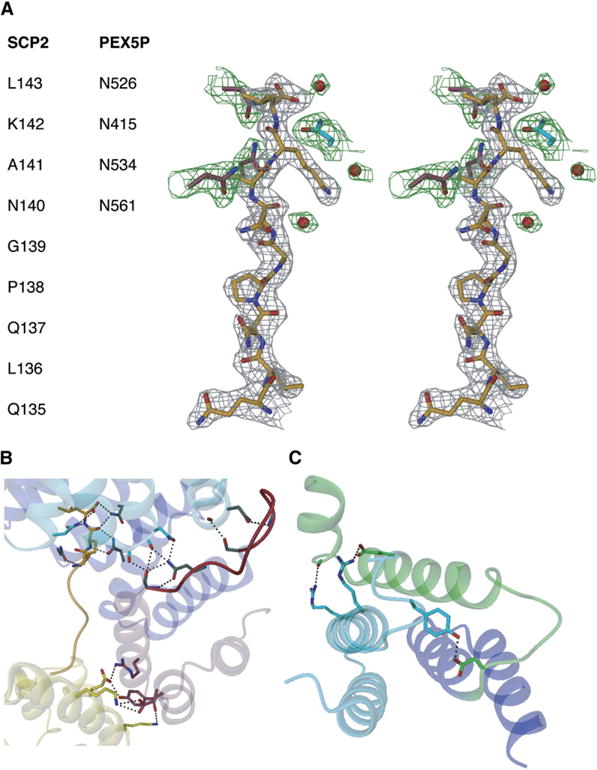
Peroxisomes require the translocation of folded and functional target proteins of various sizes across the peroxisomal membrane. We have investigated the structure and function of the principal import receptor Pex5p, which recognizes targets bearing a C-terminal peroxisomal targeting signal type 1. Crystal structures of the receptor in the presence and absence of a peroxisomal target, sterol carrier protein 2, reveal major structural changes from an open, snail-like conformation into a closed, circular conformation. These changes are caused by a long loop C terminal to the 7-fold tetratricopeptide repeat segments. Mutations in residues of this loop lead to defects in peroxisomal import in human fibroblasts. The structure of the receptor/cargo complex demonstrates that the primary receptor-binding site of the cargo is structurally and topologically autonomous, enabling the cargo to retain its native structure and function.


Figures

Figure 1
Figure 1. Sequence/Structure Relationships in Human Pex5p(C) and Human mSCP2
The positions of labeled secondary structural elements are depicted by cylinders and arrows. Color coding for Pex5p(C) in (A) is as follows: TPR1-TPR3, cyan; TPR4, green; TPR5-TPR7, blue; 7C loop, connecting TPR7 and the C-terminal helical bundle, red; and C terminus, maroon. Color coding for mSCP2 in (B) is as follows: core domain, yellow; and C terminus including PTS1 motif, orange. Residues of Pex5p and SCP2 involved in cargo and receptor binding, respectively, have been identified using the program AREAIMOL of the CCP4 suite (CCP4, 1994) and are indicated in colors matching the bound sequence segments. Conserved residues have been identified from multiple sequence alignments using BLAST/MVIEW (Brown et al., 1998). In the “cons” line, residues exhibiting 90% and 70% homology to the available sequences are indicated by upper-case and lower-case characters, respectively. Residues that were identified by Klein et al. (2001) and Shimozawa et al. (1999) as being involved in Pex5p receptor/cargo interactions are shown in red and blue colors. TPR motif signature residues according to the criteria of D’Andrea and Regan (2003) are underlined. Residue segments that function as hinge regions (496–500, 523 to 524, and 533–537), triggering the conformational changes observed for the cargo-loaded and apo-Pex5p(C) receptor, are highlighted by orange bars that have been inserted into the corresponding secondary structural elements.
Figure 2
Figure 2. Lipid Binding to mSCP2 in the Absence and Presence of Pex5p(C)
(A and B) Binding of a spin-labeled lipid molecule (5-doxylstearic acid, 5DSA) attenuates the peak intensity in 1H,15N correlation spectra due to PRE. Spectra in the presence of oxidized (i.e., paramagnetic) and reduced lipid are shown in green and black, respectively. Residues Thr105 and Gly106, which are located in the center of the lipid-binding pocket, are entirely bleached. Gly139, which is proximal to the PTS1 motif, experiences a large chemical shift perturbation in the presence of Pex5p(C). It is less bleached in the Pex5p(C) complex, consistent with the strongly reduced mobility of the C-terminal residues and the increased distance to the lipid ligand. (C and D) Comparison of the lipid-binding pocket of mSCP2 (Garcia et al., 2000), in the presence/absence of Pex5p(C). The degree of attenuation of NMR signals due to PRE is colored in green on a ribbon representation of mSCP2. Amide protons of residues with a more than 7-fold reduction in peak intensities are depicted by green spheres.
Figure 3
Figure 3. Structures of the Peroxisomal Import Receptor Pex5p(C) in the Presence and in the Absence of the Cargo mSCP2
Color coding for Pex5p(C) is as follows: TPR1-TPR3, cyan; TPR4, green; TPR5-TPR7, blue; 7C loop, connecting TPR7 and the C-terminal helical bundle, red; and C terminus, maroon. Color coding for mSCP2 is as follows: core domain, yellow; and C terminus including PTS1 motif, orange. The orientation of the receptor in (A) and (C) is identical. The ribbon of the Pex5p(C)/mSCP2 complex in (B) has been rotated by 60° around a horizontal axis within the paper plane with respect to the orientation in (A), to illustrate the mode of mSCP2 binding to the receptor. (D) Superimposed Pex5p(C) receptor structures in the presence and in the absence of mSCP2. The colors of the trace of the cargo-loaded conformation are as in (A)–(C), except that the conformational hinge regions are colored in orange. The trace of the apo-Pex5p(C) structure is in gray, except for the 7C loop, which is colored in faint red. The coordinates of TPR segments 1–4 were used for structural superposition using the program SSM (Krissinel and Henrick, 2004) (rmsd = 0.78 Å for 164 common residues). The largest structural deviations of up to 8 Å are observed at the 7C loop and adjacent regions and are indicated by a red arrow.
Figure 4
Figure 4. Surface Presentations of the Peroxisomal Import Receptor Pex5p(C) in the Presence and in the Absence of the Cargo mSCP2
The right panel structures are rotated by 45° with respect to those in the left panels by a horizontal axis within the paper plane. The color codes are as in Figure 3. While the structure of the Pex5p(C)/mSCP2 complex is shown in (A), only the structure of the cargo-loaded conformation of the Pex5p(C) receptor is displayed in (B). The PTS1- and secondary mSCP2-binding areas are mapped onto the Pex5p(C) surface in their respective colors (orange, yellow). In the structure of the apo-Pex5p(C) receptor (C), the approximate location of the PTS1-binding site, as determined from the Pex5p(C)/mSCP2 complex, is indicated by an orange circle. Conformational changes of several residues at this site lead to disappearance of the open tunnel, observed in the Pex5p(C)/mSCP2 complex (B). In the apo conformation, the 7C-loop region (red) is well separated from the remaining TPR segments of the receptor.
Figure 5
Figure 5. Structural Determinants of mSCP2 Cargo Loading onto Pex5p(C)
(A) Stereo view of the 2FO − FC electron density, using phases from the refined model and contoured at 1σ, of the PTS1 motif from mSCP2 (gray) and some interacting residues from Pex5p and ordered solvent molecules (dark green). (B) Pex5p(C)/mSCP2 complex formation by two distinct interfaces: C-terminal PTS1 motif from mSCP2 (orange)-central cavity of the circular TPR motif structure from Pex5p; secondary surface from mSCP2-C-terminal helical bundle from Pex5p. Ser600 is in a central position between the two surface patches, allowing the proper arrangement of the two cargo surface patches of Pex5p to support binding of mSCP2. The C terminus of the 7C loop (red) interacts by a few hydrogen bonds with the TPR1 segment. (C) TPR4 motif of Pex5p(C), as observed in the cargo-loaded structure of the receptor. Specific interactions between TPR3 and TPR4, generating a circular conformation of Pex5p(C), are shown. Colors are as in Figures 3 and 4, except that some of the bonds of residues from the C-terminal TPR motifs 5–7 and the 7C loop are colored in gray to allow illustrations of oxygen and nitrogen atoms. Hydrogen bonds are shown by dashed lines.
Figure 6
Figure 6. 7C-Loop Mutants Lead to Functional PTS1 Import Defects
Pex5p-deficient fibroblast cells from Zellweger patient PBD005 were cotransfected with a PTS2-tagged CacT-expressing plasmid, pEGFP-SCP2, and plasmids expressing either WT Pex5p or a range of different single-residue mutants (N382A, Q586R, S589Y, and S600W). At 24 hr after transfection, PTS2-CAcT ([A], red color) and endogenous catalase ([B], red color) were labeled by immunofluorescence, while EGFP-SCP2 was detected by direct fluorescence ([A] and [B], green color). In cells expressing WT Pex5p, both marker proteins and EGFP-SCP2 colocalized in peroxisomes ([A] and [B], yellow color). All Pex5p mutants were capable of restoring the PTS2 import defect of PEX5-deficient cells. In contrast, all mutants were impaired in catalase import and showed more or less pronounced import defects for EGFP-SCP2. The introduction of the 7C-loop mutations Q586R and S600W resulted in an inefficient SCP2 import as indicated by the diffuse cytosolic staining and only weak labeling of peroxisomes in the representative cells. Strikingly, in cells expressing the Pex5p mutants S589Y and N382A, both cytosolic and peroxisomal localizations of SCP2 were found, while the same cells were devoid of functional catalase import as indicated by the lack of a congruent punctate pattern.

References

    1. Agne B, Meindl NM, Niederhoff K, Einwachter H, Rehling P, Sickmann A, Meyer HE, Girzalsky W, Kunau WH. Pex8p: an intraperoxisomal organizer of the peroxisomal import machinery. Mol Cell. 2003;11:635–646. - PubMed
    1. Battiste JL, Wagner G. Utilization of site-directed spin labeling and high-resolution heteronuclear nuclear magnetic resonance for global fold determination of large proteins with limited nuclear overhauser effect data. Biochemistry. 2000;39:5355–5365. - PubMed
    1. Braverman N, Dodt G, Gould SJ, Valle D. An isoform of Pex5p, the human PTS1 receptor, is required for the import of PTS2 proteins into peroxisomes. Hum Mol Genet. 1998;7:1195–1205. - PubMed
    1. Brocard CB, Jedeszko C, Song HC, Terlecky SR, Walton PA. Protein structure and import into the peroxisomal matrix. Traffic. 2003;4:74–82. - PubMed
    1. Brown NP, Leroy C, Sander C. MView: a web-compatible database search or multiple alignment viewer. Bioinformatics. 1998;14:380–381. - PubMed

Publication types

Substances