. 2010 Aug 11;18(8):996-1010.
doi: 10.1016/j.str.2010.04.018.

Crystal structures of Phd-Doc, HigA, and YeeU establish multiple evolutionary links between microbial growth-regulating toxin-antitoxin systems

Affiliations

Crystal structures of Phd-Doc, HigA, and YeeU establish multiple evolutionary links between microbial growth-regulating toxin-antitoxin systems

Mark A Arbing et al. Structure. .

Abstract

Bacterial toxin-antitoxin (TA) systems serve a variety of physiological functions including regulation of cell growth and maintenance of foreign genetic elements. Sequence analyses suggest that TA families are linked by complex evolutionary relationships reflecting likely swapping of functional domains between different TA families. Our crystal structures of Phd-Doc from bacteriophage P1, the HigA antitoxin from Escherichia coli CFT073, and YeeU of the YeeUWV systems from E. coli K12 and Shigella flexneri confirm this inference and reveal additional, unanticipated structural relationships. The growth-regulating Doc toxin exhibits structural similarity to secreted virulence factors that are toxic for eukaryotic target cells. The Phd antitoxin possesses the same fold as both the YefM and NE2111 antitoxins that inhibit structurally unrelated toxins. YeeU, which has an antitoxin-like activity that represses toxin expression, is structurally similar to the ribosome-interacting toxins YoeB and RelE. These observations suggest extensive functional exchanges have occurred between TA systems during bacterial evolution.


Figures

Figure 1.
Figure 1.. Evolutionary Relationships between Components of Bacterial Toxin-Antitoxin Systems
Cladograms modeling the evolutionary relationship between a representative member of toxin (left) and antitoxin (right) families were prepared using MEGA (as described in Experimental Procedures). Structurally related proteins are grouped together into single cladograms. The double-headed arrows connect cognate toxins and antitoxins from representative TA systems. Branches of families that have a member whose structure has been experimentally determined are labeled in red.
Figure 2.
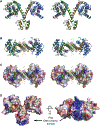
Figure 2.. The Structure of the Heterotetrameric Phd-Doc Complex
(A) Stereo view of the 2:2 Phd-Doc complex. Phd molecules are colored green and brown, while Doc molecules are colored blue and dark teal. (B) Another stereo view of the 2:2 Phd-Doc complex highlighting the Phd-Doc interactions (rotated 90° around a horizontal axis compared with the view in A). Charged residues in the Phd neutralization domain that interact with charged Doc residues are shown in stick representation. Residues implicated in Doc function (His-66, Asp-70) are colored yellow and orange for contrast and are shown in stick representation. (C) Surface representation of Doc colored according to electrostatic potential, with blue and red, respectively, representing surface potentials of 5 kT and —5 kT in 100 mM salt (oriented as in B). Charged residues in the neutralization domain of Phd are shown in stick representation. (D) Two views of the molecular surface of the Phd- Doc complex colored by electrostatic potential using the same parameters. The left panel is in the same orientation as (A), whereas the right panel is rotated 180° compared to the view in (B).
Figure 3.
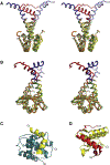
Figure 3.. Homologous Phd/YefM/NE2111 Antitoxins Neutralize Toxins with Disparate Folds
(A and B) Stereo view of the superposition of Phd with EcYefM and NE2111 antitoxins. In (A), Phd is in the same orientation as in Figure 2A, whereas in (B), the antitoxins are rotated by 90_. The DNA-binding domains are in green (Phd), yellow (EcYefM), and orange (NE2111) while the toxin neutralization domains of Phd and EcYefM are colored blue and red, respectively. (C) The interaction of the Phd neutralization domain (in yellow, residues 52–73) with Doc (colored dark teal). (D) Interaction of the EcYefM neutralization domain (in red, residues 61–92) with YoeB (colored yellow).
Figure 4.
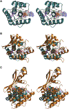
Figure 4.. Structural Similarity of Doc to the Secreted Toxin AvrB
(A) Stereo view of the putative Doc active site. Residues conserved among Fido domains (Kinch et al., 2009) are colored magenta. Additional residues identified by Magnuson and Yarmolinsky (1998) that effect Doc activity are colored green, as is the F68S mutation present in our crystal structure. Mutations at sites labeled in bold text greatly attenuate the toxicity of Doc while interfering (italics) or not interfering (nonitalics) with its regulatory activity. The F68S mutation and additional sites that are conserved among Fido domains are labeled in plain text. Structural alignment of AvrB (B and C) shows the residues conserved among Fido domains are proximal to its bound ADP molecule (shown in transparent space-filling representation). (B) Stereo pair in the same orientation as (A) showing superposition of Doc (dark teal) with AvrB (orange). The small domain in AvrB covering its mononucleotide-binding site has been omitted to improve clarity. (C) Stereo pair showing a different view of a superposition of the full AvrB structure with Doc.
Figure 5.
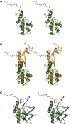
Figure 5.. Structure of the HigA Antitoxin and Comparison with the MqsA Antitoxin
(A) Stereo view of the E. coli CFT073 HigA antitoxin. Although HigA neutralizes a toxin that is homologous to YoeB, it is structurally unrelated to the YefM antitoxin that neutralizes YoeB (see Figure 1). (B) Stereo view of superimposed HigA and MqsA antitoxins. HigA and MqsA have structurally similar HTH DNA-binding domains but structurally dissimilar neutralization domains, even though they inhibit structurally homologous RelE/ YoeB-family toxins. Therefore, RelE/YoeB-family toxins are neutralized by antitoxins from at least three fold families (YefM-like, HigA, and MqsA). (C) Stereo view of a model of the HigA antitoxin in complex with DNA generated by superimposing HigA on the structure of the P22 c2 repressor protein in complex with DNA (PDB ID code 2R1J).
Figure 6.
Figure 6.. Comparison of the YeeU Antitoxin Structure to Related Structures
(A) The EcYeeU structure. The left side shows a YeeU stereopair with putative functional residues labeled and shown in stick representation. The middle panel shows the electrostatic surface potential of YeeU, with blue and red, respectively, representing surface potentials of 9 kT and —6.5 kTin 100 mM salt. The panel on the right shows a surface representation of EcYeeU colored according to sequence conservation using ConSurf (Landau et al., 2005) with a set of 20 YeeU homologs (including E. coli YafW and YfjZ) aligned by Clustal W (Larkin et al., 2007). Burgundy indicates a high degree of conservation whereas teal represents regions of variable sequence. Yellow indicates regions where degree of conservation could not be assigned with confidence. (B) Stereo pair of EcYeeU superimposed on PhRelE. Residues known to be involved in PhRelE function are labeled and shown in stick representation. The middle and right panels show the molecular surface of PhRelE colored according to electrostatic surface potential and sequence conservation, respectively, using the same parameters as for EcYeeU. (C) Stereo pair of EcYeeU superimposed on EcYoeB. Residues known to be involved in EcYoeB function are labeled and shown in stick representation. The middle and right panels show the molecular surface of EcYoeB colored according to electrostatic surface potential and sequence conservation, respectively, using the same parameters as for EcYeeU.

References

    1. Ades SE, and Sauer RT (1995). Specificity of minor-groove and majorgroove interactions in a homeodomain-DNA complex. Biochemistry 34, 14601–14608. - PubMed
    1. Altschul SF, Madden TL, Schaffer AA, Zhang J, Zhang Z, Miller W, and Lipman DJ (1997). Gapped BLAST and PSI-BLAST: a new generation of protein database search programs. Nucleic Acids Res 25, 3389–3402. - PMC - PubMed
    1. Anantharaman V, and Aravind L (2003). New connections in the prokaryotic toxin-antitoxin network: relationship with the eukaryotic nonsense-mediated RNA decay system. Genome Biol 4, R81. - PMC - PubMed
    1. Andreeva A, and Murzin AG (2006). Evolution of protein fold in the presence of functional constraints. Curr. Opin. Struct. Biol 16, 399–408. - PubMed
    1. Benach J, Lee I, Edstrom W, Kuzin AP, Chiang Y, Acton TB, Montelione GT, and Hunt JF (2003). The 2.3-Å crystal structure of the shikimate 5-dehydrogenase orthologue YdiB from Escherichia coli suggests a novel catalytic environment for an NAD-dependent dehydrogenase. J. Biol. Chem 278, 19176–19182. - PubMed

Publication types

LinkOut - more resources