. 2006 Nov 6;175(3):465-75.
doi: 10.1083/jcb.200605091.

Shigella applies molecular mimicry to subvert vinculin and invade host cells

Affiliations

Shigella applies molecular mimicry to subvert vinculin and invade host cells

Tina Izard et al. J Cell Biol. .

Abstract

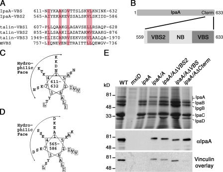
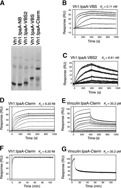
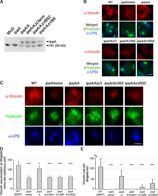
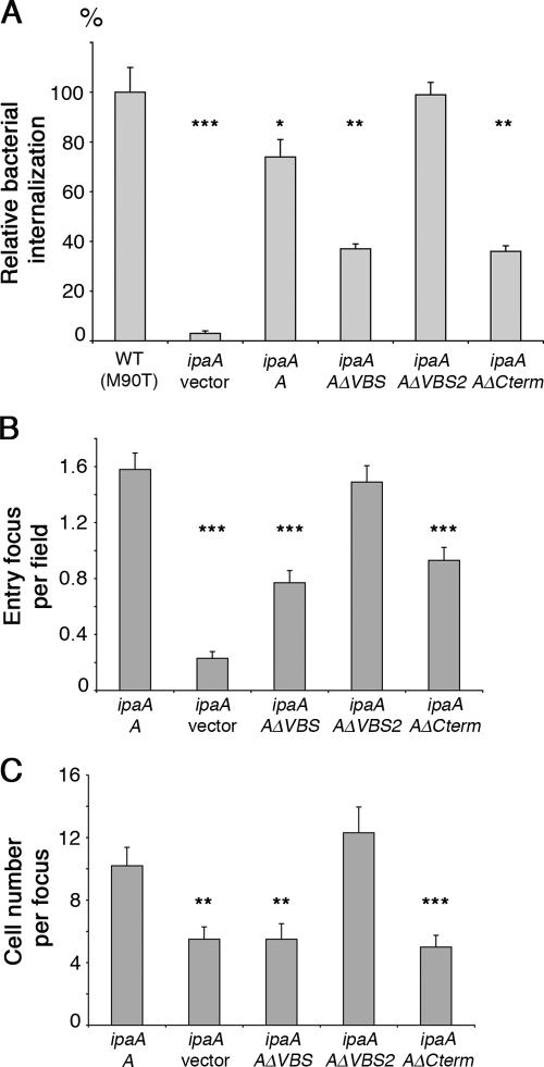
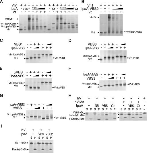
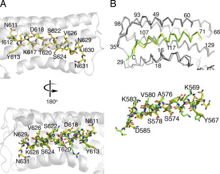
Shigella flexneri, the causative agent of bacillary dysentery, injects invasin proteins through a type III secretion apparatus upon contacting the host cell, which triggers pathogen internalization. The invasin IpaA is essential for S. flexneri pathogenesis and binds to the cytoskeletal protein vinculin to facilitate host cell entry. We report that IpaA harbors two vinculin-binding sites (VBSs) within its C-terminal domain that bind to and activate vinculin in a mutually exclusive fashion. Only the highest affinity C-terminal IpaA VBS is necessary for efficient entry and cell-cell spread of S. flexneri, whereas the lower affinity VBS appears to contribute to vinculin recruitment at entry foci of the pathogen. Finally, the crystal structures of vinculin in complex with the VBSs of IpaA reveal the mechanism by which IpaA subverts vinculin's functions, where S. flexneri utilizes a remarkable level of molecular mimicry of the talin-vinculin interaction to activate vinculin. Mimicry of vinculin's interactions may therefore be a general mechanism applied by pathogens to infect the host cell.


Figures

Figure 1.
Figure 1.
S. flexneri IpaA harbors two putative VBSs (IpaA-VBSs). (A) Structure-based sequence alignment of the complete IpaA amino acid sequence, using the Heidelberg PHD secondary-structure program, predicted that IpaA contains 66% α-helical character and identified two short regions, residues 563–589 and 612–630, which share strong similarity to the VBSs of talin and α-actinin. These putative IpaA VBSs, IpaA-VBS and -VBS2, were aligned with three of the VBSs of human talin (talin-VBS1, -VBS2, and -VBS3) and that of α-actinin (αVBS) using CLUSTAL protein software. As seen in the Vh1–αVBS structure (Bois et al., 2005), the orientation of the αVBS is inverted. Hydrophobic residues conserved in all five VBSs are shaded in pink. (B) Schematic of IpaA is shown and the C-terminal region of IpaA (residues 559–633), indicating the position and nomenclature of the peptides used in this study. (C) IpaA-VBS and (D) -VBS2 are predicted to be amphipathic α helices. The predicted IpaA-VBSs were arranged around a helical wheel, which revealed amphipathic helices having a hydrophilic basic face, and a hydrophobic face. Hydrophobic residues are boxed. (E) Either IpaA-VBS or -VBS2 is sufficient to bind to vinculin. Media from cultures of the indicated strains of S. flexneri were harvested and analyzed for the expression of S. flexneri's invasin proteins, which were detected by staining SDS-PAGE gels with Coomassie blue (top). Samples were then blotted and levels of wild-type and mutant IpaA proteins were determined by Western blotting with IpaA-specific antibody (middle). Samples were also blotted with I125-labeled vinculin using a vinculin overlay assay. Note that vinculin could bind to IpaA, IpaA-ΔVBS, and -ΔVBS2, but not to IpaA-ΔCterm, which lacks both of the VBSs of IpaA. The mxiD deletion (type III secretion system defective) does not secrete any of the invasin proteins (Perdomo et al., 1994).
Figure 2.
Figure 2.
S. flexneri's IpaA protein harbors high-affinity VBSs. (A) Native gel analyses of free Vh1, or Vh1 incubated with IpaA-NB, -VBS2, -VBS, or -Cterm. (B–E) SPR of the affinity of IpaA-VBS (B), -VBS2 (C), or -Cterm (D and E) for Vh1 (B, C, and D) or for full-length human vinculin (E). Representative sensorgrams are shown. The calculated K d is shown above each panel. (F and G) Off-rate analyses of the (F) Vh1–IpaA-Cterm and (G) vinculin–IpaA-Cterm interactions over a time course of >100 min are shown. Representative sensorgrams from three independent experiments are shown.
Figure 3.
Figure 3.
The VBSs of IpaA recruit vinculin to S. flexneri at bacterial entry sites. (A) The VBSs of S. flexneri are dispensable for translocation of IpaA into infected cells. Lysates of HeLa cells infected with equal multiplicities of infection of the indicated strains of S. flexneri were evaluated for their levels of intracellular IpaA protein using anti-IpaA antibody complexed to G–Sepharose, SDS-Page, and Western blotting with anti-IpaA antibody. IpaA protein appeared as a doublet, suggesting that some IpaA degradation occurred. IpaA and the antibody heavy chain (HC; loading control) are indicated. (B) Immunofluorescence analyses of vinculin (red), LPS (blue), and actin (phalloidin; green) in HeLa cells infected with the indicated strains of S. flexneri is shown. Vinculin is effectively recruited to wild-type and ipaA/A S. flexneri at entry foci, whereas this does not occur in HeLa cells infected with the S. flexneri ipaA deletion strain, nor in HeLa cells infected with the S. flexneri ipaA/AΔCterm (Ct), ipaA/AΔVBS, and ipaA/AΔVBS2 strains. Actin foci containing vinculin are evident in HeLa cells infected with the ipaA, ipaA/AΔCt, ipaA/AΔVBS, and ipaA/AΔVBS2 strains of S. flexneri. Note that infection of HeLa cells with wild-type S. flexneri or strains expressing the IpaA mutants did not overtly disrupt focal adhesions, which were still evident in cells stained with anti-vinculin antibody, and which are consistent with the lack of appreciable effects of the VBSs of IpaA in displacing vinculin–talin interactions (Fig. 5, C, D, and F). (C) Vinculin (red) recruitment to internalized bacteria (anti-LPS; blue) is observed in HeLa cells infected with wild-type and ipaA/A strains of S. flexneri, where vinculin (and actin) appears to coat the surface of their membranes. Reduced vinculin staining of the bacterium was observed with ipaA/AΔVBS and ipaA/AΔVBS2 S. flexneri, and was not detected in cells expressing the ipaA deletion strain or the ipaA/AΔCt strain of S. flexneri. Images correspond to “maximum” reconstructions from z planes delimitating S. flexneri entry sites using the Metamorph software (see Materials and methods). (D) Quantification of total vinculin recruitment at entry sites of S. flexneri was determined using the MetaMorph software. (E) The percentages of bacteria decorated with vinculin at entry foci were quantified. A minimum of 56 foci were scored for each strain (mean of 100). Error bars indicating the mean ± the SEM are provided together with the t test P value (*, P < 0.05; **, P < 0.005; and *** P, < 0.0005).
Figure 4.
Figure 4.
S. flexneri's IpaA-VBSs contribute to host cell entry and to dissemination of the pathogen. (A) Deletion of IpaA-VBS compromises the invasion of HeLa cells by S. flexneri. HeLa cells were challenged with the indicated strains and bacterial internalization was determined by the gentamicin protection assay (Perdomo et al., 1994). The relative percentage of bacterial internalization corresponds to values normalized to that obtained for the wild-type strain. Each value is the mean of six independent determinations. (B) Polarized colonic epithelial Caco-2 cells were challenged with the indicated mutants, and bacterial internalization was assessed by scoring entry events by fluorescent microscopy. (C) Deletion of IpaA-VBS affects S. flexneri dissemination. Polarized Caco-2 cells were challenged with the indicated mutants for 90 min, and bacterial dissemination after a 6-h incubation was determined by scoring the numbers of infected cells per focus. Error bars indicating the mean ± the SEM are provided together with the t test P value (*, P < 0.05; **, P < 0.005; and *** P, < 0.0005).
Figure 5.
Figure 5.
S. flexneri's IpaA-VBSs activate vinculin. (A) S. flexneri's IpaA-VBSs and its C-terminal domain (Ct) sever the Vh1–Vt interaction that clamps vinculin in its inactive state (Borgon et al., 2004; Izard et al., 2004). Lane 1 in both the left and right native gels show Vh1 alone, and lane 2 shows the Vh1–IpaA-VBS complex (left) or the Vh1–IpaA-Cterm complex (right). The + denotes the addition of the indicated IpaA domain to preformed Vh1–Vt complexes. Competing IpaA-VBS (left) was titrated (lanes 4–7, arrowheads) into preformed Vh1–Vt complexes at molar ratios of IpaA-VBS at ∼2-, 3.5-, 10-, and 20-fold molar ratios (Vh1–IpaA-VBS). Competing IpaA-Cterm peptide (right) was titrated (arrows) into preformed Vh1–Vt complexes at 0.9-, 1.7-, 3.4-, and 6.8-fold molar ratios (Vh1–IpaA-Cterm). As is evident, titration of even very low molar ratios of IpaA-VBS or IpaA-Cterm was sufficient to totally disrupt the Vh1–Vt complex to form novel Vh1–IpaA-VBS (left) or Vh1–IpaA-Cterm (right) complexes. Because of its basic pI, free Vt is not visible in native gels (Izard et al., 2004). (B) IpaA-VBS2 is also sufficient to disrupt the Vh1–Vt interaction. Native gel analyses of free Vh1 (lane 1), Vh1 in complex with IpaA-VBS2 (lane 2), Vh1–Vt complex (lane 3), or preexisting Vh1–Vt complexes titrated with increasing concentrations of IpaA-VBS2 (at ∼2-, 3.5-, 10-, and 20-fold molar ratios; Vh1–IpaA-VBS2). The identity of the complexes is shown. (C–G) The selective nature of the IpaA–vinculin interaction. Reciprocal VBS displacement native gel assays of Vh1 in complex with talin-VBS1 (C), talin-VBS3 (D and F), or the VBS of α-actinin (αVBS; E and G) versus when in complex with IpaA-VBS (C–E) or IpaA-VBS2 (F and G). Competing IpaA-VBS, IpaA-VBS2, talin-VBS3, talin-VBS1, or αVBS peptides were titrated (arrows) into preformed complexes at 2.2- or 10-fold molar excess (Vh1–VBS). Native gels of the indicated complexes are shown. (H and I) IpaA-VBS (H) and -VBS2 (I) promote vinculin binding to F-actin. IpaA-NB, -VBS (VBS), -VBS2 (VBS2), or -Cterm (Ct) were incubated with full-length human vinculin (hV) or human vinculin lacking the tail domain Vt (hVΔVt), which mediates binding to F-actin (Johnson and Craig, 1995). Samples were incubated with F-actin, as previously described (Bois et al., 2006), and centrifuged into pellet (containing polymerized F-actin) and supernatant fractions, and then equal volumes were analyzed by SDS-PAGE. F-actin and vinculin were visualized by staining the gels with Coomassie blue. The last lane on the right of H shows the size difference between full-length vinculin (residues 1–1,066) and the head domain of vinculin (residues 1–840).
Figure 6.
Figure 6.
The mechanism of vinculin activation by S. flexneri's IpaA protein. (A, top) The crystal structure of the Vh1–IpaA-VBS complex showing the hydrophobic face of IpaA-VBS (yellow bonds; some residues are labeled) that interacts with vinculin (transparent white helices). Vinculin is shown as a ribbon diagram in white, and the individual bonds are drawn in yellow for IpaA-VBS (oxygen atoms, red; carbon, yellow; nitrogen, blue). (bottom) Superposition of talin-VBS3 (green bonds) onto IpaA-VBS (yellow bonds) bound to vinculin (white and gray, respectively). This orientation is rotated by 180° in respect to the top image showing the hydrophilic solvent–exposed face of these VBSs. Some IpaA-VBS residues are labeled. (B) (top) The Cα trace of the Vh1 domain of vinculin bound to IpaA-VBS (yellow) superimposed onto IpaA-VBS2 (green) bound to Vh1 in gray (Vh1–IpaA-VBS2) or white (Vh1–IpaA-VBS). Some vinculin residues are labeled. (bottom) Ball-and-stick representation of the structure of IpaA-VBS (yellow) superimposed onto that of IpaA-VBS2 (green); some residues of IpaA-VBS2 are labeled. Both images are shown in the same orientation.

References

    1. Bakolitsa, C., D.M. Cohen, L.A. Bankston, A.A. Bobkov, G.W. Cadwell, L. Jennings, D.R. Critchley, S.W. Craig, and R.C. Liddington. 2004. Structural basis for vinculin activation at sites of cell adhesion. Nature. 430:583–586. - PubMed
    1. Bakolitsa, C., J.M. de Pereda, C.R. Bagshaw, D.R. Critchley, and R.C. Liddington. 1999. Crystal structure of the vinculin tail suggests a pathway for activation. Cell. 99:603–613. - PubMed
    1. Bois, P.R.J., R.A. Borgon, C. Vonrhein, and T. Izard. 2005. Structural dynamics of α-actinin-vinculin interactions. Mol. Cell. Biol. 25:6112–6122. - PMC - PubMed
    1. Bois, P.R.J., B.P. O'Hara, D. Nietlispach, J. Kirkpatrick, and T. Izard. 2006. The vinculin binding sites of talin and α-actinin are sufficient to activate vinculin. J. Biol. Chem. 281:7228–7236. - PubMed
    1. Borgon, R.A., C. Vonrhein, G. Bricogne, P.R.J. Bois, and T. Izard. 2004. Crystal structure of human vinculin. Structure. 12:1189–1197. - PubMed

Publication types

MeSH terms