Comparative Study
. 2007 Jan 9;104(2):462-6.
doi: 10.1073/pnas.0609773104. Epub 2006 Dec 26.

Structural basis for intramembrane proteolysis by rhomboid serine proteases

Affiliations
Comparative Study

Structural basis for intramembrane proteolysis by rhomboid serine proteases

Adam Ben-Shem et al. Proc Natl Acad Sci U S A. .

Abstract

Intramembrane proteases catalyze peptide bond cleavage of integral membrane protein substrates. This activity is crucial for many biological and pathological processes. Rhomboids are evolutionarily widespread intramembrane serine proteases. Here, we present the 2.3-A-resolution crystal structure of a rhomboid from Escherichia coli. The enzyme has six transmembrane helices, five of which surround a short TM4, which starts deep within the membrane at the catalytic serine residue. Thus, the catalytic serine is in an externally exposed cavity, which provides a hydrophilic environment for proteolysis. Our results reveal a mechanism to enable water-dependent catalysis at the depth of the hydrophobic milieu of the membrane and suggest how substrates gain access to the sequestered rhomboid active site.


Conflict of interest statement

The authors declare no conflict of interest.

Figures

Fig. 1.
Fig. 1.
Organization of GlpG functional and secondary structural elements. (A) Linear map of GlpG and comparison to other rhomboid proteases. Above the GlpG amino acid sequence, helices are indicated as cylinders, and the positions of the catalytic residues are marked with asterisks. Although a precise sequence alignment with rhomboid proteases from other kingdoms (Drome rhomboid = Drosophila melanogaster rhomboid, gi:201416977; Human mito PARL = Homo sapiens mitochondrial presenilins-associated rhomboid-like protein, gi:62511133; P. hirokoshii = rhomboid-like protein from the thermophilic archeon Pyrococcus hirokoshii, gi:14591283) is not possible because of low sequence similarity, predicted transmembrane regions and shared sequence motifs were used to align functional and structural regions. The starting residue numbers of the indicated sequence fragments within their full-length proteins are given. Trypsin cleavage sites in the preparation of the crystallized GlpG are indicated by arrowheads. (B) Schematic representation of GlpG helix orientation with respect to the membrane. (C) Slab of an experimental SIRAS map contoured at 1σ over a stick model of molecule B from the GlpG crystals. (D) Ribbon diagram of the GlpG crystal structure in three orientations with helices color-coded as in A and B; the top view is shown in stereo.
Fig. 2.
Fig. 2.
Distinctive structural features of GlpG. (A) Stereoview into the active-site cavity of GlpG. The side chains of polar residues lining the cavity are shown in color. The oxygen atoms of water molecules are shown as red balls. For clarity, only the phosphate group (red/orange tetrahedron) of the bound phospholipid is shown. A cluster of hydrophobic residues lining the lip of the cavity and closing the gap between TM2 and TM5 is shown in dark gray. The loop between TM3 and TM4 that leads down into the core of the enzyme and the active-site Ser-201 is in yellow, and TM4 is in cyan. (B) The membrane-embedded helix H2 lies perpendicular to the TM helices within the membrane. Some hydrogen bonding interactions stabilizing the local geometry of helices H1 and H2 are indicated as dotted lines. Hydrophobic residues from H2 facing the lipid interior are also shown.
Fig. 3.
Fig. 3.
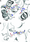
Comparison of the active sites of GlpG and trypsin. The GlpG active site viewed from the membrane (A) compared with that of the soluble serine protease trypsin (B) (PDB entry 1TLD) (23). Dashed lines indicate hydrogen bond distances. Solid lines indicate distances that demonstrate the resemblance between the positions of the oxyanion holes in the two structures.
Fig. 4.
Fig. 4.
A model for interaction of substrates with GlpG. A surface representation of GlpG is shown with the polar residues in the active-site cavity in blue. A schematic representation of a substrate TM (straight or tilted) and its external domain is shown. The top quarter of the substrate TM is portrayed in a nonhelical conformation that penetrates into the active-site cavity in between TM2 and TM5. (A) View from the membrane. (B) View from the periplasm.

Comment in

  • Membrane-embedded protease poses for photoshoot.
    Lieberman RL, Wolfe MS. Lieberman RL, et al. Proc Natl Acad Sci U S A. 2007 Jan 9;104(2):401-2. doi: 10.1073/pnas.0610236103. Epub 2007 Jan 3. Proc Natl Acad Sci U S A. 2007. PMID: 17213330 Free PMC article. No abstract available.

References

    1. Brown MS, Ye J, Rawson RB, Goldstein JL. Cell. 2000;100:391–398. - PubMed
    1. Wolfe MS, Kopan R. Science. 2004;305:1119–1123. - PubMed
    1. Marjaux E, Hartmann D, De Strooper B. Neuron. 2004;42:189–192. - PubMed
    1. Selkoe D, Kopan R. Annu Rev Neurosci. 2003;26:565–597. - PubMed
    1. Rawson RB, Zelenski NG, Nijhawan D, Ye J, Sakai J, Hasan MT, Chang TY, Brown MS, Goldstein JL. Mol Cell. 1997;1:47–57. - PubMed

Publication types

MeSH terms

Associated data