. 2007 Mar;11(3):217-27.
doi: 10.1016/j.ccr.2006.12.017.

Structures of lung cancer-derived EGFR mutants and inhibitor complexes: mechanism of activation and insights into differential inhibitor sensitivity

Affiliations

Structures of lung cancer-derived EGFR mutants and inhibitor complexes: mechanism of activation and insights into differential inhibitor sensitivity

Cai-Hong Yun et al. Cancer Cell. 2007 Mar.

Abstract

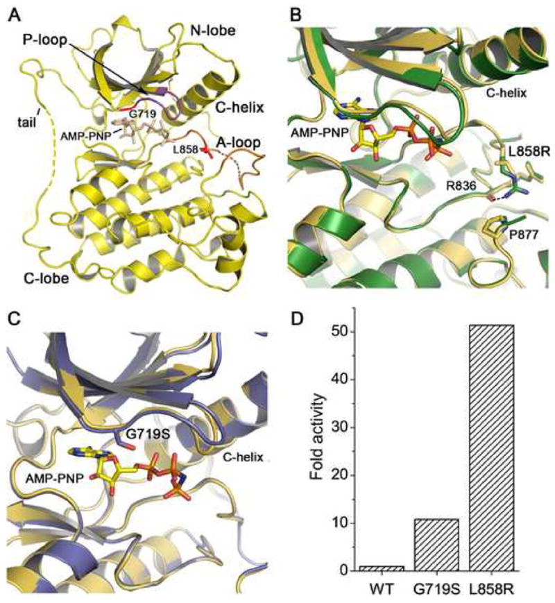
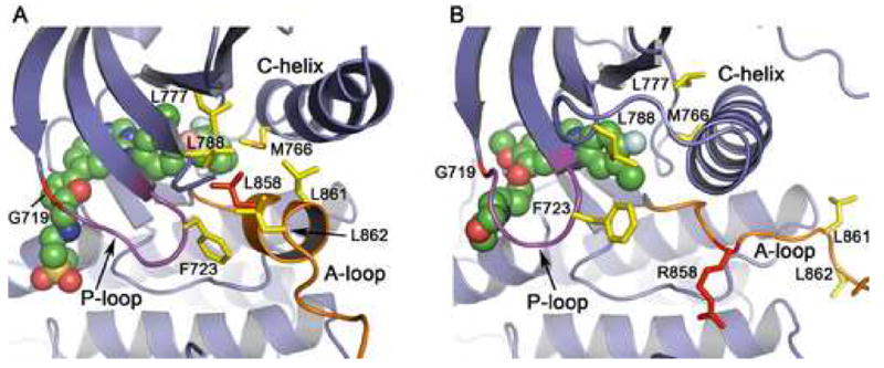
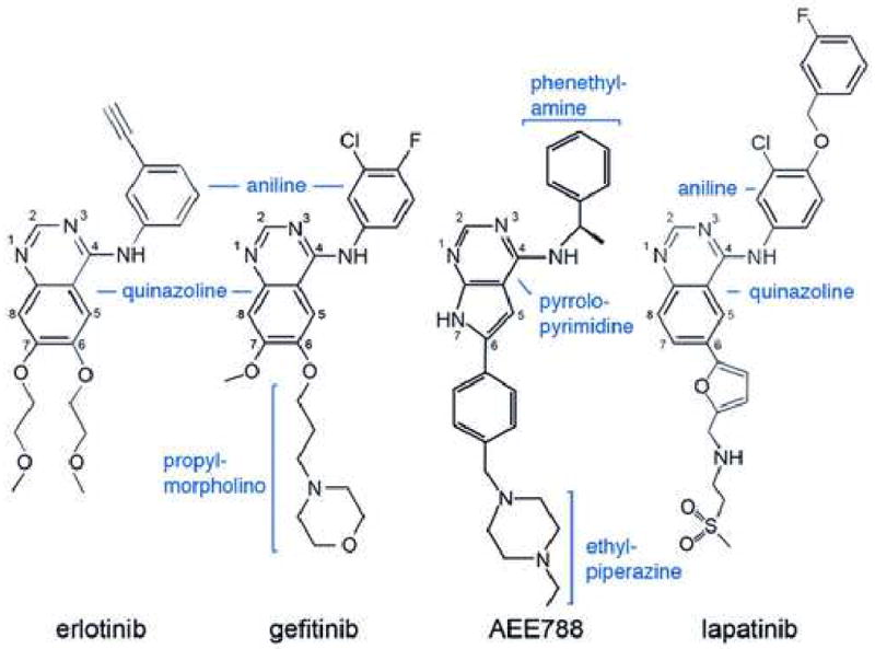
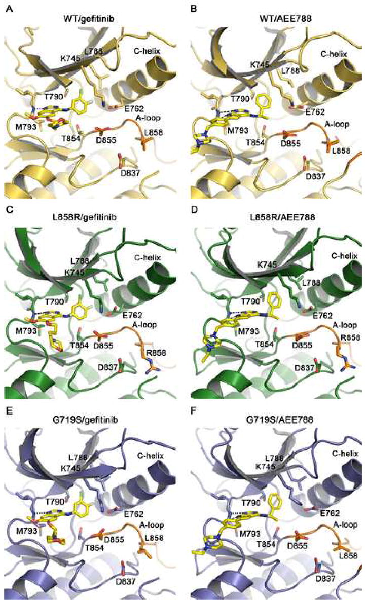
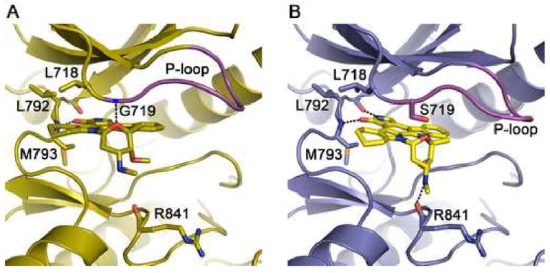
Mutations in the EGFR kinase are a cause of non-small-cell lung cancer. To understand their mechanism of activation and effects on drug binding, we studied the kinetics of the L858R and G719S mutants and determined their crystal structures with inhibitors including gefitinib, AEE788, and a staurosporine. We find that the mutations activate the kinase by disrupting autoinhibitory interactions, and that they accelerate catalysis as much as 50-fold in vitro. Structures of inhibitors in complex with both wild-type and mutant kinases reveal similar binding modes for gefitinib and AEE788, but a marked rotation of the staurosporine in the G719S mutant. Strikingly, direct binding measurements show that gefitinib binds 20-fold more tightly to the L858R mutant than to the wild-type enzyme.


Figures

Figure 1
Figure 1
Structure and activity of mutant EGFR kinases. (A) Overview of the structure of the EGFR kinase. The structure of the wild-type kinase is shown in complex with the ATP analog AMP-PNP. The locations of the L858R and G719S mutations in the activation loop (A-loop) and P-loop, respectively, are indicated. Dashed lines indicate short segments of the activation loop and C-terminal tail that are disordered in the structures reported here. Due to a difference in crystallization buffers, we observe more of the C-terminal tail than reported previously (Stamos et al., 2002), allowing us to confirm that it makes an intra- rather than inter- molecular interaction with the N-lobe of the kinase in a manner similar to that described for the inactive kinase (Wood et al., 2004). (B) The structure of the active site region of the L858R mutant (green) superimposed on the wild-type kinase (yellow). (C) The structure of the active site region of the G719S mutant (blue) superimposed on the wild-type kinase (yellow). (D) Comparison of the activity of the wild-type, G719S and L858R kinases. The fold activity of wild-type and mutant enzymes was calculated by determining the kcat for each protein with saturating ATP and poly-[Glu4Tyr1] as peptide substrate and dividing by the kcat for the wild-type enzyme.
Figure 2
Figure 2
Mechanism of activation of the L858R and G719S mutants. The structure of the inactive, wild-type enzyme in complex with lapatinib (panel A) is compared with that of the active, L858R mutant in complex with gefitinib (panel B). (A) In the inactive state, the N-terminal portion of the activation loop (shown in orange) forms a short helix that displaces the regulatory C-helix from the active site. A cluster of hydrophobic residues (shown in yellow), including Leu 858 (red), stabilize the inactive conformation. Lapatinib (shown as CPK spheres) extends into the space created by the displaced C-helix and appears to have allowed “trapping” of the inactive conformation in the crystal structure. Substitution of Leu858 with arginine is expected to destabilize this conformation, as arginine cannot be favorably accommodated in the hydrophobic pocket occupied by Leu858. Similarly, substitution of G719S with serine may destabilize the inactive conformation of the P-loop (which has a conformation favoring glycine at this position), and therefore activate the kinase. (B) In the active conformation, the activation loop (orange) is reorganized and the C-helix rotates into its active position. Note that the hydrophobic cluster (yellow) is dismantled, and Arg858 (red) is readily accomodated (see also Fig. 1B). Also, note the difference in conformation of the P-loop (purple) and orientation of Phe 723 in the inactive vs. active structures.
Figure 3
Figure 3
Schematic drawings of the EGFR inhibitors discussed here. Inhibitors are drawn in a consistent orientation approximately reflecting their conformations when bound to the EGFR kinase (see Figure 4).
Figure 4
Figure 4
Drug binding modes in the wild-type and mutant EGFR kinase. The binding modes of gefitinib (panels A, C and E) and AEE788 (panels B, D and F) are compared in the wild-type (yellow), L858R (green), and G719S (blue) kinases. Key sidechains are labeled, the inhibitors are shown in stick form with carbons colored yellow, and hydrogen bonds are indicated with dashed lines. Compare binding of different inhibitors to the same mutant within rows, and binding of the same inhibitor among wild-type and mutants within columns. Binding modes of both compounds are essentially the same in all three structures. Note also the closely corresponding orientations of the pyrrolopyrimidine scaffold in the AEE788 complexes and the quinazoline core in the gefitinib complexes. Additionally, the phenylethyl amine moeity in AEE788 occupies the same space as the aniline substituent in the gefitinib and erlotinib complexes.
Figure 5
Figure 5
The G719S mutation alters the binding mode of staurosporine analog AFN941. (A) The wild-type EGFR in complex with AFN941. Hydrogen bonds (dashed lines) are formed with the backbone amides of Met 793 in the hinge region and Gly 719 in the P-loop. (B) Structure of the G719S mutant in complex with AFN941. The serine substitution displaces and rotates the inhibitor, disrupting the hydrogen bond with the P-loop and promoting additional interactions with the backbone carbonyls of Gln 791 and Arg 841.

Comment in

  • The EGF receptor Hokey-Cokey.
    Niculescu-Duvaz D, Whittaker S, Springer C, Marais R. Niculescu-Duvaz D, et al. Cancer Cell. 2007 Mar;11(3):209-11. doi: 10.1016/j.ccr.2007.02.021. Cancer Cell. 2007. PMID: 17349577

References

    1. Amann J, Kalyankrishna S, Massion PP, Ohm JE, Girard L, Shigematsu H, Peyton M, Juroske D, Huang Y, Stuart SJ, Kim YH, Pollack JR, Yanagisawa K, Gazdar A, Minna JD, Kurie JM, Carbone DP. Aberrant epidermal growth factor receptor signaling and enhanced sensitivity to EGFR inhibitors in lung cancer. Cancer Res. 2005;65:226–235. - PubMed
    1. Arao T, Fukumoto H, Takeda M, Tamura T, Saijo N, Nishio K. Small in-frame deletion in the epidermal growth factor receptor as a target for ZD6474. Cancer Res. 2004;64:9101–9104. - PubMed
    1. Auffinger P, Hays FA, Westhof E, Ho PS. Halogen bonds in biological molecules. Proc Natl Acad Sci U S A. 2004;101:16789–16794. - PMC - PubMed
    1. Barker AJ, Gibson KH, Grundy W, Godfrey AA, Barlow JJ, Healy MP, Woodburn JR, Ashton SE, Curry BJ, Scarlett L, Henthorn L, Richards L. Studies leading to the identification of ZD1839 (IRESSA): an orally active, selective epidermal growth factor receptor tyrosine kinase inhibitor targeted to the treatment of cancer. Bioorg Med Chem Lett. 2001;11:1911–1914. - PubMed
    1. Barker SC, Kassel DB, Weigl D, Huang X, Luther MA, Knight WB. Characterization of pp60c-src tyrosine kinase activities using a continuous assay: autoactivation of the enzyme is an intermolecular autophosphorylation process. Biochemistry. 1995;34:14843–14851. - PubMed

Publication types

MeSH terms

LinkOut - more resources