. 2007 Sep;19(9):2886-97.
doi: 10.1105/tpc.106.045682. Epub 2007 Sep 21.

Molecular architecture of strictosidine glucosidase: the gateway to the biosynthesis of the monoterpenoid indole alkaloid family

Affiliations

Molecular architecture of strictosidine glucosidase: the gateway to the biosynthesis of the monoterpenoid indole alkaloid family

Leif Barleben et al. Plant Cell. 2007 Sep.

Abstract

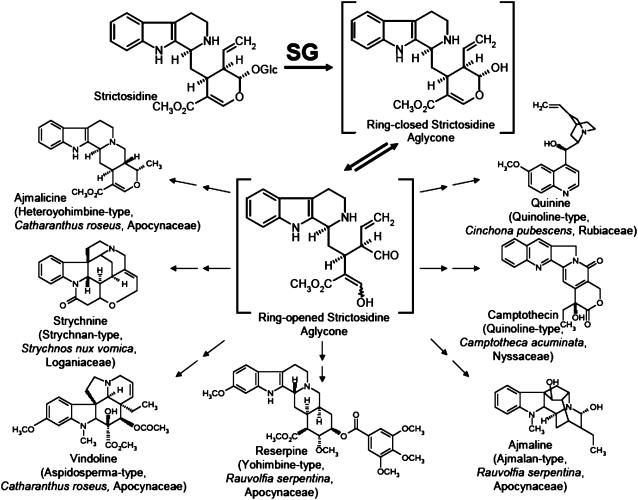
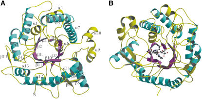
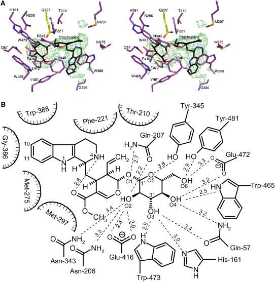
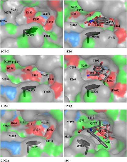
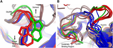
Strictosidine beta-D-glucosidase (SG) follows strictosidine synthase (STR1) in the production of the reactive intermediate required for the formation of the large family of monoterpenoid indole alkaloids in plants. This family is composed of approximately 2000 structurally diverse compounds. SG plays an important role in the plant cell by activating the glucoside strictosidine and allowing it to enter the multiple indole alkaloid pathways. Here, we report detailed three-dimensional information describing both native SG and the complex of its inactive mutant Glu207Gln with the substrate strictosidine, thus providing a structural characterization of substrate binding and identifying the amino acids that occupy the active site surface of the enzyme. Structural analysis and site-directed mutagenesis experiments demonstrate the essential role of Glu-207, Glu-416, His-161, and Trp-388 in catalysis. Comparison of the catalytic pocket of SG with that of other plant glucosidases demonstrates the structural importance of Trp-388. Compared with all other glucosidases of plant, bacterial, and archaeal origin, SG's residue Trp-388 is present in a unique structural conformation that is specific to the SG enzyme. In addition to STR1 and vinorine synthase, SG represents the third structural example of enzymes participating in the biosynthetic pathway of the Rauvolfia alkaloid ajmaline. The data presented here will contribute to deciphering the structure and reaction mechanism of other higher plant glucosidases.


Figures

Figure 1.
Figure 1.
The Key Role of the Deglucosylation of Strictosidine Catalyzed by SG in the Biosynthesis of Various Alkaloid Types Belonging to the Families of Monoterpenoide Indole and Quinoline Alkaloids. Typical plant genera and species from which corresponding alkaloids were isolated are illustrated with the appropriate plant families.
Figure 2.
Figure 2.
Overall Structure of SG from R. serpentina, Illustrating Its (β/α)8 Fold, the Groove Leading to the Catalytic Center, and the Secondary Structure Elements. (A) (β/α) repeats are illustrated in magenta and cyan, respectively. The extra helices and strands as well as connecting loops are shown in yellow. The secondary elements are labeled. (B) (β/α)8 fold is shown in complex with the substrate strictosidine in ball-and-stick representation. The structure is rotated by 170° around the y axis with respect to (A). Color code is same as depicted in (A).
Figure 3.
Figure 3.
Structural Representation of SG: Ligand Complexes. (A) Stereo view of strictosidine binding site of SG. The (Fo-Fc) SIGMAA-weighted electron density of strictosidine contoured at 4 σ is shown in green and strictosidine in black. In complex with the inactive mutant (Glu207Gln), residues within 4.1-Å distance from strictosidine are shown in violet, and Gln-207 is in yellow. (B) Hydrogen binding network between the glucosidic part of strictosidine and residues in a distance ≤4.1 Å in the ligand structure of SG inactive mutant Glu207Gln.
Figure 4.
Figure 4.
Comparison of the Catalytic Center of Crystallized Plant Glucosyl Hydrolases of Family 1. The conformation of Trp-388 is highlighted in black. Hydrophobic residues (Trp, Phe, Leu, Met, Cys, Ile, Ala, Gly, and Val), positively charged residues (His), negatively charged residues (Glu and Asp), and hydrophilic residues (Ser, Thr, Tyr, Pro, Gln, and Asn) are shown in gray, blue, red and green, respectively. 1CBG, cyanogenic β-glucosidase from T. repens; 1E56, Zm Glu1-Glu191Asp in complex with DIMBOA-β-d-glucoside from Z. mays; 1HXJ, Zmp60.1 from Z. mays; 1V03, Dhurrinase1-Glu189Asp in complex with dhurrin from S. bicolor; 2DGA, Ta Glu1b from T. aestivum; SG, strictosidine glucosidase-Glu207Gln in complex with strictosidine from R. serpentina. Equivalent residues are labeled (in 1CBG, there are nine instead of 10 residues displayed because the SG-corresponding Thr-210 is replaced by Gly, which is out of figure margins). Residues in brackets are screened from sight by other amino acids.
Figure 5.
Figure 5.
Comparison of the Region around Trp-388 with Respect to Bacteria, Archaea, and Higher Plants. (A) The three different conformations of Trp-388 in 16 β-glycosidases from bacteria (blue), archaea (brown), higher plants (red), and SG from Rauvolfia (green) described in this study. The location of the sugar binding site compared with the Trp side chains and direction to the enzyme surface are indicated by black arrows. Gly-386 and Gly-387 are labeled to indicate the direction to the N terminus, and the distance between Cα of Trp-388 in microbial and eukaryotic enzymes is illustrated by a yellow arrow. The sequence numbering is according to SG. The structures shown are as follows: 1NP2, β-glycosidase from Thermus nonproteolyticus HG102; 1UG6, β-glycosidase (TTHB087) from Thermus thermophilus HB8; 1GNX, β-glucosidase (Bgl3) from Streptomyces sp QM-B814; 1OD0, β-glucosidase A (BglA) from Thermotoga maritima MSB8; 1QOX, β-glucosidase (BglA) from Bacillus circulans subsp Alkalophilus; 1E4I, β-glucosidase A (BglA) from Paenibacillus polymyxa; 1CBG, cyanogenic β-glucosidase from T. repens; SG, strictosidine glucosidase from R. serpentina; 1E1E, Zm Glu1 from Z. mays; 1HXJ, Zmp60.1 from Z. mays; 1V02, Dhurrinase1 from S. bicolor; 2DGA, Ta Glu1b from T. aestivum; 1PBG, 6-P-β-galactosidase from Lactococcus lactis Z268; 1VFF, alkyl β-glycosidase (BGPh; PH0366) from Pyrococcus horikoshii OT3; 1GOW, β-glycosidase S (LacS;S-β-gly; SSO3019) from Sulfolobus solfataricus P2; 1QVB, β-glycosidase from Thermosphaera aggregans M11TL. (B) To illustrate the different backbone orientations of the glycosidases, (A) was rotated by 90° around the x axis (colors are as in [A]). Near Thr-372 (yellow arrow), all the main chains diverge in three groups depending on their origin (bacteria, achaea, and higher plants). Archaea and bacteria backbones merge around residue Ala-383 (yellow arrow). Microbial and plant main chains merge after Trp-388. For better recognition, Gly-387 (yellow arrow) and the substrate binding region are marked. The sequence numbering is according to SG.

References

    1. Barleben, L., Ma, X.Y., Koepke, J., Peng, G., Michel, H., and Stöckigt, J. (2005). Expression, purification, crystallization and preliminary X-ray analysis of strictosidine glucosidase, an enzyme initiating biosynthetic pathways to a unique diversity of indole alkaloid skeletons. Biochim. Biophys. Acta 1747 89–92. - PubMed
    1. Barrett, T., Suresh, C.G., Tolley, S.P., Dodson, E.J., and Hughes, M.A. (1995). The crystal structure of a cyanogenic beta-glucosidase from white clover, a family 1 glycosyl hydrolase. Structure 3 951–960. - PubMed
    1. Blechert, S., Brodschelm, W., Holder, S., Kammerer, L., Kutchan, T.M., Mueller, M.J., Xia, Z.Q., and Zenk, M.H. (1995). The octadecanoic pathway: Signal molecules for the regulation of secondary pathways. Proc. Natl. Acad. Sci. USA 92 4099–4105. - PMC - PubMed
    1. Bradford, M.M. (1976). A rapid and sensitive method for the quantitation of microgram quantities of protein utilizing the principle of protein-dye binding. Anal. Biochem. 72 248–254. - PubMed
    1. Brünger, A.T. (1993). Assessment of phase accuracy by cross validation: The free R value. Methods and applications. Acta Crystallogr. D Biol. Crystallogr. 49 24–36. - PubMed

Publication types

MeSH terms

LinkOut - more resources