. 2008 Apr;17(4):711-24.
doi: 10.1110/ps.073295308.

Structure dissection of human progranulin identifies well-folded granulin/epithelin modules with unique functional activities

Affiliations

Structure dissection of human progranulin identifies well-folded granulin/epithelin modules with unique functional activities

Dmitri Tolkatchev et al. Protein Sci. 2008 Apr.

Abstract

Progranulin is a secreted protein with important functions in several physiological and pathological processes, such as embryonic development, host defense, and wound repair. Autosomal dominant mutations in the progranulin gene cause frontotemporal dementia, while overexpression of progranulin promotes the invasive progression of a range of tumors, including those of the breast and the brain. Structurally, progranulin consists of seven-and-a-half tandem repeats of the granulin/epithelin module (GEM), several of which have been isolated as discrete 6-kDa GEM peptides. We have expressed all seven human GEMs using recombinant DNA in Escherichia coli. High-resolution NMR showed that only the three GEMs, hGrnA, hGrnC, and hGrnF, contain relatively well-defined three-dimensional structures in solution, while others are mainly mixtures of poorly structured disulfide isomers. The three-dimensional structures of hGrnA, hGrnC, and hGrnF contain a stable stack of two beta-hairpins in their N-terminal subdomains, but showed a more flexible C-terminal subdomain. Interestingly, of the well-structured GEMs, hGrnA demonstrated potent growth inhibition of a breast cancer cell line, while hGrnF was stimulatory. Poorly folded peptides were either weakly inhibitory or without activity. The functionally active and structurally well-characterized human hGrnA offers a unique opportunity for detailed structure-function studies of these important GEM proteins as novel members of mammalian growth factors.


Figures

Figure 1.
Figure 1.
SDS-PAGE analysis of the localization and purification of an expressed GEM protein. Bands indicated by arrows correspond to thioredoxin-hGrnA fusion protein (fusion, ∼25.8 kDa), thioredoxin tag protein fragment (Trx, ∼17.9 kDa), and granulin A (Grn, ∼7.9 kDa). (A) IPTG-induced Origami(DE3) cells transformed with the granulin A construct after sonication for 3, 4, and 5 min. Soluble (3S, 4S, and 5S) and insoluble fractions (3P, 4P, and 5P) were separated by centrifugation, transferred into an equal volume of a solution that was 55 mM in Tris-HCl, 70 mM DTT, 2.2% SDS, 10% glycerol, pH 6.8, and analyzed by SDS-PAGE. (B) Purification of the thioredoxin-hGrnA fusion protein on a Ni-NTA agarose column. Protein fractions were eluted progressively with a solution of 8 M urea in 0.1 M sodium phosphate buffer, 0.01 M Tris-HCl pH 6.3 (lane I), pH 5.9 (lane II), and pH 4.5 (lane III). (C) Release of the GEM peptide by enterokinase cleavage of the thioredoxin-hGrnA fusion protein. (D) Partial purification of the mixture of Grn disulfide isomers. The reaction mixture after enterokinase cleavage containing granulin A was applied onto a Waters Sep-Pak C18 cartridge and washed as specified in Materials and Methods. Granulin A was eluted with three column volumes of 35% acetonitrile.
Figure 2.
Figure 2.
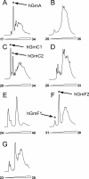
(A–G) HPLC separation profiles of the disulfide isomers of human granulin modules. Enterokinase-cleaved and SEP-Pak prepurified granulins were lyophilized, dissolved in 0.5–2.0 mL of 0.1% TFA, and loaded onto a 1.0 × 25 cm (hGrnA and hGrnC) or 0.46 × 25 cm (hGrnB, hGrnD, hGrnE, hGrnF, and hGrnG) Vydac C18 HPLC column, equilibrated with 10% acetonitrile in 0.1% TFA. The fractions were eluted with a linear 10%–70% acetonitrile gradient in 0.1% TFA, at a flow rate of 7.0 or 1.5 mL/min, respectively. Letters at the top of every HPLC profile correspond to the name of a specific human granulin module. Numbers at the bottom of each profile indicate the acetonitrile percentage.
Figure 3.
Figure 3.
Dose-response representation of GEM peptides on the prolilferation of MDA-MB-468 cells. Cell counts were determined using a hemocytometer 4 d after the addition of each peptide. The fold change was set at 1 for the controls, and all other counts were normalized to this value (n = 3 ± STD error of the mean). (A) Activity of well-folded, and full-length GEM peptides. (Inset) The dose response of hGrnA1 at nanomolar concentrations. (B) Activity of the full-length but poorly folded peptides. The identities of the peptides are labeled by one-letter codes; for example, D3, D6, D7, and D9 represent various isoforms of hGrnD.
Figure 4.
Figure 4.
(A) [15N,1H]-HSQC spectrum of 15N-labeled hGrnA in a buffered solution that was 10 mM in sodium phosphate, 150 mM NaCl, 0.2 mM EDTA, 10% D2O at pH 6.8. The concentration of hGrnA was ∼0.5 mM, and the NMR spectrum was recorded at 298 K. The backbone HSQC peaks from the hGrnA protein (i.e., the underlined residues in Table 1) are indicated by the corresponding residue numbers. (B) Schematic representation of the three-dimensional structure and disulfide-bonding pattern in cGrn1 (Hrabal et al. 1996). The disulfide bonds are shown by bold lines connecting two cysteine residues. Numbers in square brackets indicate the corresponding pairs of cysteines numbered from 1 to 12. (C) Location of cysteines in the second β-hairpin of a typical N-terminal S2H. Arrows between backbone protons indicate putative backbone–backbone NOE connectivities. (Dotted lines) Hydrogen bonds.
Figure 5.
Figure 5.
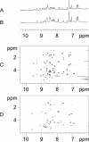
NMR spectra of paragranulin-derived peptides. (A) 1D proton spectrum of [hGrnP(C14S, C25S)] with the [Cys3–Cys13; Cys9–Cys24] disulfide-pairing pattern. (B) 1D proton spectrum of hGrnP (C14CAcm, C25CAcm) with the [Cys3–Cys13; Cys9–Cys24] disulfide-pairing pattern. (C) 2D TOCSY spectrum of hGrnP(C14CAcm, C25CAcm) with the [Cys3–Cys13; Cys9–Cys24] disulfide-pairing pattern. (D) 2D TOCSY spectrum of hGrnP peptide produced by the oxidation of hGrnP (C14CAcm, C25CAcm) with a [Cys3–Cys13; Cys14–Cys25; Cys9–Cys24] disulfide-pairing pattern. The NMR spectra were recorded on a Bruker Avance 500 MHz spectrometer at 298 K and in a buffered solution of 50 mM sodium acetate-d3, pH 5.0, 10% D2O. The concentration of the peptides was ∼0.5 mM.
Figure 6.
Figure 6.
Distribution of secondary structural elements in human granulins. (Arrows) Residues in β-strand conformation. (Broken lines) Poorly defined regions (with a local average pairwise backbone RMSD >0.5 Å based on tripeptide fragments with the residue in question as the central amino acid). β-Hairpin loops and β-turns are defined according to Sibanda and Thornton (1991).
Figure 7.
Figure 7.
The clusters of the 10 lowest-energy human granulin structures superimposed using the backbone of the residues in structurally defined regions. Regions corresponding to the third β-hairpin of cGrn1 (Hrabal et al. 1996) are consistently poorly defined and, for clarity of presentation, are not displayed. The N- and C-terminal fragments of hGrnA are superimposed over the backbone atoms of residues (2–4, 7–19, 23–28) and (45–49, 52–57), respectively. The N- and C-terminal fragments of hGrnC1/hGrnC2 are superimposed over the backbone atoms of residues (6–11, 14–20, 22–27) and (41–58), respectively. HGrnF1 is superimposed over the backbone atoms of residues (2–4, 8–11, 14–28).

References

    1. Baba, T., Hoff H.B., III, Nemoto, H., Lee, H., Orth, J., Arai, Y., Gerton, G.L. Acrogranin, an acrosomal cysteine-rich glycoprotein, is the precursor of the growth-modulating peptides, granulins, and epithelins, and is expressed in somatic as well as male germ cells. Mol. Reprod. Dev. 1993;34:233–243. - PubMed
    1. Baker, M., Mackenzie, I.R., Pickering-Brown, S.M., Gass, J., Rademakers, R., Lindholm, C., Snowden, J., Adamson, J., Sadovnick, A.D., Rollinson, S., et al. Mutations in progranulin cause tau-negative frontotemporal dementia linked to chromosome 17. Nature. 2006;442:916–919. - PubMed
    1. Bateman, A., Bennett, H.P. Granulins: The structure and function of an emerging family of growth factors. J. Endocrinol. 1998;158:145–151. - PubMed
    1. Bateman, A., Belcourt, D., Bennett, H., Lazure, C., Solomon, S. Granulins, a novel class of peptide from leukocytes. Biochem. Biophys. Res. Commun. 1990;173:1161–1168. - PubMed
    1. Belcourt, D.R., Lazure, C., Bennett, H.P. Isolation and primary structure of the three major forms of granulinlike peptides from hematopoietic tissues of a teleost fish (Cyprinus carpio) J. Biol. Chem. 1993;268:9230–9237. - PubMed

Publication types

MeSH terms