. 2011 Jul 13;19(7):945-54.
doi: 10.1016/j.str.2011.03.024.

NusA interaction with the α subunit of E. coli RNA polymerase is via the UP element site and releases autoinhibition

Affiliations

NusA interaction with the α subunit of E. coli RNA polymerase is via the UP element site and releases autoinhibition

Kristian Schweimer et al. Structure. .

Abstract

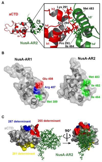
Elongating Escherichia coli RNAP is modulated by NusA protein. The C-terminal domain (CTD) of the RNAP α subunit (αCTD) interacts with the acidic CTD 2 (AR2) of NusA, releasing the autoinhibitory blockade of the NusA S1-KH1-KH2 motif and allowing NusA to bind nascent nut spacer RNA. We determined the solution conformation of the AR2:αCTD complex. The αCTD residues that interface with AR2 are identical to those that recognize UP promoter elements A nusA-ΔAR2 mutation does not affect UP-dependent rrnH transcription initiation in vivo. Instead, the mutation inhibits Rho-dependent transcription termination at phage λtR1, which lies adjacent to the λnutR sequence. The Rho-dependent λtimm terminator, which is not preceded by a λnut sequence, is fully functional. We propose that constitutive binding of NusA-ΔAR2 to λnutR occludes Rho. In addition, the mutation confers a dominant defect in exiting stationary phase.


Figures

Figure 1
Figure 1. Affinity of AR2:αCTD
A: Chemical shift changes in an expanded region of the 1H,15N HSQC of 15N labeled AR2 upon addition of unlabeled αCTD. The arrows mark the direction of signal shifts during the titration series. B: Titration curves for selected residues. Data fitting using a bimolecular two-state binding model results in a KD in a range of 1 μM - 5 μM (Leu434: 2.8 μM; Ala468: 3.4 μM; Ala480: 1.2 μM). Due to the high concentration required for the NMR experiments these results represent an estimate of the upper limit of the binding affinity.
Figure 2
Figure 2. Solution structure of the AR2:αCTD complex
A: Superposition of the structural ensemble of the AR2 (green):αCTD (red) complex. B: Ribbon representation of the lowest energy structure. Helices are labeled h1 through h4 for αCTD and h1′ through h5′ for NusA-AR2, colors as in (A).
Figure 3
Figure 3. Binding interface of the αCTD:AR2 complex
A: The hairpin of αCTD, red, interacts with h4 and h5 of AR2, green. Pro293 and Lys291 of αCTD, red, interact mainly with Met483 and Ile464 of AR2, green. B: The differences between the highly homologous domains NusA AR1 (surface representation: left) and AR2 are evident. Residues 356 to 414 of NusA-AR1 have been fitted onto residue 431 to 490 of AR2 in complex with αCTD (right; negatively charged residues, red; positively charged residues, blue; hydrophobic residues, green). C: αCTD, grey, in complex with AR2, green. The 261 determinant, yellow, interacts mainly with the σ70 factor, the 265 determinant, red, interacts mainly with UP-elements, and the 287 determinant, blue, interacts with regulatory proteins such as CAP (Benoff, Yang, et al, 2002). In the αCTD:AR2 complex, the 265 determinant is blocked by AR2, green, whereas the 287 and 261 determinants are accessible.
Figure 4
Figure 4. NusA AR2 displaces αCTD from the UP-element
A: Gel retardation experiments detecting displacement of αCTD from an UP-element by purified AR2 domain. All reactions contain 25 μM 32P-labeled wild-type UP-element DNA and an increasing amount of αCTD (up to 100 μM) and AR2 (up to 250 μM). B: Gel retardation experiments detecting displacement of αCTD from an UP-element by full-length NusA and control with NusA AR1. All reactions contain 25 μM 32P-labeled wild-type UP-element DNA, increasing amount of αCTD (up to 100 μM), NusA AR1 (up to 250 μM), and NusA wt (up to 200 μM).
Figure 5
Figure 5. NusA deletions slow the rate of exit from stationary phase
Strain MDS42 and its nusA mutant derivatives were grown overnight at 37 °C in LB or, for plasmid-bearing strains, LB + ampicillin (50 μg/ml) and then diluted 100-fold into LB or for plasmid-bearing strains, LB + ampicillin + 0.5 mM IPTG and grown at 37 °C for the times indicated. Initial OD600’s and viable counts for the 6 strains were equivalent. Strains: 10323 = wild-type (wt); 10324 = ΔnusA; 10881 = nusA-ΔAR2; 10875 = wt/ptac-NusA+; 10876 = ΔnusA/ptac-NusA+; 10889 = nusA-ΔAR2/ptac-NusA+.
Figure 6
Figure 6. Chemical shift perturbation of NusA-SKK by AR2 and nutL RNA
A: Chemical shift changes of NusA-SKK upon binding to AR2 as a function of primary sequence. B: Chemical shift changes of NusA-SKK upon binding to λ nutL RNA as a function of primary sequence; X = residues not assigned. Dotted line represents the significance level of 0.04 ppm; bars represent the three RNA binding domains.
Figure 7
Figure 7. NusA-SKK interacts with AR2 and nutL RNA
A: Binding of AR2 to SKK. Chemical shift changes of residues Arg270 and Gly249 during an NMR titration experiment of 2H, 15N labeled NusA-SKK with unlabeled AR2 demonstrate binding of AR2 to SKK. B: Release of autoinhibition. The direction of titration could be reversed by adding αCTD showing the release of NusA autoinhibition by AR2:αCTD interaction. C: KH1 is interaction domain with AR2 and nutL RNA. Because of the 52% homology of E. coli and Thermotoga maritima NusA-SKK (Worbs et al., 2001), we used the crystal structure of the latter as a template and mapped the observed chemical shift changes onto the surface, highlighting the binding interface of AR2 (C) and λ nutL RNA (D) (S1, light blue; KH1, gray; KH2 blue; Residues with resonances showing chemical shift changes of 0.04 ppm < Δδ < 0.1 ppm, green; those with Δδ > 0.1 ppm, red; unassigned resonances, yellow). Bottom: structure of nutL RNA as used in the titration experiment.

References

    1. Benoff B, Yang H, Lawson CL, Parkinson G, Liu J, Blatter E, Ebright YW, Berman HM, Ebright RH. Structural basis of transcription activation: the CAP-alpha CTD-DNA complex. Science. 2002;5586:1562–1566. - PubMed
    1. Beuth B, Pennell S, Arnvig KB, Martin SR, Taylor IA. Structure of a Mycobacterium tuberculosis NusA-RNA complex. EMBO J. 2005;20:3576–3587. - PMC - PubMed
    1. Bonin I, Muhlberger R, Bourenkov GP, Huber R, Bacher A, Richter G, Wahl MC. Structural basis for the interaction of Escherichia coli NusA with protein N of phage lambda. Proc. Natl. Acad. Sci. U. S. A. 2004;38:13762–13767. - PMC - PubMed
    1. Borukhov S, Lee J, Laptenko O. Bacterial transcription elongation factors: new insights into molecular mechanism of action. Mol. Microbiol. 2005;5:1315–1324. - PubMed
    1. Bubunenko M, Baker T, Court DL. Essentiality of ribosomal and transcription antitermination proteins analyzed by systematic gene replacement in Escherichia coli. J Bacteriol. 2007;7:2844–53. - PMC - PubMed

Publication types

MeSH terms

Associated data

LinkOut - more resources