

Review
doi: 10.1002/pro.2441.
Epub 2014 Mar 4.
Structural biology of the IL-1 superfamily: key cytokines in the regulation of immune and inflammatory responses
Affiliations
- PMID: 24677376
- PMCID: PMC4005705
- DOI: 10.1002/pro.2441
Item in Clipboard
Review
Structural biology of the IL-1 superfamily: key cytokines in the regulation of immune and inflammatory responses
Protein Sci.
2014 May.
Abstract
Interleukin-1 superfamily of cytokines (IL-1, IL-18, IL-33) play key roles in inflammation and regulating immunity. The mechanisms of agonism and antagonism in the IL-1 superfamily have been pursued by structural biologists for nearly 20 years. New insights into these mechanisms were recently provided by the crystal structures of the ternary complexes of IL-1β and its receptors. We will review here the structural biology related to receptor recognition by IL-1 superfamily cytokines and the regulation of its cytokine activities by antagonists.
Keywords: agonist; antagonist; cytokine; inflammation; interleukin; receptors.
© 2014 The Protein Society.
Figures
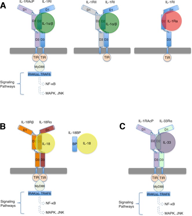
Cartoon representation of IL-1 superfamily signaling. A: IL-1 signaling is initiated by the ternary complex formation of IL-1α/β:IL-1RI:IL-1RAcP. The decoy receptor IL-1RII lacks an intracellular TIR domain and forms a non-signaling ternary complex with IL-1α/β and IL-1RAcP. The receptor antagonist IL-1Ra binds IL-1RI and forms an inhibitory binary complex that fails to recruit IL-1RAcP. B) IL-18 signaling complex involves IL-18, IL-18Rα and IL-18Rβ. Antagonism is achieved through binding of IL-18BP to IL-18. C) IL-33 signaling involves IL-33, IL-33Rα and IL-1RAcP. The ectodomains of IL-1 superfamily receptors each contains three Ig-like domains, which are labeled as D1, D2, and D3 respectively.
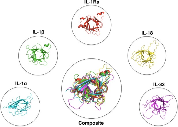
Structures of IL-1 superfamily cytokines. Cytokines of the IL-1 superfamily adopt a conserved β-trefoil fold. PDB files displayed: IL-1α:2KKI, IL-1β:1I1B, IL-1Ra:1ILR, IL-18:1J0S, IL-33:2KLL.
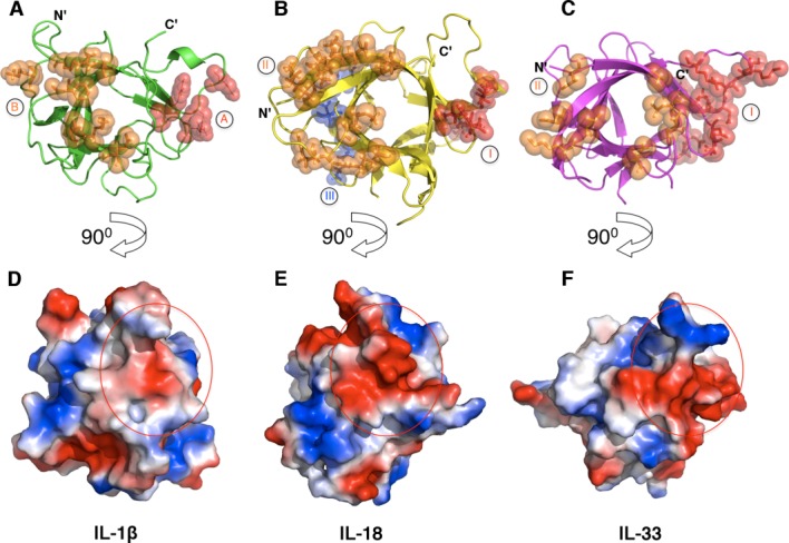
Receptor binding sites on IL-1β, IL-18, and IL-33. Depicted are the secondary structures (top) and the electropotential surfaces (bottom) of IL-1β (A and D, PDB ID 1ITB), IL-18 (B and E, PDB ID 4EKX) and IL-33 (C and F, PDB ID 4KC3). Residues that have been shown to interact with their respective receptors are shown as spheres and colored in red and orange for site A (site I for IL-18 and IL-33) and site B (site II for IL-18 and IL-33), respectively. A third putative receptor-binding site (site III) on IL-18 is shown as blue spheres. The surface area of the binding site A (site I) is indicated as a red circle on each cytokine (bottom).
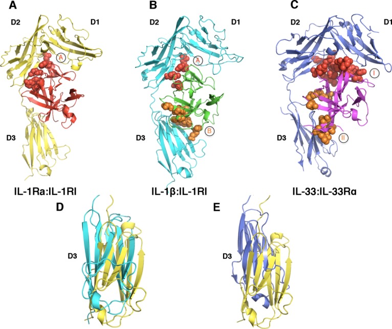
A common binding mode for receptor:ligand binary complexes. A: IL-1Ra (red) binds IL-1RI (yellow) predominantly at site A, with minimal interactions with the D3 domain of IL-1RI (PDB ID 1IRA). B) The binary complex of IL-1β (green):IL-1RI (cyan) revealed two binding sites, shown as spheres and colored in red (site A) and orange (site B), respectively (PDB ID 1ITB). C) The binary complex of IL-33 (magenta):IL-33Rα (blue) (PDB ID 4KC3). The receptor binding sites I and II on IL-33 are shown as red and orange spheres, respectively. D) and E) Rotation of the D3 domains of IL-1RI in different binary complexes. D) Superimposition of the structures of IL-1Ra:IL-1RI and IL-1β:IL-1RI reveals an approximate 200 rotation between the D3 domains of IL-1RI in the respective structures. E) The D3 domains of the respective receptors in IL-33:IL-33Rα and IL-1Ra:IL-1RI complexes display an approximate 100 rotation. The coloring schemes for the D3 domains of IL-1RI and IL-33Rα are same as above.
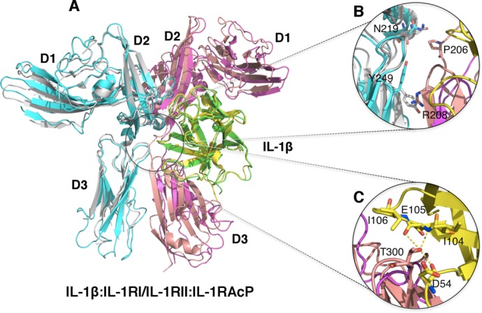
Ternary complex structures of IL-1β and its receptors. A) Superimposition of the non-signaling complex [IL-1β (Green):IL-1RII (Magenta):IL-1RAcP (Cyan), PDB ID:3O4O] and the signaling Complex [IL-1β (Yellow):IL-1RI (Pink):IL-1RAcP (Silver) PDB ID:4DEP]. B) Zoom-in view of the D3-D3 domain interactions of IL-1RAcP:IL-1RI and IL-1RAcP:IL-1RII. IL-1β is removed for clarity. The signaling complex showed additional interactions (residues shown in sticks) between IL-1RAcP with IL-1RI due to further rotation of the D3 domain of IL-1RI. These interactions were not observed between IL-1RAcP and IL-1RII in the non-signaling complex. C) Zoom-in view of the additional interactions between IL-1β and IL-1RAcP in the signaling ternary complex. Residue T300 of IL-1RAcP is involved in exquisite interactions with residues D54, I104, E105 and I106 of IL-1β.
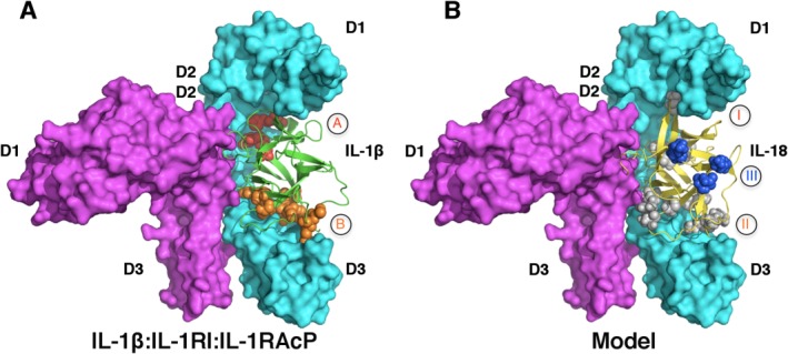
Unresolved questions regarding IL-18 subfamily signaling. A) IL-1R signaling ternary complex. IL-1β, IL-1RI and IL-1RAcP are colored in green, cyan and magenta (PDB ID 4DEP). Binding sites A and B are shown in red and orange spheres, respectively. B) A model of IL-18 (yellow):IL-18Rα (cyan):IL-18Rβ (magenta) based on the structure of IL-1β:IL-1RI:IL-1RAcP ternary complex. Putative receptor binding sites I (grey), II (grey) and III (blue) on IL-18 are shown as spheres. Notice site III is not involved in binding of either IL-18R in this model.
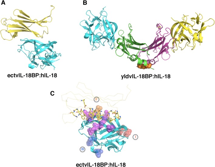
Mechanism of antagonism of IL-18 signaling by IL-18BP. A) The inhibitory complex of ectromelia virus (ectv) IL-18BP (yellow) and human IL-18 (hIL-18, cyan) in a 1:1 stoichiometry (PDB ID 3F62). B) The inhibitory complex of yaba-like disease virus (yldv) IL-18BP (green and magenta) and hIL-18 (yellow and cyan) in a 2:2 stoichiometry (PDB ID 4EKX). There is an intra-molecular disulfide bond within the yldv-IL-18BP homodimer as shown in orange spheres. C) Ectv-IL-18BP blocks the putative receptor-binding site II. Ectv-IL-18BP is shown as yellow ribbon with the key residues at the interface highlighted shown as sticks. Putative receptor binding sites I, II, and III on hIL-18 (cyan) are shown as spheres and colored in red, orange, and blue, respectively. Residues shared for binding with both IL-18Rα and ectvIL-18BP are shown as purple spheres.
References
-
- Dinarello C, Arend W, Sims J, Smith D, Blumberg H, O'Neill L, Goldbach-Mansky R, Pizarro T, Hoffman H, Bufler P, Nold M, Ghezzi P, Mantovani A, Garlanda C, Boraschi D, Rubartelli A, Netea M, van der Meer J, Joosten L, Mandrup-Poulsen T, Donath M, Lewis E, Pfeilschifter J, Martin M, Kracht M, Muehl H, Novick D, Lukic M, Conti B, Solinger A, Kelk P, van de Veerdonk F, Gabel C. IL-1 family nomenclature. Nat Immunol. 2010;11:973. - PMC - PubMed
-
- Dinarello CA. Immunological and inflammatory functions of the interleukin-1 family. Annu Rev Immunol. 2009;27:519–550. - PubMed
-
- Arend WP, Palmer G, Gabay C. IL-1, IL-18, and IL-33 families of cytokines. Immunol Rev. 2008;223:20–38. - PubMed
-
- O'Neill LA. The interleukin-1 receptor/Toll-like receptor superfamily: 10 years of progress. Immunol Rev. 2008;226:10–18. - PubMed
-
- Heguy A, Baldari CT, Macchia G, Telford JL, Melli M. Amino acids conserved in interleukin-1 receptors (IL-1Rs) and the Drosophila toll protein are essential for IL-1R signal transduction. J Biol Chem. 1992;267:2605–2609. - PubMed
Publication types
MeSH terms
Substances
Associated data
- Actions
- Actions
- Actions
- Actions
- Actions
- Actions
- Actions
- Actions
- Actions
- Actions
- Actions
- Actions
Grants and funding
LinkOut - more resources
Full Text Sources
Other Literature Sources
Miscellaneous
