. 2020 Oct 2;295(40):13927-13939.
doi: 10.1074/jbc.RA120.014244. Epub 2020 Aug 11.

The chemokine X-factor: Structure-function analysis of the CXC motif at CXCR4 and ACKR3

Affiliations

The chemokine X-factor: Structure-function analysis of the CXC motif at CXCR4 and ACKR3

Michael J Wedemeyer et al. J Biol Chem. .

Abstract

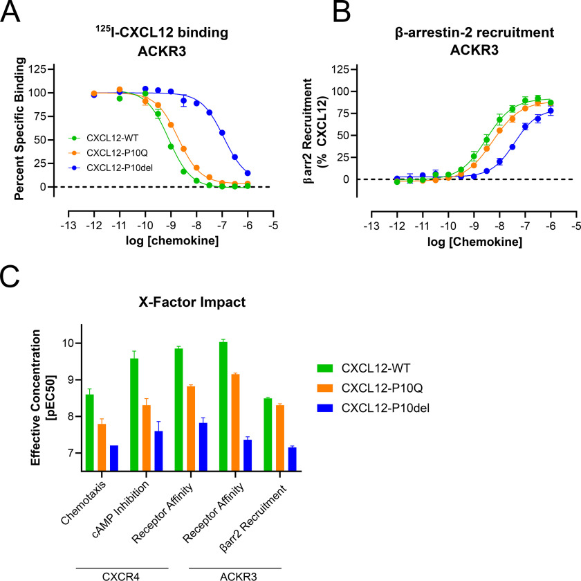
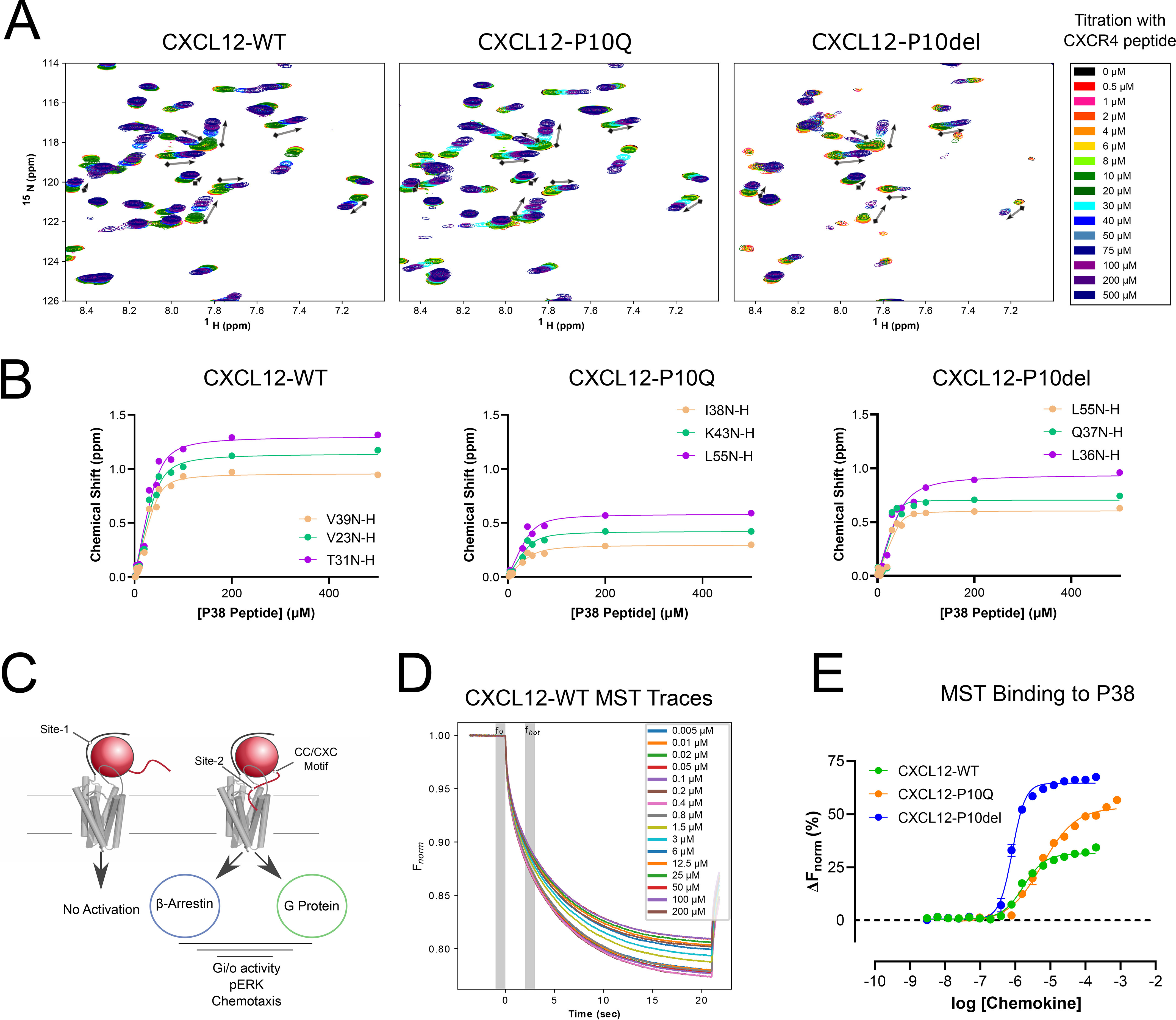
The human chemokine family consists of 46 protein ligands that induce chemotactic cell migration by activating a family of 23 G protein-coupled receptors. The two major chemokine subfamilies, CC and CXC, bind distinct receptor subsets. A sequence motif defining these families, the X position in the CXC motif, is not predicted to make significant contacts with the receptor, but instead links structural elements associated with binding and activation. Here, we use comparative analysis of chemokine NMR structures, structural modeling, and molecular dynamic simulations that suggested the X position reorients the chemokine N terminus. Using CXCL12 as a model CXC chemokine, deletion of the X residue (Pro-10) had little to no impact on the folded chemokine structure but diminished CXCR4 agonist activity as measured by ERK phosphorylation, chemotaxis, and Gi/o-mediated cAMP inhibition. Functional impairment was attributed to over 100-fold loss of CXCR4 binding affinity. Binding to the other CXCL12 receptor, ACKR3, was diminished nearly 500-fold. Deletion of Pro-10 had little effect on CXCL12 binding to the CXCR4 N terminus, a major component of the chemokine-GPCR interface. Replacement of the X residue with the most frequent amino acid at this position (P10Q) had an intermediate effect between WT and P10del in each assay, with ACKR3 having a higher tolerance for this mutation. This work shows that the X residue helps to position the CXCL12 N terminus for optimal docking into the orthosteric pocket of CXCR4 and suggests that the CC/CXC motif contributes directly to receptor selectivity by orienting the chemokine N terminus in a subfamily-specific direction.

Keywords: CC/CXC motif; G protein-coupled receptor (GPCR); cell migration; chemokine; chemokine network; nuclear magnetic resonance (NMR); signal transduction; structure-function.


Conflict of interest statement

Conflict of interest—B. F. V. and F. C. P. have ownership interests in Protein Foundry, LLC.

Figures

Figure 1.
Figure 1.
Selectivity in the chemokine network between CC and CXC chemokines. A, the chemokine network is illustrated with chemokine receptors labeled inside the membrane and the chemokines that bind that receptor labeled outside. The inner phylogenetic tree describes the evolutionary relationships between chemokine receptors. Colors represent each chemokine family: CC (blue), CXC (green), CX3C (red), XC (orange), and atypical (purple). B, the CXC receptors are shown linked with the X residues of their cognate ligands. Each one-letter code is colored by amino acid property: hydrophilic (green), hydrophobic (pink), positive charge (blue), conformationally special (purple), aromatic (yellow), or negative charge (red). The 17 CXC chemokines are represented below the corresponding X residue. C, unique solution-state NMR structures of CXC chemokines were analyzed to determine intramolecular contacts with the side chain of the X residue. These data are illustrated on the structure of CXCL12 (PDB entry 2KED). D, the average lengths of the β1- and β2-strands are shorter in CC chemokines than CXC chemokines, as calculated from 27 solution-state NMR structures. This is demonstrated by structures of CXCL12 (PDB entry 2KED) and CCL20 (PDB entry 2JYO). The X residue in CXCL12 (Pro-10) makes contacts with Leu-29, Thr-31, and Gln-37 to extend secondary structure into the 30s loop. E, each conformer in CC and CXC NMR structures were separated and aligned via the conserved chemokine core. Vectors describing the N terminus of each structure were averaged by family to construct the CC or CXC mean axes, which are separated by 45°. F, the structure of vMIP-II bound to CXCR4 (PDB entry 4RWS) is overlaid with the coordinate system of D. The CXC mean axis is near the N terminus of vMIP-II in the orthosteric pocket, whereas the CC mean axis is outside of the receptor.
Figure 2.
Figure 2.
Sequence and structure variation between CC and CXC chemokines. A, the sequences of the activation (site-1) and binding (site-2) motifs were compared between CC and CXC chemokines. Both the chemokine and receptor N termini are unstructured in the unbound state and thus are aligned to conserved cysteine residues. The X residue in the CXC motif unequivocally divides the CC and CXC chemokines. The next deterministic position directly precedes the first cysteine and is negatively charged in 76% CXC chemokines and only 4% of CC chemokines. No difference in average N-terminal length was observed. B, constructs of CXCL12-WT, -P10Q, and -P10del are shown. Glutamine was chosen for substitution as it is the most common X residue. C, structural models of CXCL12-P10del were constructed using the solution-state NMR structure 2KED. Deviation from the WT structure was observed at the N terminus and 30s loop. D, to understand the dynamic impact, 100-ns MD simulations were performed with CXCL12-WT and CXCL12-P10del in explicit solvent. Orientation of the proximal N terminus consistently differs between CC and CXC final states shown overlaid with the CXCR4 co-crystal structure (PDB entry 4RWS). The position of Arg-8 in CXCL12-WT is near the expected binding partner CXCR4:Asp-262, whereas Arg-8 in CXCL12-P10del is facing the opposite side of the pocket. E, each construct was examined by NMR HSQC. Uniform peak distribution supports tertiary folding. Unequal peak intensities in CXCL12-P10del result from an increase in dynamics. Overlays with CXCL12-WT demonstrate altered peak position. F, the intrinsic fluorescence of each chemokine was measured throughout thermal denaturation. The plotted first derivative illustrates the Tm of CXCL12-WT (91.1 °C), CXCL12-P10Q (88.3 °C), or CXCL12-P10del (86.3 °C) performed in triplicate.
Figure 3.
Figure 3.
Modification of the CXC X-factor reduces signaling and binding to CXCR4. A, displacement of [125I]CXCL12-WT from CXCR4 was measured for each construct via scintillation after a 4-h equilibration with membrane suspensions at room temperature. Measurements were performed in duplicate from three independent experiments. B, inhibition of cAMP was measured via the GloSensor assay after stimulation with forskolin. Measurements were performed in quadruplicate from at least three independent experiments. C, HEK-293 cells were stimulated with 10 nm chemokine for 5 min, and ERK phosphorylation was determined by Western blotting. Quantification of ERK phosphorylation relative to vehicle is shown to the right. D, transwell chemotaxis assays were performed over 2 h with THP1 cells. Raw counts of migrated cells display the full chemotactic curve (left). The first half of the biphasic curve (right) shows a loss in efficacy and potency in CXCL12-P10del. Measurements were performed in quadruplicate from at least three independent experiments.
Figure 4.
Figure 4.
Effects of CXC mutants on another chemokine receptor, ACKR3. A, displacement of [125I]CXCL12-WT from ACKR3 was measured for each construct via scintillation after a 4- h equilibration with membrane suspensions at room temperature. Measurements were performed in duplicate from at least three independent experiments. B, recruitment of β-arrestin was measured via the Tango assay performed in quadruplicate from at least three independent experiments. C, summary of the X-factor impact in each signaling and binding experiment. CXCL12-P10del requires a significantly higher concentration to reach 50% effectiveness. CXCL12-P10Q is intermediate between WT and P10del.
Figure 5.
Figure 5.
Receptor N-terminal binding is maintained through X-mutation and deletion. A, the NMR HSQC spectra over a 16-point titration series shows specific peak movement with the addition of a 38-residue CXCR4 peptide. Directionality of peak movement is consistent for each protein. B, example nonlinear fits for three residues in each titration series. Apparent affinity was calculated for CXCL12-WT (2 μm), CXCL12-P10Q (2 μm), and CXCL12-P10del (7 μm) from averaging the 15 largest shifting residues. C, a cartoon demonstrates the two-site model of chemokine interaction. The peptide used in the NMR titration series corresponds to site-1 binding. D, MST traces plot the resulting fluorescence (Fnorm) at each concentration (0.006–200 μm) of chemokine mixed with 25 nm P38-Cy5 normalized to the 1.0-s equilibration f0. Time region fhot corresponds to the 1.0-s data collection interval (1.5–2.5 s). Sample conditions were optimized to prevent aggregation, adsorption, photobleaching, and insufficient fluorescence (signal/noise > 20.0). E, dose-response curve plot calculated ΔFnorm (%), with error bars showing S.E. with n > 5 runs for each chemokine. Dissociation constants were calculated for CXCL12-WT (2.0 ± 0.5 μm), CXCL12-P10Q (5.0 ± 1.0 μm), and CXCL12-P10del (0.9 ± 0.4 μm).
Figure 6.
Figure 6.
Impact of the chemokine X-factor. A, the two determined co-crystal structures of CC and CXC receptors display unique chemokine binding orientations. The CC chemokine body interacts closely with the receptor core and includes penetration of the 30s loop into the orthosteric pocket. The CXCR4 co-crystal complex has vMIP-II above the extracellular vestibule with few interactions between the receptor and the 30s loop. B, a proposed model describing the potential influence of the X-factor between CC and CXC chemokines. The X residue directly interacts with the β1-β2 hairpin to stabilize this region in an extended conformation. The loss of the X position pulls the 30s loop into a narrow conformation that favors the increased CC binding depth. Additionally, the presence of the X position constrains the orientation of the chemokine N terminus in solution. The more linear N terminus found in CXC structures matches with the chemokine position above the receptor pocket in the co-crystal structure.

References

    1. Thomas M. A., Kleist A. B., and Volkman B. F. (2018) Decoding the chemotactic signal. J. Leukoc. Biol. 104, 359–374 10.1002/JLB.1MR0218-044 - DOI - PMC - PubMed
    1. Bachelerie F., Ben-Baruch A., Burkhardt A. M., Combadiere C., Farber J. M., Graham G. J., Horuk R., Sparre-Ulrich A. H., Locati M., Luster A. D., Mantovani A., Matsushima K., Murphy P. M., Nibbs R., Nomiyama H., et al. (2014) International Union of Pharmacology. LXXXIX. Update on the extended family of chemokine receptors and introducing a new nomenclature for atypical chemokine receptors. Pharmacol. Rev. 66, 1–79 10.1124/pr.113.007724 - DOI - PMC - PubMed
    1. Alexander S. P. H., Mathie A., and Peters J. A. (2011) Guide to receptors and channels (GRAC), 5th Ed. Br. J. Pharmacol. 164, S1–S2 10.1111/j.1476-5381.2011.01649_1.x - DOI - PMC - PubMed
    1. Stone M. J., Hayward J. A., Huang C., Huma Z. E., and Sanchez J. (2017) Mechanisms of regulation of the chemokine-receptor network. Int. J. Mol. Sci. 18, 342 10.3390/ijms18020342 - DOI - PMC - PubMed
    1. Mantovani A. (1999) The chemokine system: redundancy for robust outputs. Immunol. Today 20, 254–257 10.1016/s0167-5699(99)01469-3 - DOI - PubMed

Publication types

MeSH terms

Associated data

LinkOut - more resources