. 2014 Dec 12;289(50):34953-64.
doi: 10.1074/jbc.M114.599878. Epub 2014 Oct 23.

Copsin, a novel peptide-based fungal antibiotic interfering with the peptidoglycan synthesis

Affiliations

Copsin, a novel peptide-based fungal antibiotic interfering with the peptidoglycan synthesis

Andreas Essig et al. J Biol Chem. .

Abstract

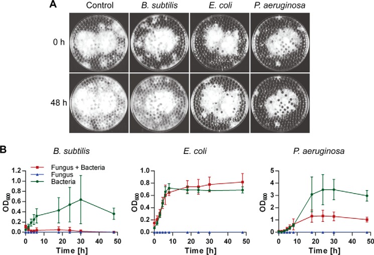
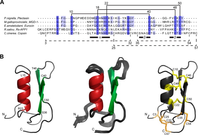
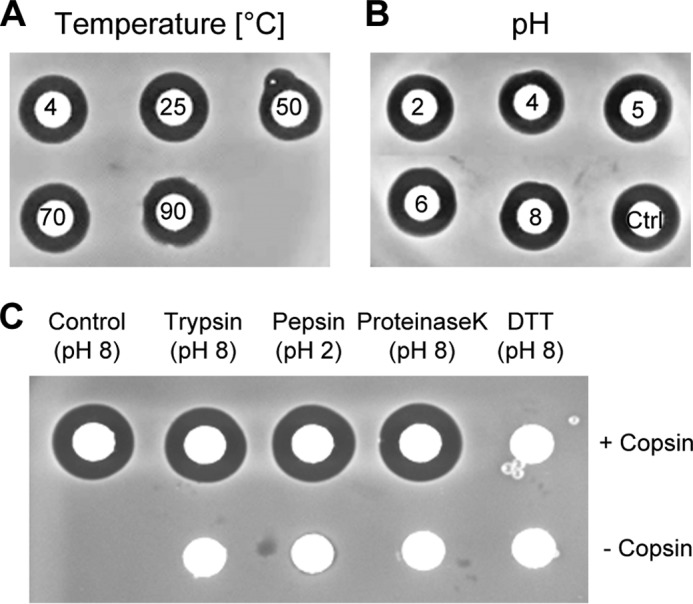
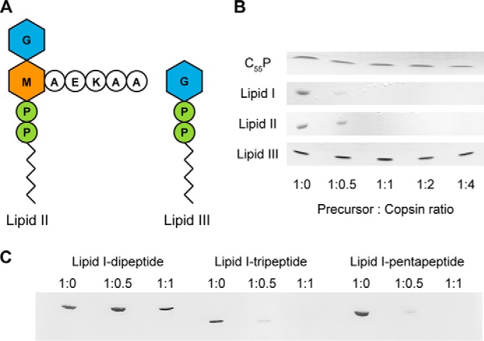
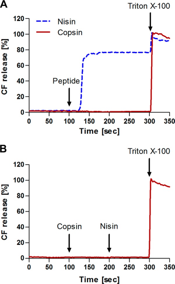
Fungi and bacteria compete with an arsenal of secreted molecules for their ecological niche. This repertoire represents a rich and inexhaustible source for antibiotics and fungicides. Antimicrobial peptides are an emerging class of fungal defense molecules that are promising candidates for pharmaceutical applications. Based on a co-cultivation system, we studied the interaction of the coprophilous basidiomycete Coprinopsis cinerea with different bacterial species and identified a novel defensin, copsin. The polypeptide was recombinantly produced in Pichia pastoris, and the three-dimensional structure was solved by NMR. The cysteine stabilized α/β-fold with a unique disulfide connectivity, and an N-terminal pyroglutamate rendered copsin extremely stable against high temperatures and protease digestion. Copsin was bactericidal against a diversity of Gram-positive bacteria, including human pathogens such as Enterococcus faecium and Listeria monocytogenes. Characterization of the antibacterial activity revealed that copsin bound specifically to the peptidoglycan precursor lipid II and therefore interfered with the cell wall biosynthesis. In particular, and unlike lantibiotics and other defensins, the third position of the lipid II pentapeptide is essential for effective copsin binding. The unique structural properties of copsin make it a possible scaffold for new antibiotics.

Keywords: Antibiotic Resistance; Antibiotics; Antimicrobial Peptide (AMP); Bacterial-Fungal Interaction (BFI); Fungal Secretome; Fungi; Lipid II.


Figures

FIGURE 1.
FIGURE 1.
Interactions between C. cinerea and bacteria. A, vegetative mycelium of C. cinerea was grown on submerged glass beads in minimal medium. After 60 h, 4 ml of medium was replaced by a suspension of B. subtilis, E. coli, or P. aeruginosa. 0 and 48 h after addition of the bacteria, the plates were photographed. Fungal growth is indicated by the white mycelium. B, bacterial growth was monitored by measuring A600 (OD600) over 48 h. As controls, bacteria and fungus were grown independently in the beads system. All data points were acquired in three biological replicates and are displayed with the standard deviation. C. cinerea exhibited a bactericidal effect in competition with B. subtilis and was strongly inhibited by P. aeruginosa.
FIGURE 2.
FIGURE 2.
Identification of an AMP in the secretome of C. cinerea. A, proteins were extracted from the unchallenged C. cinerea secretome, digested with proteinase K, and the remaining proteins were fractionated on a cation exchange column. The effluent was monitored at 280 nm (solid line), and the conductivity was measured (dashed line). B, fractions collected (0.5 ml) during the run were spotted in a disk diffusion assay against B. subtilis. Activity was detected from 7 to 8.5 ml of elution volume. C, proteins in the active fractions and flow through (FT) were identified by electrospray ionization-MS/MS and quantified by spectral counting. The relative counts corresponding to the protein CC1G_13813 (copsin) were best correlating to the diameter of the inhibition zones displayed against B. subtilis.
FIGURE 3.
FIGURE 3.
Three-dimensional structure of copsin and similarity to other AMPs. A, a sequence alignment of copsin with AMPs exhibited the highest sequence identities with defensins from plants, fungi, and invertebrates (5, 28, 41, 45). Disulfide bonds shown in solid lines form the core structural motif CSαβ. Disulfide bonds in dashed lines were additionally detected in copsin. Regions of the α-helix and the two β-strands as well as the positions of the Cys residues are indicated according to the sequence of copsin. The alignment was performed with the ClustalW algorithm and visualized with the Jalview software (46, 47). B, cartoon representation of the structure of copsin determined by NMR spectroscopy. Left panel, ribbon diagram of the structure with the lowest energy after energy refinement with AMBER (16), highlighting secondary structure elements: α-helical region (red) and the β-sheet (green). The N and C termini are denoted by N and C, respectively, and selected residues are indicated with the residue type and sequence number. Middle panel, bundle of 20 conformers after energy refinement. Right panel, Cys residues are denoted by the sequence position and are colored in yellow for the conserved disulfide bonds and in orange for the three additional disulfide bonds of copsin.
FIGURE 4.
FIGURE 4.
Temperature, pH, and protease stability of copsin. A and B, shown are disk diffusion assays on B. subtilis with copsin that was exposed to different temperatures (4, 25, 50, 70, 90 °C) and in a pH range of 2–8 for 1 h at 25 °C. C, to evaluate the protease resistance, copsin/protease mixtures in a ratio of 10:1 (w/w) were incubated at pH 8 for trypsin and proteinase K and at pH 2 for pepsin for 3 h. Copsin was also subjected to a treatment with 5 mm DTT. Additionally, the reaction mixtures were spotted without copsin. As control, untreated copsin was loaded on a paper disk. The activity of copsin was retained in the whole temperature and pH range tested, and it showed no sensitivity in a treatment with proteases. The only way to delete the activity of copsin was by disrupting the disulfide bonds.
FIGURE 5.
FIGURE 5.
Killing kinetics of copsin. B. subtilis grown in MHB at pH 6 and 7.3 and incubated with and without (Ctrl) 4 μg/ml copsin (4× MIC). The A600 is shown above the spectrum, measured at 0, 2, and 5 h.
FIGURE 6.
FIGURE 6.
Co-localization of copsin and vancomycin. B. subtilis and S. carnosus cells in the exponential growth phase were stained with TAMRA-copsin (red) and subsequently stained with BODIPY-vancomycin (green). Cells showed a co-localization of copsin with vancomycin at the cell septa. Scale bars, 2 μm.
FIGURE 7.
FIGURE 7.
Binding of copsin to cell wall precursors. A, schematic of the peptidoglycan precursor lipid II and the wall teichoic acid precursor lipid III. M, MurNAc; G, GlcNAc; P, phosphate. B, purified cell wall precursors were incubated with increasing molar ratios of copsin. After extraction with n-butanol/pyridine acetate (pH 4.2), the samples were analyzed by TLC, which displayed a binding of copsin to lipids I and II. C, truncated versions of lipid I (lipid I-dipeptide and lipid I-tripeptide) as well as lipid I-pentapeptide were synthesized, using purified enzymes from S. aureus. Copsin was added in increasing molar ratios to the lipid I versions. After incubation for 20 min at room temperature, samples were extracted as described and analyzed by TLC. The result showed that copsin binds to the third position of the pentapeptide.
FIGURE 8.
FIGURE 8.
Carboxyfluorescein efflux from lipid II containing liposomes. Activity of copsin and nisin against unilamellar liposomes made of 1,2-dioleoyl-sn-glycero-3-phosphocholine supplemented with 0.1 mol % lipid II. Peptide-induced marker release from liposomes with entrapped CF was measured. The 100% leakage level was determined by addition of Triton X-100 after 300 s. A, 1 μm copsin (solid line) or nisin (dashed line) was added after 100 s. B, first, 1 μm copsin was added (100 s) following the addition of 1 μm nisin (200 s) to the same sample. Copsin did not permeabilize the membrane, but it blocked the action of nisin.

References

    1. Frey-Klett P., Burlinson P., Deveau A., Barret M., Tarkka M., Sarniguet A. (2011) Bacterial-fungal interactions: hyphens between agricultural, clinical, environmental, and food microbiologists. Microbiol. Mol. Biol. Rev. 75, 583–609 - PMC - PubMed
    1. Scherlach K., Graupner K., Hertweck C. (2013) Molecular bacteria-fungi interactions: effects on environment, food, and medicine. Annu. Rev. Microbiol. 67, 375–397 - PubMed
    1. Fleming A. (1929) On the antibacterial action of cultures of a Penicillium, with special reference to their use in the isolation of B. influenzae. Br. J. Exp. Pathol. 10, 226–236 - PubMed
    1. Zasloff M. (2002) Antimicrobial peptides of multicellular organisms. Nature 415, 389–395 - PubMed
    1. Mygind P. H., Fischer R. L., Schnorr K. M., Hansen M. T., Sönksen C. P., Ludvigsen S., Raventós D., Buskov S., Christensen B., De Maria L., Taboureau O., Yaver D., Elvig-Jørgensen S. G., Sørensen M. V., Christensen B. E., Kjaerulff S., Frimodt-Moller N., Lehrer R. I., Zasloff M., Kristensen H. H. (2005) Plectasin is a peptide antibiotic with therapeutic potential from a saprophytic fungus. Nature 437, 975–980 - PubMed

Publication types

MeSH terms

Associated data