. 2007 Oct 12;28(1):41-56.
doi: 10.1016/j.molcel.2007.08.024.

Structures of GRP94-nucleotide complexes reveal mechanistic differences between the hsp90 chaperones

Affiliations

Structures of GRP94-nucleotide complexes reveal mechanistic differences between the hsp90 chaperones

D Eric Dollins et al. Mol Cell. .

Abstract

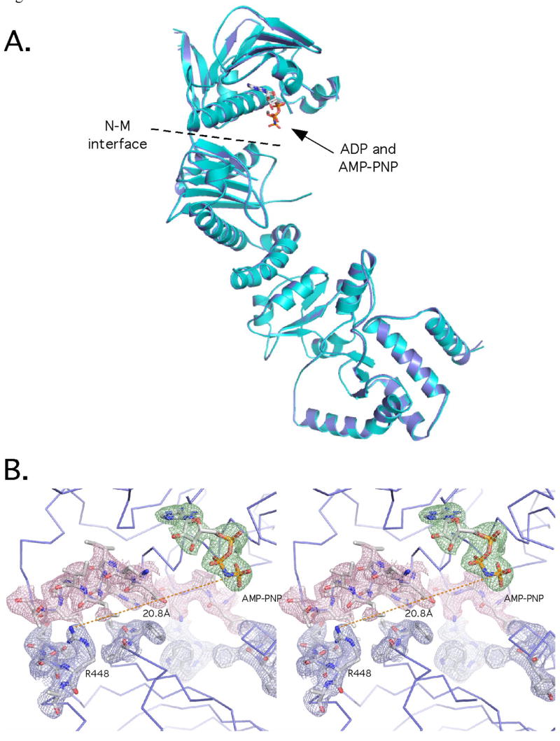
GRP94, an essential endoplasmic reticulum chaperone, is required for the conformational maturation of proteins destined for cell-surface display or export. The extent to which GRP94 and its cytosolic paralog, Hsp90, share a common mechanism remains controversial. GRP94 has not been shown conclusively to hydrolyze ATP or bind cochaperones, and both activities, by contrast, result in conformational changes and N-terminal dimerization in Hsp90 that are critical for its function. Here, we report the 2.4 A crystal structure of mammalian GRP94 in complex with AMPPNP and ADP. The chaperone is conformationally insensitive to the identity of the bound nucleotide, adopting a "twisted V" conformation that precludes N-terminal domain dimerization. We also present conclusive evidence that GRP94 possesses ATPase activity. Our observations provide a structural explanation for GRP94's observed rate of ATP hydrolysis and suggest a model for the role of ATP binding and hydrolysis in the GRP94 chaperone cycle.


Figures

Figure 1
Figure 1
Overview of GRP94-NMC and comparison with GRP94-NM and GRP94-MC. The two protomers are shown in blue and cyan. A) Ribbon drawing of side and top views. The AMPPNP is shown as a stick model. In the top view, the positions of helix N1, which mediate N-domain dimer interactions, are indicated. B) Stereo surface view of the GRP94-NMC dimer. The N-domains do not contact each other. C) Comparison of GRP94-NMC, GRP94-NM, and GRP94-MC structures. Domain boundaries are indicated by dashed lines. The same relative orientation is shown for each structure. Only a single protomer from the GRP94-NMC and GRP94-MC dimers is shown. D) Overlay of the 3 structures. Colors are as in panel C.
Figure 2
Figure 2
Comparison of GRP94-NMC with yeast Hsp82 and HtpG. A) Ribbon diagrams of each structure. Hsp82 coordinates are from PDB code 2CG9, and HtpG coordinates are from PDB code 2IOQ. GRP94-NMC is AMPPNP-bound. Hsp82 is AMPPNP bound and co-chaperone Sba1 has been removed for clarity. HtpG is unliganded. B) Comparison of N- and M domain orientations in GRP94-NMC and Hsp82. GRP94 (blue) and Hsp82 (green) were superimposed using the middle and C-terminal domains for alignment. Alpha helices are shown as cylinders, and the AMPPNP is shown as sticks. The helix 4/5 lid of Hsp82 is colored pink. C) The N-middle interaction dictates the positioning of the catalytic residues. Views of the N-middle interaction in Hsp82 (green) and GRP94 (blue) with the N domains aligned. The catalytic E33 and R380 in Hsp82, and the equivalent E103 and R448 are indicated.
Figure 3
Figure 3
GRP94 N domain conformations. A,B) Structures of the isolated N domain determined in the absence (A) or presence (B) of bound adenosine nucleotide. The core of the domain is colored gray, and the lid components, helices N1, N4, and N5, along with strand 1 are colored magenta or yellow. C) N domain as found in the AMPPNP-bound GRP94-NMC structure. The orientation of the molecule is the same as in panels A and B. The likely trajectory of the disordered portion of the helix N4/N5 lid is shown in dashed lines. D) Details of the backbones of the 3 N domains in the vicinity of the nucleotide binding pocket. Only the alpha carbons are shown. Colors are as in A, B, and C. The ball denotes the position of the Gly197 alpha carbon, and shows the backbone transition observed in the N domain when adenosine nucleotides bind. E) Close-up overlay as in D) showing the movement of helix N1 upon nucleotide binding. F) Superposition of yeast Hsp82-NMC (PDB code 2CG9) with GRP94-NMC. Hsp82 is in green, and GRP94 is in blue. The N domains from each structure were the basis for the alignment. The helix N4/N5 lid from Hsp82 is shown in pink. The molecular surface of the GRP94 M domain is shown.
Figure 4
Figure 4
C-terminal domain of GRP94 has additional dimer interactions. A) The C-terminal domain. Helices comprising the strap residues are shown in gold. B) A potential client binding surface composed of Met-Met pairs and hydrophobic residues.
Figure 5
Figure 5
ADP and AMPPNP-bound GRP94 have identical conformations. A) Overlay of the two nucleotide bound complexes. The AMPPNP complex is in blue, and the ADP complex is in cyan. Only one protomer of the GRP94 dimer is shown. B) Stereo view of the bound AMPPNP. SA omit electron density contoured at 1.3σ is shown with a 2 Å carve radius. The density for the nucleotide is shown in green, for the residues of the N domain in pink, and the M domain in blue. The position of Arg448 is indicated, and the distance to the gamma phosphate of the AMPPNP is shown with a dashed line.
Figure 6
Figure 6
GRP94 ATPase assays. Activity was monitored by the detection of free phosphate and represents the average of at least 3 independent measurements. A) GRP94 (white) and Hsp82 (shaded) constructs and chimeras used in these assays. S1 refers to strand N1, and H1 to helix N1. The GRP94 charged region segment is indicated by horizontal lines and that of Hsp82 by vertical lines. B,C,D) Specific activity of GRP94 and selected GRP94 mutants. Constructs used in each assay are indicated next to each reaction curve. E) Comparison of yeast Hsp82, GRP94-NMC, and Hsp82/GRP94 chimera ATPase rates. F) Summary of relative ATPase rates for GRP94 and Hsp82. Activity is given as a percentage of the Hsp82 rate.
Figure 7
Figure 7
Model of GRP94 ATP hydrolysis mechanism. The apo N domains of the “Twisted V” dimer (cartoon diagram in blue and cyan, helices represented as cylinders), undergo local conformational changes in the N mobile domain elements strand N1 and helix N1 (orange), and the Helix N4/N5 lid (green) upon binding nucleotide (shown as sticks). Each protomer of ATP bound “Twisted V” dimer, undergoes an ~90° rotation around the N/M interface aligning the nucleotide with the catalytic residues of the M domain and transiently dimerizing the N domains. The ATP bound “catalytically competent” closed dimer form is stabilized by cross-protomer interactions between strand N1 and helix N1 (orange) from the opposing chain, those elements previously rearranged upon nucleotide binding, as well a bridging effect between the terminal phosphate of the nucleotide and the catalytic residue of the M domain. The helix N4/N5 lid is closed over the nucleotide, trapping it in preparation for hydrolysis. After hydrolysis, there is a concomitant disassembly of the transient N dimer, rotation around the N/M interface, and re-structuring of the mobile domain elements of the N domain.

Comment in

  • A Grp on the Hsp90 mechanism.
    Richter K, Reinstein J, Buchner J. Richter K, et al. Mol Cell. 2007 Oct 26;28(2):177-9. doi: 10.1016/j.molcel.2007.10.007. Mol Cell. 2007. PMID: 17964255 Review.

References

    1. Ali MM, Roe SM, Vaughan CK, Meyer P, Panaretou B, Piper PW, Prodromou C, Pearl LH. Crystal structure of an Hsp90-nucleotide-p23/Sba1 closed chaperone complex. Nature. 2006;440:1013–1017. - PMC - PubMed
    1. Ban C, Junop M, Yang W. Transformation of MutL by ATP binding and hydrolysis: a switch in DNA mismatch repair. Cell. 1999;97:85–97. - PubMed
    1. Chadli A, Bouhouche I, Sullivan W, Stensgard B, McMahon N, Catelli MG, Toft DO. Dimerization and N-terminal domain proximity underlie the function of the molecular chaperone heat shock protein 90. Proc Natl Acad Sci U S A. 2000;97:12524–12529. - PMC - PubMed
    1. Chiosis G. Targeting chaperones in transformed systems--a focus on Hsp90 and cancer. Expert Opin Ther Targets. 2006;10:37–50. - PubMed
    1. Cochran AG, Skelton NJ, Starovasnik MA. Tryptophan zippers: stable, monomeric beta -hairpins. Proc Natl Acad Sci U S A. 2001;98:5578–5583. - PMC - PubMed

Publication types

MeSH terms