. 2015 Oct 9;290(41):24902-13.
doi: 10.1074/jbc.M115.680389. Epub 2015 Aug 28.

Structural Basis for the Discriminative Recognition of N6-Methyladenosine RNA by the Human YT521-B Homology Domain Family of Proteins

Affiliations

Structural Basis for the Discriminative Recognition of N6-Methyladenosine RNA by the Human YT521-B Homology Domain Family of Proteins

Chao Xu et al. J Biol Chem. .

Abstract

N(6)-Methyladenosine (m(6)A) is the most abundant internal modification in RNA and is specifically recognized by YT521-B homology (YTH) domain-containing proteins. Recently we reported that YTHDC1 prefers guanosine and disfavors adenosine at the position preceding the m(6)A nucleotide in RNA and preferentially binds to the GG(m(6)A)C sequence. Now we systematically characterized the binding affinities of the YTH domains of three other human proteins and yeast YTH domain protein Pho92 and determined the crystal structures of the YTH domains of human YTHDF1 and yeast Pho92 in complex with a 5-mer m(6)A RNA, respectively. Our binding and structural data revealed that the YTH domain used a conserved aromatic cage to recognize m(6)A. Nevertheless, none of these YTH domains, except YTHDC1, display sequence selectivity at the position preceding the m(6)A modification. Structural comparison of these different YTH domains revealed that among those, only YTHDC1 harbors a distinctly selective binding pocket for the nucleotide preceding the m(6)A nucleotide.

Keywords: N6-methyladenosine; RNA binding protein; RNA methylation; RNA-protein interaction; crystal structure; isothermal titration calorimetry (ITC).


Figures

FIGURE 1.
FIGURE 1.
Overall structure of the YTHDF1 YTH domain. A, sequence alignment of the YTH domains of human YTHDF1-3, YTHDC1-2, and yeast Pho92 by ESPript 3. B, overall structure of the YTHDF1 YTH domain (amino acids 361–559) shown in orange cartoon. The invisible residues in the apo structure are denoted with dashes. C, topology of the YTH domain of YTHDF1 with marked secondary structure elements.
FIGURE 2.
FIGURE 2.
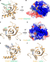
Complex structures of the YTHDF1 YTH domain with GG(m6A)CU and the Pho92 YTH domain with UG(m6A)CU. A, overall structure of the YTHDF1 YTH domain (amino acids 365–554) with a 5-mer RNA GG(m6A)CU. The four visible nucleotides (GG(m6A)C) are shown in green cartoon. m6A is shown in a green stick model. The protein is shown in orange cartoon. B, electrostatic surface representation of the YTHDF1 complex structure by PyMOL. C, overall structure of the Pho92 YTH domain in complex with a 5-mer RNA UG(m6A)CU, with only m6A visible and shown as a green stick model). D, electrostatic surface representation of the Pho92 complex structure. E, the simulated annealing m|Fo| − D|Fc| omit map of the RNA (green) is contoured at 2.8σ. The protein is shown in orange cartoon. F, the simulated annealing m|Fo| − D|Fc| omit map of the m6A (green) is contoured at 2.8σ. The protein is shown in orange cartoon.
FIGURE 3.
FIGURE 3.
Detailed interactions between the YTH domain of YTHDF1 (orange) and the 5-mer RNA −2GG(m6A)CU+2 (green). Top left panel, overall view. Nucleotides and residues involved in binding to RNA are shown in stick mode. The intermolecular hydrogen bonds are shown as black dashes. Bottom left panel, G-1 interactions. Bottom right panel, the m6A pocket. Top right panel, C+1 interactions.
FIGURE 4.
FIGURE 4.
YTHDF1 specifically recognizes m6A RNA without sequence selectivity. A–D, the representative ITC binding curves of YTHDF1 binding to different 16-mer m6A RNAs. A, 16-mer GG(m6A)CU. B, 16-mer GA(m6A)CU. C, 16-mer GC(m6A)CU. D, 16-mer GU(m6A)CU. E–G, the methylated nucleotide is shown as green sticks. The residues involved in binding the m6A are shown as orange sticks. E, the m6A binding pocket of wild type YTHDF1. F, the hypothetic model of the m6A binding pocket of YTHDF1 with the D401N mutation. A hydrogen bond could be formed between the side chain of Asp401 and the N1 atom of the m6A nucleotide. Asp401 is marked in black. G, the m6A binding pocket of the Pho92 YTH domain.
FIGURE 5.
FIGURE 5.
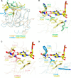
YTHDC1 has a selective binding pocket at the −1 position to accommodate the nucleotide preceding m6A, distinct from other YTH domains.A, superposition of the crystal structures of the YTHDF1 (orange cartoon)-GG(m6A)CU (green cartoon) and YTHC1 (blue cartoon)-GG(m6A)CU (yellow cartoon) (PDB entry 4R3I). The m6A nucleotides are shown in stick mode. B–D, the G-1 binding pocket of the YTHDC1 YTH domain is superimposed with the corresponding pocket of the YTHDF1 YTH domain (B), the Pho92 YTH domain (C), and the YTHDC2 YTH domain (D, PDB entry 2YU6). The G-1 nucleotide is shown as a yellow stick model. The Leu380, Val382, Asn383, and Met438 of YTHDC1 and their corresponding residues in the YTH domains of YTHDF1, YTHDC2, and Pho92, are shown as sticks.
FIGURE 6.
FIGURE 6.
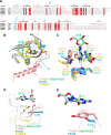
Structure comparison betweenYTHDF1, Pho92, and YTHDC2 (PDB entry 2YU6). A, sequence alignment of the YTH domains of human YTHDC1, YTHDC2, and YTHDF1-3. The secondary structures of the YTHDC1 YTH domain are shown at the top of the sequences according to Ref. . The two critical G-1 binding residues of YTHDC1, Leu380 and Met438, which are not conserved in other YTH family members, are marked in blue. B, superposition of the crystal structures of YTHDF1 (salmon cartoon)-GG(m6A)CU (green cartoon), YTHDC2 (yellow cartoon), and Pho92 (blue cartoon). The m6A nucleotide is shown as green sticks. C, superposition of the m6A binding pockets of the YTH domains of YHTDF1 (salmon stick), YTHDC2 (yellow stick), and Pho92 (blue stick). D, the binding pocket for the nucleotide at the −1 position in the YTHDC1 (gray ribbon) and GG(m6A)CU (green stick for G-1) complex with YTHDF1 (salmon ribbon), YTHDC2 (yellow ribbon), and Pho92 (blue ribbon) superimposed on it. Met438 of YHDC1 and its corresponding residues in the YTH domains of YTHDF1, YTHDC2, and Pho92 are shown as stick models. E, the binding pocket for the nucleotide at the −1 position in the YTHDF1 (salmon ribbon) and GG(m6A)CU (green stick for G-1) complex with Pho92 (blue ribbon) superimposed on it. The Tyr397 and its corresponding residues in Pho92 are shown as stick models.
FIGURE 7.
FIGURE 7.
The YTH fold is similar to that of the EVE domain in prokaryotes.A, superposition of the YTHDF1(orange cartoon)-GG(m6A)CU (green cartoon) complex and MJECL36 (PDB entry 2P5D, blue cartoon). The m6A nucleotide is shown as green sticks. B, MJECL36 (PDB entry 2P5D, blue ribbon) has a similar pocket to the m6A binding pocket in the YTHDF1 YTH domain (orange ribbon). One hydrogen bond is formed between Tyr397 of the YTHDF1 YTH domain and the N3 atom of the m6A, in contrast, no hydrogen bond is formed between Asn12 of the MJECL36 and the N3 atom of the m6A. The m6A nucleotide and the residues forming the pockets are shown as sticks.

References

    1. Kouzarides T. (2007) Chromatin modifications and their function. Cell 128, 693–705 - PubMed
    1. Wu H., Zhang Y. (2014) Reversing DNA methylation: mechanisms, genomics, and biological functions. Cell 156, 45–68 - PMC - PubMed
    1. Machnicka M. A., Milanowska K., Osman Oglou O., Purta E., Kurkowska M., Olchowik A., Januszewski W., Kalinowski S., Dunin-Horkawicz S., Rother K. M., Helm M., Bujnicki J. M., Grosjean H. (2013) MODOMICS: a database of RNA modification pathways: 2013 update. Nucleic Acids Res. 41, D262–D267 - PMC - PubMed
    1. Bokar J. A. (2005) Fine-tuning of RNA Functions by Modification and Editing (Grosjean H., ed) pp. 141–177, Springer-Verlag, Berlin
    1. Dominissini D., Moshitch-Moshkovitz S., Schwartz S., Salmon-Divon M., Ungar L., Osenberg S., Cesarkas K., Jacob-Hirsch J., Amariglio N., Kupiec M., Sorek R., Rechavi G. (2012) Topology of the human and mouse m6A RNA methylomes revealed by m6A-seq. Nature 485, 201–206 - PubMed

Publication types

MeSH terms