Comparative Study
. 2007 Aug 31;282(35):25801-16.
doi: 10.1074/jbc.M703268200. Epub 2007 Jun 25.

Modulation of androgen receptor activation function 2 by testosterone and dihydrotestosterone

Affiliations
Comparative Study

Modulation of androgen receptor activation function 2 by testosterone and dihydrotestosterone

Emily B Askew et al. J Biol Chem. .

Abstract

The androgen receptor (AR) is transcriptionally activated by high affinity binding of testosterone (T) or its 5alpha-reduced metabolite, dihydrotestosterone (DHT), a more potent androgen required for male reproductive tract development. The molecular basis for the weaker activity of T was investigated by determining T-bound ligand binding domain crystal structures of wild-type AR and a prostate cancer somatic mutant complexed with the AR FXXLF or coactivator LXXLL peptide. Nearly identical interactions of T and DHT in the AR ligand binding pocket correlate with similar rates of dissociation from an AR fragment containing the ligand binding domain. However, T induces weaker AR FXXLF and coactivator LXXLL motif interactions at activation function 2 (AF2). Less effective FXXLF motif binding to AF2 accounts for faster T dissociation from full-length AR. T can nevertheless acquire DHT-like activity through an AR helix-10 H874Y prostate cancer mutation. The Tyr-874 mutant side chain mediates a new hydrogen bonding scheme from exterior helix-10 to backbone protein core helix-4 residue Tyr-739 to rescue T-induced AR activity by improving AF2 binding of FXXLF and LXXLL motifs. Greater AR AF2 activity by improved core helix interactions is supported by the effects of melanoma antigen gene protein-11, an AR coregulator that binds the AR FXXLF motif and targets AF2 for activation. We conclude that T is a weaker androgen than DHT because of less favorable T-dependent AR FXXLF and coactivator LXXLL motif interactions at AF2.


Figures
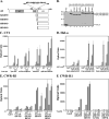
FIGURE 1
FIGURE 1. AF2 activity in the AR LBD
A, schematic diagram of AR LBD deletion mutants. Full-length human AR (amino acid residues 1–919) contains activation function 1 (AF1, amino acid residues 142–337), DNA binding domain (DBD residues 559–623), hinge region residues 624–670, and LBD residues 671–919 that includes AF2. AR hinge region residues 624–639 contain the carboxyl-terminal portion of the bipartite AR nuclear targeting signal residues Arg-629, Lys-630, Lys-632, and Lys-633 (underlined) (38). AR-(624–919), -(640–919), and -(658–919) with WT and mutant sequence were expressed as GAL4 DNA binding domain fusion proteins. AR residues 663–919 and H874Y mutant were expressed for crystallography as His6-tagged fusion proteins with intervening thrombin cleavage site. B, similar expression of GAL-AR-LBD fusion proteins. COS cells were transfected with 10 μg of GAL-0 (lane 1), GAL-AR-(658–919) (lanes 2–5), and GAL-AR-(624–919) (lanes 6–9) with WT or mutant sequence. Protein extracts (60 μg of protein/lane) were separated on a 10% acrylamide gel containing SDS and the blot probed using an anti-GAL antibody. C, androgen-dependent activity of GAL-AR-(624–919), GAL-AR-(658–919) WT and H874Y mutants in CV1 cells requires coexpression of TIF2. CV1 cells plated in 6-cm dishes were transfected by calcium phosphate DNA precipitation with 5 μg of 5XGAL4Luc3 reporter vector and 0.1 μg of GAL-AR-(624–919) or GAL-AR-(658–919) with WT or H874Y sequence in the absence and presence of 2 μg of pSG5-TIF2. Cells were treated with and without increasing concentrations of DHT (D) and T as indicated and luciferase activity was determined. Data are representative of three independent experiments. D, inhibition of the AR N/C and coactivator interactions by AR hinge residues 624–639. HeLa cells were transfected using FuGENE 6 by adding per well 0.1μg of 5XGAL4Luc, 50 ng of VP16, VP-AR-(1–660), or VP-TIF2-(624–1287) with 0.1 μg of GAL-AR-(624–919), -(640–919), or -(658–919). Cells were incubated with and without 0.1–10 nM DHT for 24 h as indicated and assayed for luciferase activity. Data are representative of three independent experiments. E, androgen-dependent transcriptional activity of GAL-AR-(624–919), GAL-AR-(658–919), and H874Y mutants in CWR-R1 cells. CWR-R1 cells (2 × 10⁵/well) were transfected using Effectene by adding per well 0.1 μg of GAL-AR-(624–919), GAL-AR-(658–919), or H874Y mutants and 0.25 μg of 5XGAL4Luc3. Cells were treated with and without increasing concentrations of T and DHT for 24 h as indicated, and luciferase activity was determined. Data are representative of three independent experiments. F, androgen-dependent AF2 activity of GAL-AR-(658–919) in CWR-R1 cells. CWR-R1 cells (2 × 10⁵/well) were transfected using Effectene by adding per well 0.1 μg of GAL-AR-(658–919) or H874Y, K720A, or E897K mutants and 0.25 μg of 5XGAL4Luc3. Lys-720 and Glu-897 are charge clamp residues in AF2. Cells were incubated with and without 0.1, 1, and 10 nM T for 24 h, and luciferase activity was determined. Data are representative of at least three independent experiments.
FIGURE 2
FIGURE 2. Increased AR-H874Y LBD AF2 activity response to T
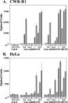
A, CWR-R1 prostate cancer cells (2 × 10⁵/well) were transfected using Effectene by adding per well 0.1 μg of GAL-0, GAL-AR-(658–919), or H874Y mutant and 0.25 μg of 5XGAL4Luc3. Cells were incubated in the absence and presence of 0.1, 1, and 10 nM 5α-androstane-3α,17β-diol (Dl, Diol), androstenedione (Dn, Dione), T and DHT for 24 h as indicated, and luciferase activity was determined. B, HeLa cells were transfected using FuGENE 6 by adding per well 0.1 μg of GAL-0, GAL-AR-(658–919), or H874Y mutant and 0.25 μg of 5XGAL4Luc3. Transfected cells were incubated with and without androgen as indicated for 24 h and assayed for luciferase activity. Data in A and B are representative of at least three independent experiments.
FIGURE 3
FIGURE 3. Increased transcriptional activity of AR-H874Y by T and adrenal androgens
A, CWR-R1 cells (1.6 × 10⁵ cells/well) were transfected with 0.1 μg of MMTV-Luc/well of 12-well plates using Effectene. Cells were incubated in the absence and presence of increasing concentrations of DHT, T, androstenedione (Dione), and androstanediol (Diol) as indicated for 24 h and assayed for luciferase activity. B, HeLa cells were transfected using FuGENE 6 by adding per well 10 ng of pCMV5 empty vector (p5), pCMVhAR, or the H874Y mutant and 0.25 μg of PSA-Enh-Luc. Cells were incubated with and without 0.001 to 10 nM DHT (D) or T, and 0.1 to 10 nM androstenedione (Dn, Dione) or 5α-androstane-3α,17β-diol (Dl, Diol) for 24 h before luciferase activity was determined. Data in A and B are representative of three independent experiments.
FIGURE 4
FIGURE 4. MAGE-11 increases AR-H874Y activity response to T and DHT
CV1 cells plated in 6-cm dishes were transfected using calcium phosphate DNA precipitation by adding per dish 0.1 μg of WT pCMVhAR (AR-WT) or H874Y mutant and 5 μg of PSA-Enh-Luc reporter in the absence and presence of 2 μg of pSG5-TIF2 and/or 2 μg of pSG5-MAGE-11–(1–429) (MAGE) as indicated. Cells were incubated with and without 0.1 nM T for 48 h before luciferase activity was determined. Data are representative of three independent experiments.
FIGURE 5
FIGURE 5. Fluorescence binding isotherms
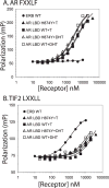
A, increasing concentrations of AR LBD and AR-H874Y LBD purified in the presence of 10 μM T or DHT, and ERβ LBD in the presence of 40 μM estradiol were incubated for 1 h at room temperature without further ligand addition with fluorescein-labeled AR FXXLF peptide as described under “Experimental Procedures.” B, increasing concentrations of purified WT AR LBD, AR-H874Y LBD, and ERβ LBD purified in the presence of 10 μM ligand were incubated for 1 h at room temperature in the presence of 40 μM T, DHT, or estradiol and fluorescein-labeled TIF2 LXXLL peptide as described under “Experimental Procedures.” Affinity binding constants for AR FXXLF and TIF2 LXXLL peptides are summarized in Table 1. The data are the mean ± S.E. expressed as millipolarization units (mP) versus purified receptor LBD concentration.
FIGURE 6
FIGURE 6. Kinetics of T and DHT dissociation from AR and AR-H874Y
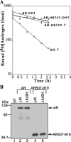
A, dissociation rates of [3H]T and [3H]DHT were determined as described under “Experimental Procedures” by transient transfection of COS cells with 2 μg of pCMVhAR (AR) and 2 μg of pCMVhAR-H874Y (AR-H874Y). Transfected cells in culture were incubated for 2 h at 37 °C in the presence of 5 nM [3H]T and 3 nM [3H]DHT followed by a chase period with unlabeled 50 μM T or 50 μM R1881 and assayed at 30-min intervals up to 2.5 h. Pseudo-first order ligand dissociation allowed use of unlabeled R1881 to prevent rebinding of [3H]DHT and avoid the complications of low water solubility of DHT. Dissociation halftimes were calculated as the time required at 37 °C to reduce specific binding by 50%. Data are representative of three independent experiments. B, immunoblot of WT and H874Y AR and AR-(507–919) expression levels in COS cells. Cells transfected with 10 μg of pCMVhAR, pCMVhAR-(507–919), and the corresponding H874Y mutants were incubated in serum-free medium in the absence of hormone. Protein extracts (20 μg protein/lane) were separated on a 10% acrylamide gel containing SDS and the transferred protein blot probed using anti-AR antibody AR-52.
FIGURE 7
FIGURE 7. Crystal structures of WT and H874Y AR LBD bound with T and AR FXXLF or TIF2 LXXLL peptide
A, global front view of superimposed structures of WT and H874Y AR LBD bound to T and AR-(20–30) FXXLF or TIF2-(740–752) LXXLL peptide. Shown are WT AR LBD-T-AR FXXLF peptide (tan, LBD ribbon; magenta, peptide), WT AR LBD-T-TIF2 LXXLL peptide (lime green, LBD ribbon; cyan, peptide), H874Y AR LBD-T-AR FXXLF peptide (yellow, LBD ribbon; green, peptide) and H874Y AR LBD-T-TIF2 LXXLL peptide (lavender, LBD ribbon; blue, peptide), and T (LBD ribbon color carbon; red, oxygen). Human AR helix (H) and β-strand (BS) amino acid residues are H1 673–680; H2 not assigned; H3 697–721; H3 725–727; H4 730–739; H5 741–756; BS3 761–765; BS4 768–771; H6 772–776; H7 780–797; H8 801–812; BS5 815–817; H9 824–842; H10 851–882; H11 884–887; H12 893–908; BS6 911–913. B, rotated view of A looking toward the carboxyl-terminal end of helix-5 and NH2-terminal ends of helices 6 and 8. Residues 843–850 between H9 and H10 were devoid of electron density. C, detailed view of the T-bound ligand binding pocket and surrounding residues of WT and H874Y AR LBD bound with AR-(20–30) FXXLF or TIF2-(740–752) LXXLL peptide. In all four T-bound structures the ligand binding pockets are essentially identical to each other. A single conformation was observed for all the displayed side chains except for Leu-712 (50%, A and B) and Gln-711 (80%, A and 20%, B in the WT AR LBD-T-LXXLL and AR-H874Y LBD-T-FXXLF structures). A buffer-derived glycerol molecule (not shown) near Gln-711 was present in all four WT and H874Y AR LBD-T structures. Color scheme as in A with nitrogen atoms in blue and oxygen atoms in red; orange dashed lines designate potential interactions with neighboring polar atoms. D, detailed view to display the molecular architecture from the ligand binding pocket to the AF2 peptide-binding site and i + 1 side chain of Phe-23 of bound AR-(20 –30) FXXLF peptide and Leu-745 of bound TIF2 LXXLL peptide. Different conformers of Met-734 and Tyr-739 correlate with the induced fit binding of the FXXLF or LXXLL motif. The WT and H874Y AR LBD bound to T and AR FXXLF or TIF2 LXXLL peptide are superimposed and use the color scheme of A. Side chains for Leu-712 and Met-895 are distributed equally into two rotamers.
FIGURE 8
FIGURE 8. Potential A-ring and water mediated H-bonding schemes for T and DHT
Predicted A-ring H-bond distances and angles are shown based on the tetrahedral geometry of conserved structural water HOH1 (see Footnote and see Table 4). Arrowhead with black dashed lines indicate the direction of donated H-bonds and orange dashed lines designate potential interactions with neighboring polar atoms of WT AR LBD bound to T and AR-(20–30) FXXLF peptide (tan) (A); WT AR LBD bound to DHT and GRIP-1-(740–752) LXXLL peptide (green) (42) (B); and the superimposition of A and B (C). Superior hydrophilic properties and a shorter distance are thought to enhance the HOH1 to T 3-keto O H-bond over that in DHT.
FIGURE 9
FIGURE 9. Comparison of WT AR LBD-T-AR FXXLF and WT AR LBD-DHT-LXXLL
Detailed view shows nearly identical molecular architecture of T and DHT-bound AR LBD from the ligand binding pocket to AF2 peptide-binding site. Shown here for WT AR LBD-T-AR-(20–30) FXXLF peptide (tan) but seen in all of our T-bound AR LBD structures are the two side chain conformations for Leu-712 and Met-895. By comparison with previously reported WT AR LBD-DHT (green) with GRIP-1-(740–752) LXXLL peptide (IT63, 42) or FXXLF (not shown, ITR7, 18), those side chains were conformed into a single rotamer. The i + 1 motif residues, Phe-23 (magenta) of the AR FXXLF peptide and Leu-745 (cyan) of the GRIP-1 LXXLL peptide, are shown. Orange dashed lines designate potential interactions with neighboring polar atoms. Portions of the LBD backbone are transparently displayed.
FIGURE 10
FIGURE 10. Detailed crystal structure comparison of WT and H874Y AR LBD bound to T and AR FXXLF peptide
A, detailed view of WT AR LBD bound to T and AR-(20–30) FXXLF peptide showing a water-mediated H-bond network from His-874 (tan) nitrogen (blue) through HOH3 (red sphere) to the helix-5 Met-742 amide and HOH4 to the helix-4 Tyr-739 backbone carbonyl. These conserved receptor core structural waters link His-874, which lies beneath helix-12, to Tyr-739. Arrowhead with black dashed lines indicate the direction of donated H-bonds; orange dashed lines designate potential interactions with neighboring polar atoms; interatomic distances are reported in Angstroms; T rings are labeled A–D. B, detailed view of AR-H874Y LBD-T-AR-(20–30) FXXLF peptide. Note the extended phenolic hydroxyl of AR H874Y prostate cancer mutation Tyr-874 displaces structural water HOH3 and provides direct H-bonds to the backbone amide of helix-5 Met-742 and the carbonyl of helix-4 Tyr-739 (yellow). H-bonds labeled as in A with HOH4 shown as blue sphere. C, superposition of A and B showing how the phenolic hydroxyl group of helix-10 mutant Tyr-874 of AR-H874Y nearly extends to the same position as HOH3 in the WT AR LBD structure with conservation of backbone positions for Tyr-739, Trp-741, and Arg-752. D, superposition of WT AR LBD (tan) bound to T and AR-(20–30) FXXLF (i + 1 Phe-23, green) and AR-H874Y LBD (yellow) bound to T and AR-(20–30) FXXLF (i + 1 Phe-23, magenta). TIF2-(740–752) LXXLL i + 1 residue Leu-745 (cyan) is shown for comparison. Direct H-bonding by Tyr-874 to the backbone of Tyr-739 displaces HOH3 and could further stabilize another H-bonding network represented with orange dashed lines that links Met-734 CO to Gln-738, Gln-902, and Lys-905.
FIGURE 11
FIGURE 11. Structural differences between the steroid and nonsteroidal ligand binding pockets
Superimposition of WT AR LBD crystal structures bound with an FXXLF peptide and T (brown), DHT (green) (18), R1881 (magenta) (4), and S-1 bicalutamide agonist analog (cyan) (43) and AR-H874Y LBD bound to the AR FXXLF peptide and T (yellow). The C-19 bridgehead methyl group on T and DHT forces the Met-745 and Trp-741 side chains away from the steroid A-ring. For R1881 and S-1, the absence of an equivalent methyl group allows these side chains to adopt different rotamers that fill the vacated space above ring A. The para-fluoro phenyl group on S1 extends into the space between helix-12 Met-895 and helix-5 Met-742 and directs Trp-741 to a third unique conformation. Appropriate para-phenyl substituents are thought to stabilize the AR LBD core by interacting with HOH3 (43).

References

    1. Wilson EM, French FS. J Biol Chem. 1976;251:5620–5629. - PubMed
    1. Simental JA, Sar M, Lane MV, French FS, Wilson EM. J Biol Chem. 1991;266:510–518. - PubMed
    1. Warnmark A, Treuter E, Wright AP, Gustafsson JA. Mol Endocrinol. 2003;17:1901–1909. - PubMed
    1. He B, Gampe RT, Kole AJ, Hnat AT, Stanley TB, An G, Stewart EL, Kalman RI, Minges JT, Wilson EM. Mol Cell. 2004;16:425–438. - PubMed
    1. He B, Kemppainen JA, Wilson EM. J Biol Chem. 2000;275:22986–22994. - PubMed

Publication types

MeSH terms