. 2007 Dec;14(12):1214-20.
doi: 10.1038/nsmb1322. Epub 2007 Nov 11.

Autotransporter structure reveals intra-barrel cleavage followed by conformational changes

Affiliations

Autotransporter structure reveals intra-barrel cleavage followed by conformational changes

Travis J Barnard et al. Nat Struct Mol Biol. 2007 Dec.

Abstract

Autotransporters are virulence factors produced by Gram-negative bacteria. They consist of two domains, an N-terminal 'passenger' domain and a C-terminal beta-domain. beta-domains form beta-barrel structures in the outer membrane while passenger domains are translocated into the extracellular space. In some autotransporters, the two domains are separated by proteolytic cleavage. Using X-ray crystallography, we solved the 2.7-A structure of the post-cleavage state of the beta-domain of EspP, an autotransporter produced by Escherichia coli strain O157:H7. The structure consists of a 12-stranded beta-barrel with the passenger domain-beta-domain cleavage junction located inside the barrel pore, approximately midway between the extracellular and periplasmic surfaces of the outer membrane. The structure reveals an unprecedented intra-barrel cleavage mechanism and suggests that two conformational changes occur in the beta-domain after cleavage, one conferring increased stability on the beta-domain and another restricting access to the barrel pore.


Figures
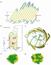
Figure 1
Figure 1
Structure of the EspP β-domain. β-strands are colored yellow, loops are green, and the α-helix is red. (a) Topology diagram of EspP. Regions missing in the structure are shown in blue. Filled (yellow) squares indicate residues whose side chains point toward the lipid bilayer. Unfilled (white) squares indicate residues whose side chains point toward the barrel pore. (b) Ribbon diagram of the β-domain. Sidechains for the aromatic girdle are shown in purple. Extracellular loops (L1 - L6) and the linker loop are labeled. (c) The EspP β-domain viewed down the barrel axis from the extracellular surface. Extracellular loop 5 makes 10 hydrogen bonds with 7 strands of the β-barrel; 5 of these interactions are shown. (d) Space filling representation of the β-domain viewed from the extracellular space. (e) Space filling representation of the β-domain viewed from the periplasm.
Figure 2
Figure 2
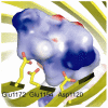
Interactions between the luminal α-helix and acidic cluster. (a) Sidechains of the residues forming the acidic cluster are shown in stick representation. The α-helix and linker loop are shown as an electrostatic surface. Blue and red regions denote areas of positive and negative charge, respectively. White surfaces are neutral.
Figure 3
Figure 3
Conformational changes occur upon cleavage of the passenger domain. (a) Cartoon representation of the pre-cleavage state of EspP. Like NalP, EspP is predicted to contain a long α-helix prior to passenger domain cleavage. (b) EspP during cleavage and release of the passenger domain. Autoproteolysis separates the β-domain from the passenger domain. Arrow 1 indicates passenger domain release from the barrel pore. Arrow 2 indicates the subsequent repositioning of the N-terminal α-helix. Arrow 3 indicates the folding of loop 5 into the barrel pore. (c) EspP β-domain after cleavage.
Figure 4
Figure 4
Effect of point mutations and deletions on the stability of the EspP β-domain. AD202 was transformed with a plasmid encoding the indicated EspP derivative and autotransporter synthesis was induced by the addition of IPTG. Cell extracts were heated in SDS-PAGE sample buffer at various temperatures and the EspP fragment denoted by the shaded bar on the right was detected by Western blot using a C-terminal antipeptide antiserum. The faster migrating (lower) band corresponds to a compactly folded form of the fragment. Asterisks show the approximate positions of point mutations. The temperature at which 50% of each EspPΔ1 derivative was denatured was determined by first normalizing the signal in the upper and lower bands and then calculating the temperature at which the intensity of the two bands would be equal using Odyssey quantitation software.
Figure 5
Figure 5
Effect of mutations in the ‘acidic cluster’ on EspP passenger domain cleavage. AD202 was transformed with a plasmid encoding EspPΔ1, EspP*Δ1 or the indicated EspPΔ1 mutant, and autotransporter synthesis was induced by the addition of IPTG. Passenger domain cleavage was then analyzed by Western blot using a C-terminal antipeptide antiserum.
Figure 6
Figure 6
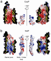
Electrostatic properties of the EspP and NalP β-domains. (a) The electrostatic properties of the EspP barrel pore and luminal α-helix with its linker loop. The panel on the right is rotated by 180°. The barrel pore is strikingly acidic. (b) The electrostatic properties of the NalP barrel pore and luminal α-helix with its linker loop. The panel on the right is rotated by 180°. Unlike the interior of the EspP β-barrel, the interior of the NalP β-barrel shows an asymmetric charge distribution, basic toward the periplasm, and acidic / neutral toward the extracellular surface.

References

    1. Henderson IR, Nataro JP. Virulence functions of autotransporter proteins. Infect Immun. 2001;69:1231–43. - PMC - PubMed
    1. Henderson IR, Navarro-Garcia F, Desvaux M, Fernandez RC, Ala'Aldeen D. Type V protein secretion pathway: the autotransporter story. Microbiol Mol Biol Rev. 2004;68:692–744. - PMC - PubMed
    1. Jacob-Dubuisson F, Fernandez R, Coutte L. Protein secretion through autotransporter and two-partner pathways. Biochim Biophys Acta. 2004;1694:235–57. - PubMed
    1. Thanassi DG, Stathopoulos C, Karkal A, Li H. Protein secretion in the absence of ATP: the autotransporter, two-partner secretion and chaperone/usher pathways of gram-negative bacteria (review) Mol Membr Biol. 2005;22:63–72. - PubMed
    1. Cotter SE, Surana NK, Geme JW., 3rd Trimeric autotransporters: a distinct subfamily of autotransporter proteins. Trends Microbiol. 2005;13:199–205. - PubMed

Publication types

MeSH terms

Associated data