Comparative Study
. 2016 Jan 15;291(3):1267-76.
doi: 10.1074/jbc.M115.688010. Epub 2015 Oct 29.

A Combination of Structural and Empirical Analyses Delineates the Key Contacts Mediating Stability and Affinity Increases in an Optimized Biotherapeutic Single-chain Fv (scFv)

Affiliations
Comparative Study

A Combination of Structural and Empirical Analyses Delineates the Key Contacts Mediating Stability and Affinity Increases in an Optimized Biotherapeutic Single-chain Fv (scFv)

Chao Tu et al. J Biol Chem. .

Abstract

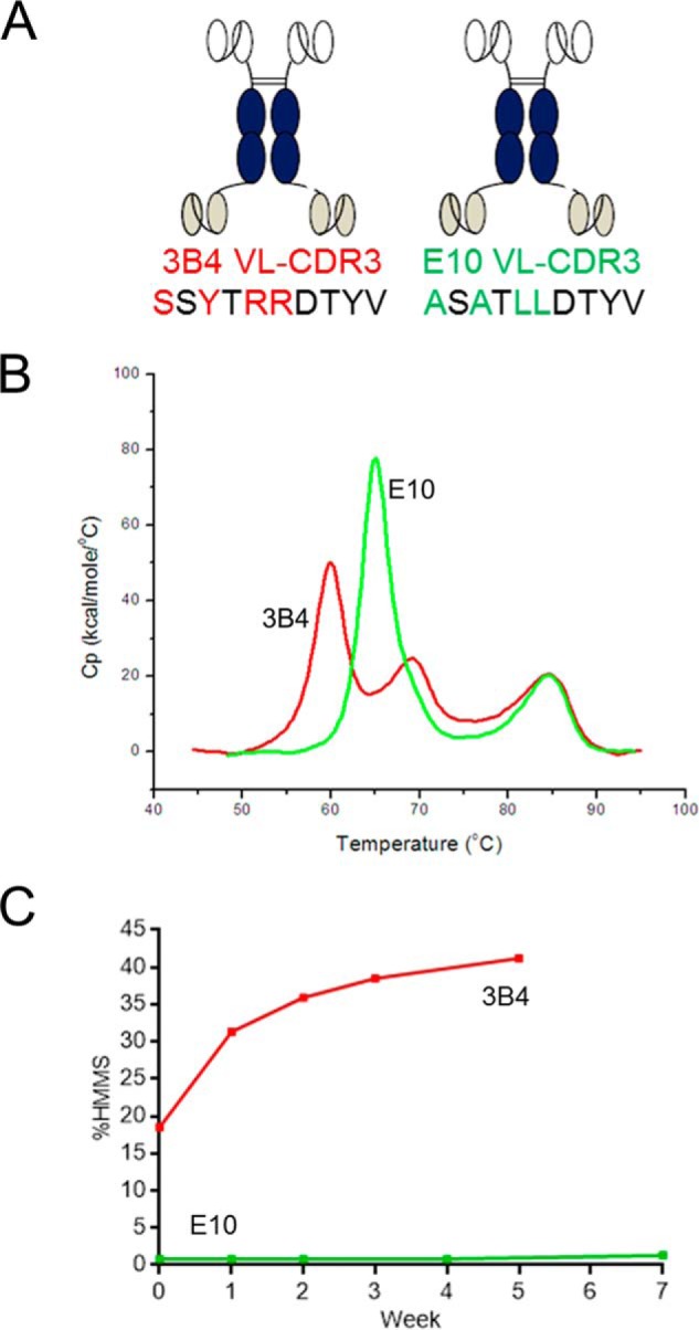
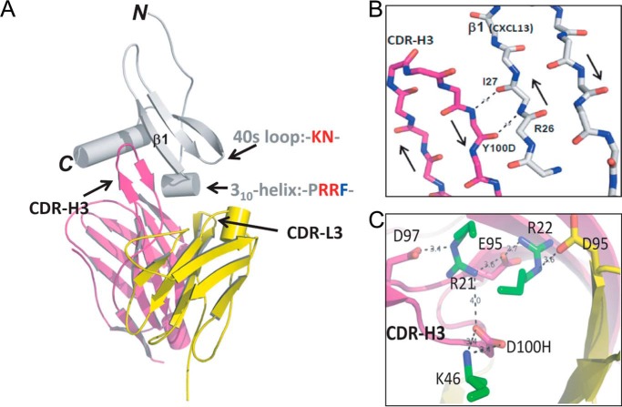
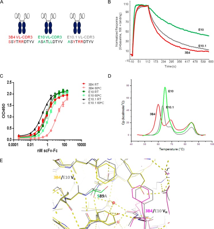
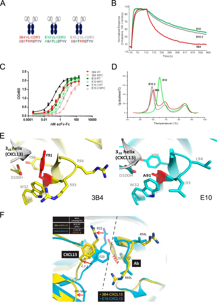
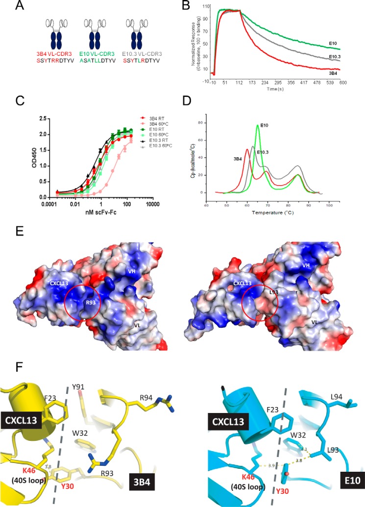
Fully-human single-chain Fv (scFv) proteins are key potential building blocks of bispecific therapeutic antibodies, but they often suffer from manufacturability and clinical development limitations such as instability and aggregation. The causes of these scFv instability problems, in proteins that should be theoretically stable, remains poorly understood. To inform the future development of such molecules, we carried out a comprehensive structural analysis of the highly stabilized anti-CXCL13 scFv E10. E10 was derived from the parental 3B4 using complementarity-determining region (CDR)-restricted mutagenesis and tailored selection and screening strategies, and carries four mutations in VL-CDR3. High-resolution crystal structures of parental 3B4 and optimized E10 scFvs were solved in the presence and absence of human CXCL13. In parallel, a series of scFv mutants was generated to interrogate the individual contribution of each of the four mutations to stability and affinity improvements. In combination, these analyses demonstrated that the optimization of E10 was primarily mediated by removing clashes between both the VL and the VH, and between the VL and CXCL13. Importantly, a single, germline-encoded VL-CDR3 residue mediated the key difference between the stable and unstable forms of the scFv. This work demonstrates that, aside from being the critical mediators of specificity and affinity, CDRs may also be the primary drivers of biotherapeutic developability.

Keywords: antibody engineering; chemokine; crystal structure; mutagenesis in vitro; protein stability.


Figures

FIGURE 1.
FIGURE 1.
Comparative biophysical analysis of parental 3B4 scFv-Fc-scFv and optimized E10 scFv-Fc-scFv. A, schematic representation of the two bispecific proteins highlighting the four amino acid differences between the two clones in VL-CDR3. B, DSC analysis showing the 5 °C increase in Tm1 between E10 (green) and 3B4 (red). C, comparative size-exclusion chromatography analysis showing time-dependent increases in formation of HMMS after storage of a 100 mg/ml solution of 3B4 (red) and E10 (green) bispecific molecules at 4 °C.
FIGURE 2.
FIGURE 2.
Structure of the anti-CXCL13 scFv E10 in complex with CXCL13. A, graphic view showing the complex. scFv heavy chain (VH) and light chain (VL) are shown in magenta and yellow, respectively. CXCL13 is shown in gray. B, detail showing the four-stranded antiparallel β-sheet formed between the ascending and descending strands of VH-CDR3 and β1-sheet of CXCL13. Hydrogen bonds between side chains and backbone are shown as dashed lines. C, detail illustrating the charge-charge interactions between negatively charged residues in VH-CDR3 (Glu95, Asp97, D100H shown in magenta) and positively charged residues in the 310-helix and 40S loop of CXCL13 (Arg21, Arg23, Lys46).
FIGURE 3.
FIGURE 3.
Characterization of mutant E10.1, S89A. A, graphic representation of 3B4, E10, and E10.1 scFv-Fc fusion proteins highlighting VL-CDR3 amino acid content (red, 3B4-specific residues; green, E10-specific residues). B, off-rate analysis as measured by Biacore comparing 3B4 (red), E10 (green), and E10.1 (gray). C, thermal stability ELISA comparing behavior of 3B4 (red), E10 (green), and 10.1 (gray) fusion proteins before (light color) and after (dark color) incubation for 60 min at 60 °C. OD450, optical density at 450 nm; RT, room temperature. Error bars indicate ± S.E. D, DSC analysis comparing 3B4 (red), E10 (green), and E10.1 (gray). E, superposition of VH-VL interface of 3B4 (yellow for VL and magenta for VH) and E10 (gray) in the region of VL residue 89 (shown in “green” for 3B4). Waters are shown as red spheres. The hydrogen-bonding network for 3B4 is shown with dashed yellow lines. The side chain-OH of 3B4 VL Ser89 has no hydrogen-bonding partners.
FIGURE 4.
FIGURE 4.
Characterization of mutant E10.2, Y91A. A, graphic representation of 3B4, E10, and E10.2 scFv-Fc fusion proteins highlighting VL-CDR3 amino acid content (red, 3B4-specific residues; green, E10-specific residues). B, off-rate analysis as measured by Biacore comparing 3B4 (red), E10 (green), and E10.2 (gray). C, thermal stability ELISA comparing behavior of 3B4 (red), E10 (green), and 10.2 (gray) fusion proteins before (light color) and after (dark color) incubation for 60 min at 60 °C. OD450, optical density at 450 nm; RT, room temperature. Error bars indicate ± S.E. D, DSC analysis comparing 3B4 (red), E10 (green), and E10.2 (gray). E, juxtaposition of 3B4 VL-CDR3 (yellow) and E10 VL-CDR3 (blue) in complex with the 310-helix of CXCL13 (gray). The position 91 side chain is marked in red for clarity in both cases. F, the impact of the Y91A mutation is made clearer with a superposition of the 3B4-CXCL13 (yellow) and E10-CXCL13 (blue) complexes. Mutation at this position to alanine removes steric occlusion at the interface increasing the surface area of interaction by ∼200 Å2.
FIGURE 5.
FIGURE 5.
Characterization of mutant E10.3, R93L. A, schematic representation of 3B4, E10, and E10.3 scFv-Fc fusion proteins highlighting VL-CDR3 amino acid content (red, 3B4-specific residues; green, E10-specific residues). B, off-rate analysis as measured by Biacore comparing 3B4 (red), E10 (green), and E10.3 (gray). C, thermal stability ELISA comparing the behavior of 3B4 (red), E10 (green), and 10.1 (gray) fusion proteins before (light color) and after (dark color) incubation for 60 min at 60 °C. OD450, optical density at 450 nm; RT, room temperature. Error bars indicate ± S.E. D, DSC analysis comparing 3B4 (red), E10 (green), and E10.3 (gray). E, surface charge representation shows that the R93L mutation removes a repulsive charge-charge interaction between Arg93 in 3B4 and Lys46 in the 40S loop of CXCL13. F, juxtaposition of 3B4-CXCL13 (yellow) and E10-CXCL13 (blue) complexes highlighting the impact of R93L on both stability and affinity. Leu93 in E10 VL CDR3 interacts with Tyr30 and Trp32 in Vl-CDR1, as indicated by dashed lines. It also facilitates a more favorable interaction between Tyr30, which can adopt an alternative rotamer in E10, and Lys46 in the 40S loop of CXCL13.

References

    1. Chan A. C., and Carter P. J. (2010) Therapeutic antibodies for autoimmunity and inflammation. Nat. Rev. Immunol. 10, 301–316 - PubMed
    1. Bargou R., Leo E., Zugmaier G., Klinger M., Goebeler M., Knop S., Noppeney R., Viardot A., Hess G., Schuler M., Einsele H., Brandl C., Wolf A., Kirchinger P., Klappers P., Schmidt M., Riethmüller G., Reinhardt C., Baeuerle P. A., and Kufer P. (2008) Tumor regression in cancer patients by very low doses of a T cell-engaging antibody. Science 321, 974–977 - PubMed
    1. Fitzgerald J. B., Johnson B. W., Baum J., Adams S., Iadevaia S., Tang J., Rimkunas V., Xu L., Kohli N., Rennard R., Razlog M., Jiao Y., Harms B. D., Olivier K. J. Jr., Schoeberl B., Nielsen U. B., and Lugovskoy A. A. (2014) MM-141, an IGF-IR- and ErbB3-directed bispecific antibody, overcomes network adaptations that limit activity of IGF-IR inhibitors. Mol. Cancer Ther. 13, 410–425 - PubMed
    1. Liddy N., Bossi G., Adams K. J., Lissina A., Mahon T. M., Hassan N. J., Gavarret J., Bianchi F. C., Pumphrey N. J., Ladell K., Gostick E., Sewell A. K., Lissin N. M., Harwood N. E., Molloy P. E., Li Y., Cameron B. J., Sami M., Baston E. E., Todorov P. T., Paston S. J., Dennis R. E., Harper J. V., Dunn S. M., Ashfield R., Johnson A., McGrath Y., Plesa G., June C. H., Kalos M., Price D. A., Vuidepot A., Williams D. D., Sutton D. H., and Jakobsen B. K. (2012) Monoclonal TCR-redirected tumor cell killing. Nat. Med. 18, 980–987 - PubMed
    1. Moore P. A., Zhang W., Rainey G. J., Burke S., Li H., Huang L., Gorlatov S., Veri M. C., Aggarwal S., Yang Y., Shah K., Jin L., Zhang S., He L., Zhang T., Ciccarone V., Koenig S., Bonvini E., and Johnson S. (2011) Application of dual affinity retargeting molecules to achieve optimal redirected T-cell killing of B-cell lymphoma. Blood 117, 4542–4551 - PubMed

Publication types

MeSH terms

LinkOut - more resources