. 2007 Nov 29;450(7170):741-4.
doi: 10.1038/nature05998.

Inhibition of the EGF receptor by binding of MIG6 to an activating kinase domain interface

Affiliations

Inhibition of the EGF receptor by binding of MIG6 to an activating kinase domain interface

Xuewu Zhang et al. Nature. .

Abstract

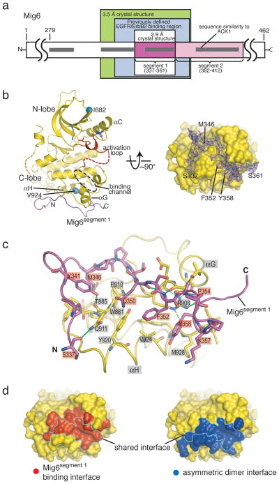
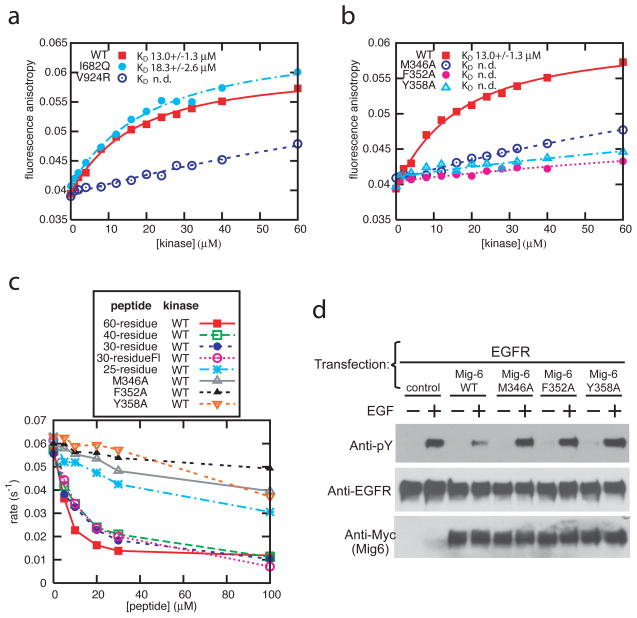
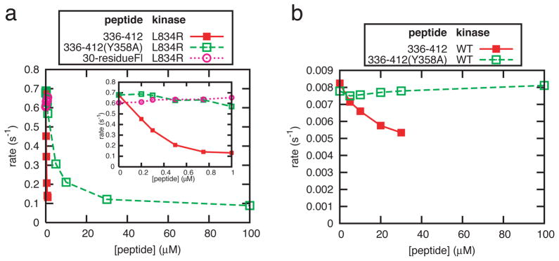
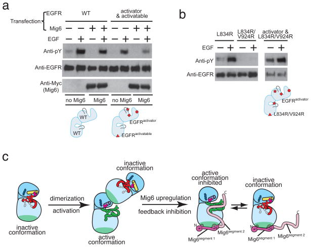
Members of the epidermal growth factor receptor family (EGFR/ERBB1, ERBB2/HER2, ERBB3/HER3 and ERBB4/HER4) are key targets for inhibition in cancer therapy. Critical for activation is the formation of an asymmetric dimer by the intracellular kinase domains, in which the carboxy-terminal lobe (C lobe) of one kinase domain induces an active conformation in the other. The cytoplasmic protein MIG6 (mitogen-induced gene 6; also known as ERRFI1) interacts with and inhibits the kinase domains of EGFR and ERBB2 (refs 3-5). Crystal structures of complexes between the EGFR kinase domain and a fragment of MIG6 show that a approximately 25-residue epitope (segment 1) from MIG6 binds to the distal surface of the C lobe of the kinase domain. Biochemical and cell-based analyses confirm that this interaction contributes to EGFR inhibition by blocking the formation of the activating dimer interface. A longer MIG6 peptide that is extended C terminal to segment 1 has increased potency as an inhibitor of the activated EGFR kinase domain, while retaining a critical dependence on segment 1. We show that signalling by EGFR molecules that contain constitutively active kinase domains still requires formation of the asymmetric dimer, underscoring the importance of dimer interface blockage in MIG6-mediated inhibition.


Figures

Figure 1
Figure 1. Structure of the EGFR kinase domain/Mig6segment 1
a, Schematic diagram of human Mig6 primary structure. Regions of interest, including the previously defined EGFR/ErbB2 binding region,,, are boxed and labeled. b, Two orthogonal views of the EGFR kinase domain/Mig6segment 1 complex. A channel which peptide inhibitors of some other kinases are docked is indicated,. The electron density around Mig6segment 1 in the right panel is contoured at 3σ and is from a simulated annealing omit map with coefficients (|Fo|-|Fc|)eC, where the calculated structure factors are generated from a model that does not contain Mig6. c, Detailed view of the interface between the EGFR kinase domain and Mig6segment 1. Hydrogen bonds are represented by dashed lines. d, Comparison of the Mig6segment 1 binding interface and the kinase domain asymmetric dimer interface on the distal surface of the kinase C-lobe. A large portion of the surface is shared by the two interfaces (outlined), and it is clear that binding of the EGFR kinase domain by Mig6segment 1 would block the formation of the asymmetric activating dimer. (c) and (d) are in similar orientations as that in the right panel of (b).
Figure 2
Figure 2. Binding and inhibition of EGFR by Mig6segment 1
a, Titrations of the wild type EGFR kinase domain and the V924R and I682Q mutants to the 30-residue (residues 334–363) fluorescein-labeled Mig6 peptide. b, Titrations of the wild type EGFR kinase domain to the wild type and three mutant 30-residue fluorescein-labeled peptides. n.d. denotes “not determined” (KD values cannot be determined reliably). c, Inhibition of the activity of the EGFR kinase domain by peptides spanning Mig6segment 1 in the vesicle-based kinase assay. The 60-, 40- and 30-residue peptides contain the entire binding epitope of segment 1, while the 25-residue peptide lacks the N-terminal 3 residues. The mutations were introduced in the 30-residue peptide. See Supplemental Table 1 for the residue boundaries. Fl: fluorescein. d, Cell-based assay showing that Mig6 inhibits full-length EGFR autophosphorylation, whereas mutations in segment 1 abolishes the inhibition.
Figure 3
Figure 3. Inhibition of the EGFR kinase activity by Mig6segment 1–2
a, Inhibition of the L834R mutant kinase in solution by peptides 336–412, 336–412(Y358A) (containing both segment 1 and 2). The 30-residue peptide (containing segment 1 only) is used as a control. The insert shows an expanded view at low peptide concentrations. b, Inhibition of the wild type kinase in solution by peptides 336–412 and 336–412(Y358A). Titration of peptide 336–412 beyond 20 μM leads to unreliable results due to precipitation of the protein and peptide (See Method).
Figure 4
Figure 4. A double-headed mechanism for EGFR inhibition by Mig6
a, Co-transfection experiment showing that EGFRactivator can activate EGFRactivatable, and that Mig6 can inhibit this activation. b, Co-transfection experiments showing that full-length EGFR containing the L834R/V924R double mutation only shows autophosphorylation when co-transfected with EGFRactivator. Co-transfection combinations in (a) and (b) are represented by the cartoons in the respective lower panels for clarity. The I682Q, D813N, L834R and V924R mutations are denoted in the cartoons by a circle, diamond, star and triangle, respectively. c, a schematic diagram showing the double-headed mechanism for EGFR inhibition by Mig6 involving both segment 1 and segment 2.

References

    1. Hynes NE, Lane HA. ERBB receptors and cancer: the complexity of targeted inhibitors. Nat Rev Cancer. 2005;5 (5):341–354. - PubMed
    1. Zhang X, et al. An allosteric mechanism for activation of the kinase domain of epidermal growth factor receptor. Cell. 2006;125 (6):1137–1149. - PubMed
    1. Hackel PO, Gishizky M, Ullrich A. Mig-6 is a negative regulator of the epidermal growth factor receptor signal. Biol Chem. 2001;382 (12):1649–1662. - PubMed
    1. Xu Dazhong, Makkinje Anthony, Kyriakis John M. Gene 33 Is an Endogenous Inhibitor of Epidermal Growth Factor (EGF) Receptor Signaling and Mediates Dexamethasone-induced Suppression of EGF Function. J Biol Chem. 2005;280 (4):2924–2933. - PubMed
    1. Fiorentino L, et al. Inhibition of ErbB-2 mitogenic and transforming activity by RALT, a mitogen-induced signal transducer which binds to the ErbB-2 kinase domain. Mol Cell Biol. 2000;20 (20):7735–7750. - PMC - PubMed

Publication types

Substances