. 2009 Jul 22;28(14):2128-42.
doi: 10.1038/emboj.2009.165. Epub 2009 Jun 25.

Structure and function of a complex between chorismate mutase and DAHP synthase: efficiency boost for the junior partner

Affiliations

Structure and function of a complex between chorismate mutase and DAHP synthase: efficiency boost for the junior partner

Severin Sasso et al. EMBO J. .

Abstract

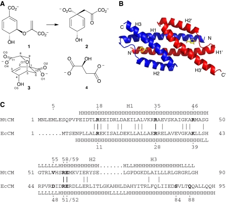
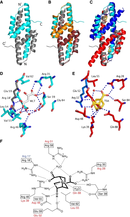
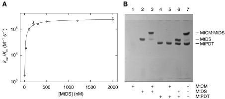
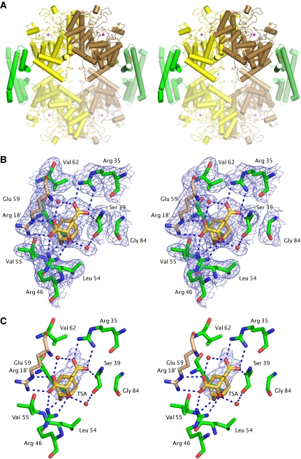
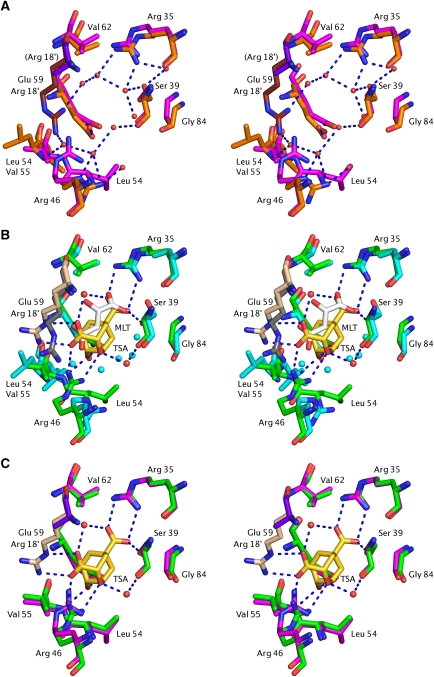
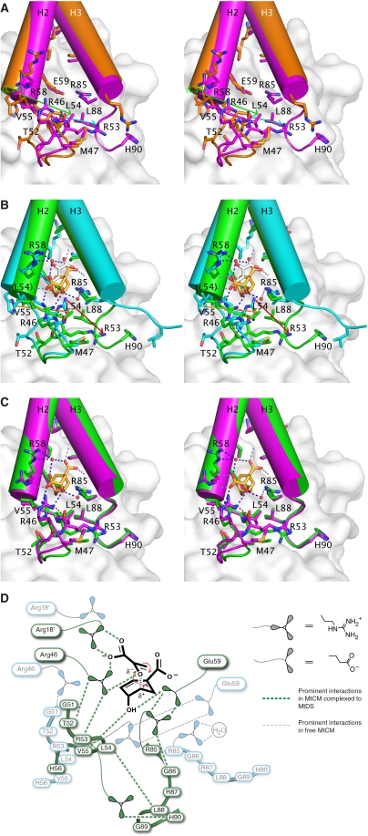
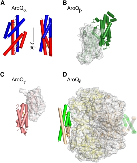
Chorismate mutase catalyzes a key step in the shikimate biosynthetic pathway towards phenylalanine and tyrosine. Curiously, the intracellular chorismate mutase of Mycobacterium tuberculosis (MtCM; Rv0948c) has poor activity and lacks prominent active-site residues. However, its catalytic efficiency increases >100-fold on addition of DAHP synthase (MtDS; Rv2178c), another shikimate-pathway enzyme. The 2.35 A crystal structure of the MtCM-MtDS complex bound to a transition-state analogue shows a central core formed by four MtDS subunits sandwiched between two MtCM dimers. Structural comparisons imply catalytic activation to be a consequence of the repositioning of MtCM active-site residues on binding to MtDS. The mutagenesis of the C-terminal extrusion of MtCM establishes conserved residues as part of the activation machinery. The chorismate-mutase activity of the complex, but not of MtCM alone, is inhibited synergistically by phenylalanine and tyrosine. The complex formation thus endows the shikimate pathway of M. tuberculosis with an important regulatory feature. Experimental evidence suggests that such non-covalent enzyme complexes comprising an AroQ(delta) subclass chorismate mutase like MtCM are abundant in the bacterial order Actinomycetales.


Conflict of interest statement

The authors declare that they have no conflict of interest.

Figures

Figure 1
Figure 1
Catalytic reaction, structure, and sequence of AroQ chorismate mutases (CMs). (A) CMs convert chorismate (1) to prephenate (2) through a transition state that resembles inhibitor 3 (Bartlett and Johnson, 1985), and to some extent L-malate (4). (B) Structure of the AroQ prototype EcCM with transition-state analogue 3 bound to its active sites (Lee et al, 1995a). (C) Sequence alignment and secondary structure (H=helix; L=loop) of MtCM (subclass AroQδ) and EcCM (AroQα), according to the crystal structures of MtCM (PDB ID: 2VKL) and EcCM (1ECM) (Lee et al, 1995a). Sequence identity is 25.3%; bold, active-site residues in EcCM and their homologues in MtCM.
Figure 2
Figure 2
Multiple sequence alignment of representative AroQδ proteins. A PSI-BLAST search (Altschul et al, 1997) was carried out to identify close MtCM homologues. Hit sequences were retained if they, compared with EcCM, had a shortened C-terminus and an arginine at the position corresponding to EcCM Lys 39. The alignment shows an arbitrarily selected sequence from each genus. The sequences were directly adopted from database entries and may not necessarily identify the correct start position. In this work, shorter versions were used for Mtu (MtCM) and Cgl (CgCM), as indicated by an N-terminal extension in grey and adjustments in the numbering. Three possible start methionines for MtCM are underlined. Although circumstantial evidence from frameshift experiments in Mycobacterium smegmatis was interpreted in favour of the originally predicted (Cole et al, 1998) 105-amino acid MtCM variant (Rv0948c) (Schneider et al, 2008), this study focused on the 90-residue version starting with the second methionine (residue numbers on top; also corresponding to MtCM in Figure 1C). This decision was based on the inspection of possible Shine–Dalgarno sequences (Doran et al, 1997), lack of sequence homology in the N-terminal region of predicted AroQδ proteins (even observed when comparing M. smegmatis and M. tuberculosis variants), and the similarity of the catalytic parameters (unpublished results; Kim et al, 2008) for the longest version and MtCM used here. Important active-site residues in EcCM with counterparts in AroQδ enzymes are identified below the alignment. Residues are boxed according to sequence identity within the AroQδ subgroup from yellow (⩾33%), orange (⩾50%), bright red (⩾75%), to dark red (100% identity). Mtu, M. tuberculosis; Ace, Acidothermus cellulolyticus; Ate, Actinoplanes teichomyceticus; Str, Salinospora tropica; Nfa, Nocardia farcinica; Rho, Rhodococcus sp.; Cgl, Corynebacterium glutamicum; Sco, Streptomyces coelicolor; Fal, Frankia alni.
Figure 3
Figure 3
Structure of MtCM and active-site details. Ligands are shown in stick representation (carbons in white for malate, in gold for inhibitor 3). (A) Overall structure of the malate-binding MtCM dimer (PDB ID: 2VKL). The two protomers are represented in cyan and grey, respectively; N- and C-termini are labelled. (B) Superimposition of MtCM as in (A) with unliganded MtCM dimer in orange/ruby (2QBV) (Kim et al, 2008). (C) Superimposition of MtCM as in (A) with EcCM liganded to 3 (1ECM) (Lee et al, 1995a). EcCM protomers are highlighted in blue and red. (D) Active-site details of malate (MLT)-bound MtCM. An apostrophe identifies residues contributed by the other protomer. Red spheres and broken lines represent water molecules and key hydrogen bonds, respectively. Arg 58 was omitted for clarity. (E) Active-site details of EcCM with bound 3 (TSA). Arg 51, which coordinates to the water molecule bridging the carboxylates of 3, was omitted for clarity. (F) Scheme of MtCM key active-site residues (boxed) with bound 3 (heavy lines; interactions according to 2W1A, see below). Homologous EcCM residues are listed next to the boxes.
Figure 4
Figure 4
Interaction between MtCM and MtDS. (A) Titration of MtCM with MtDS. The catalytic efficiency (kcat/Km) of the CM reaction was determined at different MtDS concentrations in 50 mM BTP (pH 7.5) at 30°C. The MtCM concentration was held constant at 10 nM (100 nM in the absence of MtDS). At each MtDS concentration, Michaelis–Menten kinetics with ⩾5 different chorismate concentrations were measured. Error bars correspond to the s.d. of ⩾2 independent Michaelis–Menten kinetics. (B) Band-shift assay. Purified proteins were mixed according to the scheme below the gel (at 10 μM final concentration each) and separated on a 12.5% native polyacrylamide gel at 15°C with 0.25 M Tris and 0.88 M L-alanine (pH 8.8) as the running buffer (further details in the Supplementary data). Proteins were stained with Coomassie Blue. The band shifts in lanes 3 and 7 compared with lanes 2 and 6 indicate an interaction between MtCM and MtDS. MtPDT is prephenate dehydratase from M. tuberculosis. The absence of an MtCM band when loaded alone (lane 1) is possibly due to a lack of a net charge at the pH of the running buffer, resulting in diffusion of MtCM at the site of sample application.
Figure 5
Figure 5
Structure of the MtCM–MtDS complex. (A) Stereo cartoon of the entire MtCM–MtDS hetero-octameric complex. Green, MtCM; yellow/brown, MtDS dimer; red spheres, manganese atoms; sticks, inhibitor 3. Protomers placed by exploiting two-fold crystallographic symmetry are shown in faded colour. (B) Stereo figure showing the active site of MtCM with ligand 3 (gold carbons) of the MtCM–MtDS complex (simulated annealed composite OMIT map contoured at 1.5 σ). Arg 18′ (wheat carbons) is provided from the second MtCM protomer. For comparison, the electron density for the binary MtCM–MtDS complex is shown as Supplementary Figure S3. (C) Stereo figure as in (B), showing changes in the active site on binding of 3 with an Fo–Fo map (contoured at 2 σ). The electron density is carved out around 3. Additional density differences caused by a 1 Å shift in crystal cell parameters are not shown for clarity.
Figure 6
Figure 6
Conformational changes in MtCM active site on MtDS binding. (A) Unliganded MtCM (PDB ID 2QBV, orange/ruby carbons) (Kim et al, 2008) and MtCM–MtDS complex without ligand (2W19, magenta/purple carbons). Arg 18′ in parentheses indicates that this residue (purple) has been modelled as an alanine in the unliganded MtCM–MtDS complex due to lack of electron density. (B) Ternary complex of MtCM–MtDS (PDB ID 2W1A, green/wheat carbons) with 3 (TSA, carbons in gold) and MtCM binding malate (2VKL; cyan/grey carbons). Key hydrogen bonds between MtDS-complexed MtCM and inhibitor 3 are shown by broken blue lines; red and cyan spheres represent water molecules in the ternary and the malate complex, respectively. Glu 59, which stretches from helix H2 into the active site (behind the TSA), hydrogen bonds to the hydroxyl group of inhibitor 3 in the ternary complex. (C) Ligand-free MtCM–MtDS complex superimposed onto the ternary complex, represented as in panels (A) and (B), respectively.
Figure 7
Figure 7
Global conformational changes in MtCM on binding to MtDS. (A) Stereo figure of ligand-free MtCM (2QBV, orange carbons) superimposed onto the ligand-free MtCM–MtDS complex (2W19, magenta carbons). Residue labels concern the MtCM–MtDS complex (except for Leu 54 in parentheses belonging to the MtCM structure). Green and cyan arrows highlight the difference in position for Leu 54 and Arg 53, respectively (MtCM → MtCM–MtDS). (B) Stereo figure of superimposed ligand complexes of MtCM (2VKL, cyan carbons; malate, white carbons) and the MtCM–MtDS complex (2W1A, green carbons; inhibitor 3, gold carbons). Residue labelling and arrows (orange and red for shifts of Leu 54 and Arg 53, respectively) are as in (A). (C) Stereo figure of the superimposed complexes of ligand-free MtCM–MtDS (2W19, magenta carbons) and MtCM–MtDS with bound inhibitor 3 (2W1A, green carbons). In panels (A) to (C), MtDS is shown in the background in faded surface representation. Residues in helix H1 (i.e., preceding Met 47) and part of the loop to H2 have been omitted from MtCM for clarity. Dotted lines (in B and C) mark hydrogen bonds, with those between the Arg 46 side chain and the backbone carbonyls of Thr 52 and Arg 53 highlighted in red. (D) Simplified representation of prominent and catalytically relevant conformational changes observed on complex formation (free MtCM in cyan, MtDS-complexed MtCM in green). Important residues and the reacting substrate chorismate are depicted together with the network of critical interactions (broken lines). Except for Arg 53 and Arg 85, the residues in the 50's loop and at the C-terminus (Arg 85–His 90) are shown without side chains.
Figure 8
Figure 8
Overview of known AroQ subclasses and their quaternary structure. (A) EcCM dimer [PDB ID 1ECM (Lee et al, 1995a), AroQα]. (B) ScCM dimer [4CSM (Xue et al, 1994), AroQβ]. (C) *MtCM dimer [2FP2 (Ökvist et al, 2006), AroQγ]. (D) MtCM–MtDS hetero-octamer [2W1A, AroQδ]. Representative CM active sites in (B), (C), and (D) are shown from the same angle as the right-hand view in (A). These sites are formed either by one or two protomers, as indicated by the colour of the solid helices that pack around the bound transition-state analogue 3 (sticks with golden carbons). AroQ protomers that do not contribute residues to these active sites are shown in faded surface representation in (B) and (C), like MtDS in (D). All structures are drawn to scale.

References

    1. Ahmad S, Jensen RA (1986) The evolutionary history of two bifunctional proteins that emerged in the purple bacteria. Trends Biochem Sci 11: 108–112
    1. Altschul SF, Madden TL, Schäffer AA, Zhang J, Zhang Z, Miller W, Lipman DJ (1997) Gapped BLAST and PSI-BLAST: a new generation of protein database search programs. Nucleic Acids Res 25: 3389–3402 - PMC - PubMed
    1. Andrews PR, Smith GD, Young IG (1973) Transition-state stabilization and enzymic catalysis. Kinetic and molecular orbital studies of the rearrangement of chorismate to prephenate. Biochemistry 12: 3492–3498 - PubMed
    1. Bartlett PA, Johnson CR (1985) An inhibitor of chorismate mutase resembling the transition-state conformation. J Am Chem Soc 107: 7792–7793
    1. Bradford MM (1976) A rapid and sensitive method for the quantitation of microgram quantities of protein utilizing the principle of protein-dye binding. Anal Biochem 72: 248–254 - PubMed

Publication types

MeSH terms