. 2010 Jan 12;107(2):686-91.
doi: 10.1073/pnas.0911839107. Epub 2009 Dec 22.

The quorum-quenching N-acyl homoserine lactone acylase PvdQ is an Ntn-hydrolase with an unusual substrate-binding pocket

Affiliations

The quorum-quenching N-acyl homoserine lactone acylase PvdQ is an Ntn-hydrolase with an unusual substrate-binding pocket

Marcel Bokhove et al. Proc Natl Acad Sci U S A. .

Abstract

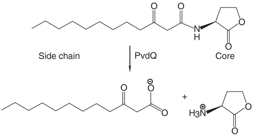
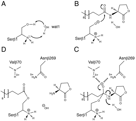
In many Gram-negative pathogens, their virulent behavior is regulated by quorum sensing, in which diffusible signals such as N-acyl homoserine lactones (AHLs) act as chemical messaging compounds. Enzymatic degradation of these diffusible signals by, e.g., lactonases or amidohydrolases abolishes AHL regulated virulence, a process known as quorum quenching. Here we report the first crystal structure of an AHL amidohydrolase, the AHL acylase PvdQ from Pseudomonas aeruginosa. PvdQ has a typical alpha/beta heterodimeric Ntn-hydrolase fold, similar to penicillin G acylase and cephalosporin acylase. However, it has a distinct, unusually large, hydrophobic binding pocket, ideally suited to recognize C12 fatty acid-like chains of AHLs. Binding of a C12 fatty acid or a 3-oxo-C12 fatty acid induces subtle conformational changes to accommodate the aliphatic chain. Furthermore, the structure of a covalent ester intermediate identifies Serbeta1 as the nucleophile and Asnbeta269 and Valbeta70 as the oxyanion hole residues in the AHL degradation process. Our structures show the versatility of the Ntn-hydrolase scaffold and can serve as a structural paradigm for Ntn-hydrolases with similar substrate preference. Finally, the quorum-quenching capabilities of PvdQ may be utilized to suppress the quorum-sensing machinery of pathogens.


Conflict of interest statement

The authors declare no conflict of interest.

Figures

Fig. 1.
Fig. 1.
The hydrolysis of 3-oxo-dodecanoic homoserine lactone performed by PvdQ.
Fig. 2.
Fig. 2.
3D structure of PvdQ. (A) A solvent-accessible surface area representation shows the heart-shape with the α-chain in orange and the β-chain in blue. (B) A secondary structure representation. The α-chain is depicted with magenta β-strands and yellow α-helices while the β-chain has light blue β-strands and orange α-helices. (C) A front view showing the three conserved disulfide bridges. All disulfide bridges (indicated in green) lie on the periphery of the enzyme. The N-terminal nucleophile is located in the center of the enzyme. The σA-weighted electron density, contoured at 1.2σ, shows that no density continues from the newly formed N-terminus after processing, indicating that the enzyme underwent complete autoproteolysis (inset).
Fig. 3.
Fig. 3.
Comparison of liganded and unliganded PvdQ. Surface slice-throughs of PvdQ showing the conformational changes upon substrate binding. (A) Apo-enzyme and (B) Dodecanoic acid bound. The substrate-binding site of PvdQ is built up from mainly bulky hydrophobic residues. Upon binding of 3-oxo-dodecanoic acid (C) (light green model) or dodecanoic acid (D) (light green model) residues move with respect to the apo-enzyme (dark blue). The weaker σA-weighted density corresponding to 3-oxo-C12 is contoured at 1σ while C12 is contoured at 1.2σ. The carboxylates of the ligands form hydrogen bonding interactions with the N-terminal nucleophile and the oxyanion hole residues. Wat1 bridges the Serβ1 Oγ and the free α-amino group, which in turn is coordinated by Asnβ269 and Hisβ23.
Fig. 4.
Fig. 4.
Covalent acyl-enzyme intermediate between PvdQ and dodecanoic acid when soaked shortly at pH 5.5. This ester intermediate is stabilized by hydrogen bonds with the oxyanion hole at the bottom of the figure. Wat1 is bound closely to nucleophile to hydrolyze the acyl-enzyme intermediate. The σA-weighted omit density is contoured at 1σ.
Fig. 5.
Fig. 5.
The mechanism of acyl-enzyme intermediate formation. (A) Wat1 is involved in the activation of the Serβ1 nucleophile by relaying the proton from the Oγ to the α-amino group. (B) Upon nucleophile activation the N-terminal nucleophile attacks the carbonyl carbon of the scissile bond in the substrate. (C) The transition state is stabilized by the oxyanion hole formed by a backbone amide and a side-chain amide. Upon protonation of the α-amino group by wat1 the transition state collapses into an ester intermediate (D).

References

    1. Bassler BL, Losick R. Bacterially speaking. Cell. 2006;125:237–246. - PubMed
    1. Eberhard A, et al. Structural identification of autoinducer of Photobacterium fischeri luciferase. Biochemistry. 1981;20:2444–2449. - PubMed
    1. Pearson JP, Passador L, Iglewski BH, Greenberg EP. A second N-acylhomoserine lactone signal produced by Pseudomonas aeruginosa. Proc Natl Acad Sci USA. 1995;92:1490–1494. - PMC - PubMed
    1. Whitehead NA, et al. Quorum-sensing in Gram-negative bacteria. FEMS Microbiol Rev. 2001;25:365–404. - PubMed
    1. Dong YH, et al. Quenching quorum-sensing-dependent bacterial infection by an N-acyl homoserine lactonase. Nature. 2001;411:813–817. - PubMed

Publication types

LinkOut - more resources