. 2011 May 6;286(18):16163-73.
doi: 10.1074/jbc.M110.171082. Epub 2011 Feb 16.

Allosteric modulation of hormone release from thyroxine and corticosteroid-binding globulins

Affiliations

Allosteric modulation of hormone release from thyroxine and corticosteroid-binding globulins

Xiaoqiang Qi et al. J Biol Chem. .

Abstract

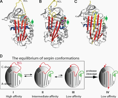
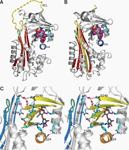
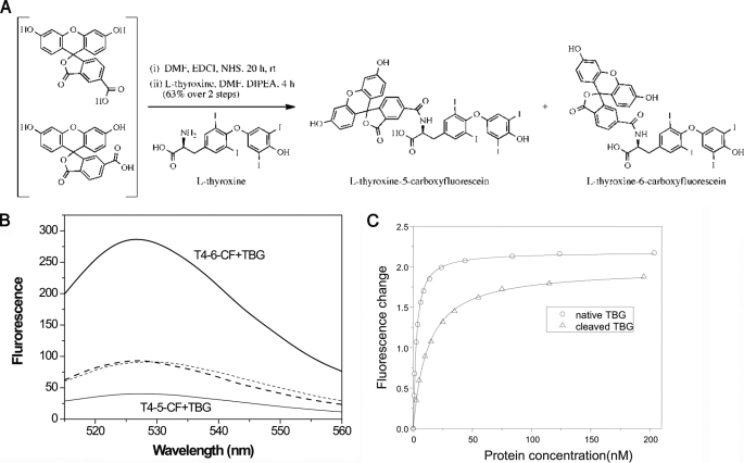
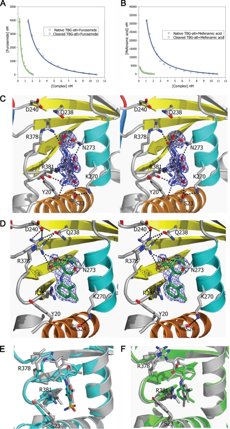
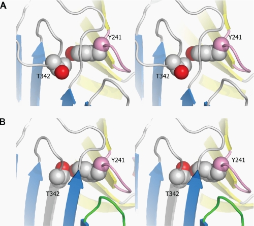
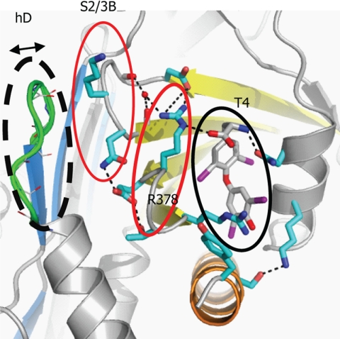
The release of hormones from thyroxine-binding globulin (TBG) and corticosteroid-binding globulin (CBG) is regulated by movement of the reactive center loop in and out of the β-sheet A of the molecule. To investigate how these changes are transmitted to the hormone-binding site, we developed a sensitive assay using a synthesized thyroxine fluorophore and solved the crystal structures of reactive loop cleaved TBG together with its complexes with thyroxine, the thyroxine fluorophores, furosemide, and mefenamic acid. Cleavage of the reactive loop results in its complete insertion into the β-sheet A and a substantial but incomplete decrease in binding affinity in both TBG and CBG. We show here that the direct interaction between residue Thr(342) of the reactive loop and Tyr(241) of the hormone binding site contributes to thyroxine binding and release following reactive loop insertion. However, a much larger effect occurs allosterically due to stretching of the connecting loop to the top of the D helix (hD), as confirmed in TBG with shortening of the loop by three residues, making it insensitive to the S-to-R transition. The transmission of the changes in the hD loop to the binding pocket is seen to involve coherent movements in the s2/3B loop linked to the hD loop by Lys(243), which is, in turn, linked to the s4/5B loop, flanking the thyroxine-binding site, by Arg(378). Overall, the coordinated movements of the reactive loop, hD, and the hormone binding site allow the allosteric regulation of hormone release, as with the modulation demonstrated here in response to changes in temperature.


Figures

FIGURE 1.
FIGURE 1.
The allosteric mechanism of hormone binding and release. Native CBG (A) has a fully exposed reactive center loop (RCL) with a connecting loop on top of helix D (green, arrow) in a helical conformation. This loop is unwound following partial insertion of the reactive loop as seen in the native TBG structure (B) or full insertion into the central β-sheet A in the reactive loop-cleaved CBG structure (C). D, the changes in flexibility of hD resulting from the dynamic flip-flop movement of the reactive loop are transmitted to the hormone binding pocket. I, native CBG, with a reactive loop four residues shorter than that of TBG, crystallizes in the fully exposed loop conformation with a high binding affinity. II, native TBG crystallizes in the partly inserted loop conformation with an intermediate affinity for thyroxine but equilibrates toward frame I with higher affinity when the reactive loop is shortened or toward III with lower affinity when the loop is extended (29). Both native TBG and CBG can equilibrate among these loop-sheet configurations, and the changes in the binding affinity result from the changes of the relative populations of each conformer of different loop-sheet configurations. IV, protease cleavage of the reactive loop results in its incorporation into the central β-sheet A to form, typically irreversibly, a stable relaxed conformation with the lowest binding affinity.
FIGURE 2.
FIGURE 2.
The thyroxine-binding site. A, the binding site of the native TBG-T4 complex. B, TBG undergoes the typical S-to-R transition with the exposed reactive loop (yellow) on cleavage being fully inserted into the central Aβ-sheet (red). The cleaved form retains the ability to bind thyroxine (in spheres with carbon atoms in gray, oxygen atoms in red, iodine atoms in purple, and nitrogen atoms in blue) in the same binding site as seen in the native TBG-thyroxine complex (supplemental Fig. S1). C, binding is mainly through hydrophobic interactions stabilized by a network of hydrogen bonds surrounding the binding site. The connecting loop on top of hD (in green) interacts with the s2/3B loop (purple) though Lys243. Arg378 of s4/5B loop is stabilized by forming salt bridges with Asp240 of s2/3B and directly with thyroxine (carbon atoms shown in dark gray sticks). Arg378 is further linked to the main chain of s2/3B loop by forming a hydrogen bond with a water molecule (red ball). Salt bridges and hydrogen bonds are shown as black dashed lines.
FIGURE 3.
FIGURE 3.
Thyroxine fluorophores. A, fluorophores were conjugated to thyroxine through chemical synthesis (as described under “Experimental Procedures”). B, the conjugates have similar basal fluorescence spectra (dashed lines) when excited at 490 nm, which differ however on binding to TBG. TBG binding of T4–6-CF (thick line) results in a near 200% enhancement in fluorescence at 525 nm, whereas binding of T4–5-CF (thin line) results in a 75% decrease in fluorescence signal. C, the binding affinities of native TBG (circles) and reactive loop-cleaved TBG (triangles) were obtained by titrating TBG into solutions of T4–6-CF with fluorescence changes plotted against protein concentrations. The curves were fitted with the quadratic equilibrium binding equation I.
FIGURE 4.
FIGURE 4.
Binding of T4 analogues to TBG. The binding affinities of furosemide (A) or mefenamic acid (B) toward TBG conformers were measured by competitive assay with titration of the analogues into a TBG/T4–6-CF mixed solution with the fluorescence signal monitored and fitted using the competitive binding equations II and III. The fitted curves are shown in blue (cTBG) or green (native TBG). C and D, stereo-view of the binding site of cTBG-furosemide complex (C) and cTBG-mefenamic acid (D). Electron density, contoured at 2.5 times the root mean square of the map for furosemide and 1 root mean square for mefenamic acid, is shown in blue mesh. Key residues are shown in sticks with carbon atoms are shown in green, nitrogen atoms are shown in blue, and oxygen atoms are shown in red. Furosemide (C) forms four hydrogen bonds with surrounding residues, but it has no interaction with Arg378. The side chain of Arg378 is largely solvent exposed with little direct interaction with the binding site. Whereas in the cTBG-mefenamic acid structure (D), Arg378 exiting in alternative conformations, forms ionic interactions with both the ligand and Asp240, similar to that of thyroxine (Fig. 2C). E and F, when these two structures are superimposed with that of cTBG-thyroxine (gray), it is apparent that the furosemide-binding site (E, light blue) is slightly smaller and the side chain of Arg378 shifts away from the s2/3B loop and will thus be insensitive to its shifts. The side chain of Arg378 of the cTBG-mefenamic acid structure (F, green), however, retains a similar position and hence responsiveness to the S-to-R change as that of cTBG-thyroxine.
FIGURE 5.
FIGURE 5.
The direct interactions between the reactive loop and the hormone-binding site in stereo. A, in the native TBG-T4 complex, the reactive loop is partially inserted in the central β-sheet A (blue) with P14 residue (Thr342) impeded from further insertion by the underlying Tyr241 from the S2/3B connecting loop (purple). B, in the cleaved TBG-T4 structure, the reactive loop is fully inserted into the central β-sheet A with the side chain of Thr342 forming a direct contact with the OH group of Tyr. The β-sheet B is colored in yellow.
FIGURE 6.
FIGURE 6.
Key interactions of TBG in regulating hormone binding and release. Hormone binding and release involves coherent movements of three closely associated elements: the connecting loops of hDs2A (dashed black line), s2/3B (thin red line), and s4/5B (thick red line). Thus, the binding of thyroxine will be destabilized by slight changes within any of these elements including increases in the flexibility of the hDs2A loop in the S-to-R transition, removal of residues from the top of hD, loosening the packing between hD and s2/3A by the K243G mutation, or the removal the side chain of Arg378. Replacing the homologous residue 243 in CBG, a glycine, with a bulkier valine will similarly affect the packing and also lead to decreased hormone binding affinity (28). Also, only the ligands that are fitted perfectly in the hormone-binding site and form direct interactions with Arg378 can be efficiently released following the S-to-R transitions of TBG.

References

    1. Flink I. L., Bailey T. J., Gustafson T. A., Markham B. E., Morkin E. (1986) Proc. Natl. Acad. Sci. U.S.A. 83, 7708–7712 - PMC - PubMed
    1. Hammond G. L., Smith C. L., Goping I. S., Underhill D. A., Harley M. J., Reventos J., Musto N. A., Gunsalus G. L., Bardin C. W. (1987) Proc. Natl. Acad. Sci. U.S.A. 84, 5153–5157 - PMC - PubMed
    1. Huber R., Carrell R. W. (1989) Biochemistry 28, 8951–8966 - PubMed
    1. Silverman G. A., Bird P. I., Carrell R. W., Church F. C., Coughlin P. B., Gettins P. G., Irving J. A., Lomas D. A., Luke C. J., Moyer R. W., Pemberton P. A., Remold-O'Donnell E., Salvesen G. S., Travis J., Whisstock J. C. (2001) J. Biol. Chem. 276, 33293–33296 - PubMed
    1. Irving J. A., Pike R. N., Lesk A. M., Whisstock J. C. (2000) Genome Res. 10, 1845–1864 - PubMed

Publication types

MeSH terms