. 2010 Dec 16;468(7326):988-91.
doi: 10.1038/nature09580. Epub 2010 Dec 5.

The mechanism of sodium and substrate release from the binding pocket of vSGLT

Affiliations

The mechanism of sodium and substrate release from the binding pocket of vSGLT

Akira Watanabe et al. Nature. .

Abstract

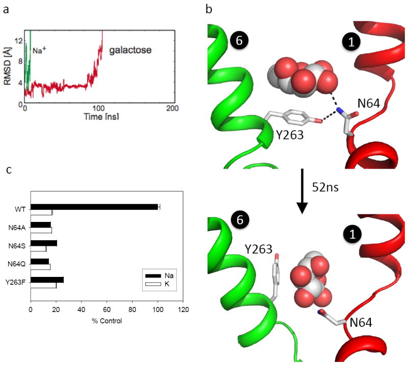
Membrane co-transport proteins that use a five-helix inverted repeat motif have recently emerged as one of the largest structural classes of secondary active transporters. However, despite many structural advances there is no clear evidence of how ion and substrate transport are coupled. Here we report a comprehensive study of the sodium/galactose transporter from Vibrio parahaemolyticus (vSGLT), consisting of molecular dynamics simulations, biochemical characterization and a new crystal structure of the inward-open conformation at a resolution of 2.7 Å. Our data show that sodium exit causes a reorientation of transmembrane helix 1 that opens an inner gate required for substrate exit, and also triggers minor rigid-body movements in two sets of transmembrane helical bundles. This cascade of events, initiated by sodium release, ensures proper timing of ion and substrate release. Once set in motion, these molecular changes weaken substrate binding to the transporter and allow galactose readily to enter the intracellular space. Additionally, we identify an allosteric pathway between the sodium-binding sites, the unwound portion of transmembrane helix 1 and the substrate-binding site that is essential in the coupling of co-transport.


Figures

Figure 1
Figure 1. Structure of the inward-open and an overlay with the inward-occluded structure
a, A cartoon representation of vSGLT. The core domain of the inward-open conformation (TMs 1-10) is coloured by specific helix bundles involved in the transition from inward-occluded to inward-open conformation. The ‘hash motif’ formed from TMs3, 4, 8, and 9 is blue; the ‘sugar bundle’ formed from TMs2, 6, and 7 is green; TM1 is red; and TMs5 and 10 are magenta. The periphery TMs (-1,11,12,13) are yellow. The inset shows an overlay between the inward-open (coloured as above) and inward-occluded (grey) conformations illustrating the coordination at the Na2- and galactose-binding sites. b, c, Overlay of the inward-open and inward-occluded conformations with the same colouring as (a). Conformational changes in the inward-open structure reveals a ~13° kink in the unwound segment of TM1 preventing sodium coordination at the Na2-site (b). In the absence of galactose, the galactose-binding residue N64 hydrogen bonds to E88 and Y263 maintaining an open pathway from the intracellular space to the substrate-binding site (c).
Figure 2
Figure 2. Mechanism of galactose release
a, Sodium and galactose exit vSGLT. The RMSD of Na+ (green) rapidly increases at 9 ns indicating exit from the Na2-site followed by the release of galactose (red) at 110 ns. b, Y263 adopts two rotamers. (Upper panel) Y263 is shown in the conformation observed in the inward-occluded structure in which it blocks substrate exit through a hydrogen bond with N64 on TM1. (Lower panel) At 52 ns, Y263 adopts a rotamer conformation that expands the exit pathway. c, D-galactose uptake by WT and vSGLT mutants in proteoliposomes. Results are expressed as % uptake of WT protein in either 100mM NaCl or KCl showing the mutants N64A, N64S, N64Q and Y263F severely impair sodium-dependent transport.
Figure 3
Figure 3. The potential of mean force for galactose unbinding
a, After Na+ exit from vSGLT, we observe galactose exit at 110 ns. Snapshots along this natural pathway are shown in different colours. Red corresponds to galactose in the binding site, and purple corresponds to a snapshot near 110 ns just as galactose is exiting the hydrophilic cavity to the bulk water. b, Galactose binding energy to vSGLT in the absence of Na+. Umbrella sampling along the natural, equilibrium pathway shown in (a) was used to determine the binding free energy. The distance from the binding site in the x-ray structure, along the pathway is shown along the x-axis. The coloured arrows correspond to the galactose snapshots shown in (a). Distance values greater than 15 Å correspond to galactose in the inner hydrophilic cavity. The largest barrier is ~2 kcal/mol at 5 Å, which corresponds to galactose interaction with residues in the kink region of TM1.
Figure 4
Figure 4. Conformational changes in the transition from inward-occluded to inward-open structure
a, TM1 superimposed between the inward-open (red) and inward-occluded (grey) structures showing a ~13° kink in TM1. b, Overlay of the inward-open (coloured as in Fig.1) and inward-occluded (grey) conformations. 3° rigid body rotations of the ‘hash motif’ and ‘sugar bundle’ in opposite directions expose the substrate-binding site to the intracellular environment. c, Accessibility cavity of the inward-occluded conformation is coloured blue. d, Accessibility cavity of the inward-open conformation is coloured gold. The conformational changes from TM1, hash motif, and sugar bundle cause an increase of ~1400 Å3 in the accessible volume of the inward-open conformation aiding galactose release.

References

    1. Abramson J, Wright EM. Structure and function of Na(+)-symporters with inverted repeats. Curr Opin Struct Biol. 2009;19:425–432. - PMC - PubMed
    1. Krishnamurthy H, Piscitelli CL, Gouaux E. Unlocking the molecular secrets of sodium-coupled transporters. Nature. 2009;459:347–355. - PMC - PubMed
    1. Jardetzky O. Simple allosteric model for membrane pumps. Nature. 1966;211:969–970. - PubMed
    1. Wright EM, Hirayama BA, Loo DF. Active sugar transport in health and disease. J Intern Med. 2007;261:32–43. - PubMed
    1. Isaji M. Sodium-glucose cotransporter inhibitors for diabetes. Curr Opin Investig Drugs. 2007;8:285–292. - PubMed

Publication types