. 2017 Mar 24;292(12):5031-5042.
doi: 10.1074/jbc.M116.767616. Epub 2017 Feb 7.

The Startle Disease Mutation E103K Impairs Activation of Human Homomeric α1 Glycine Receptors by Disrupting an Intersubunit Salt Bridge across the Agonist Binding Site

Affiliations

The Startle Disease Mutation E103K Impairs Activation of Human Homomeric α1 Glycine Receptors by Disrupting an Intersubunit Salt Bridge across the Agonist Binding Site

Fatemah Safar et al. J Biol Chem. .

Abstract

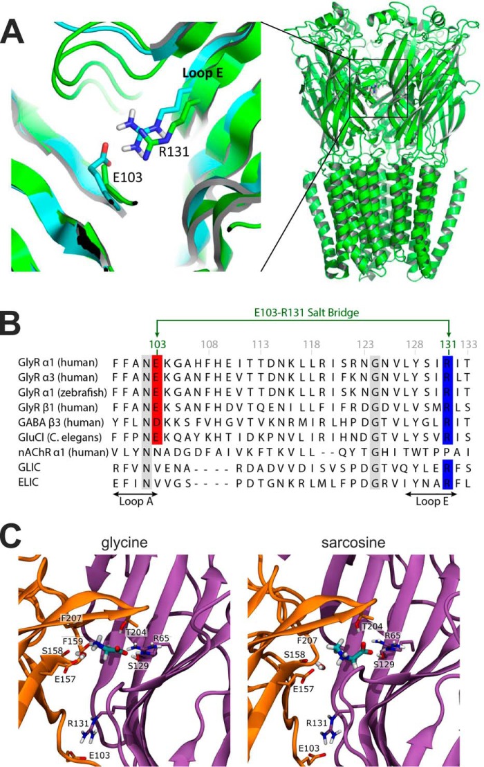
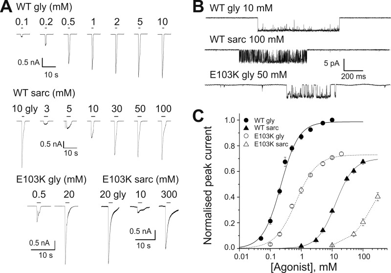
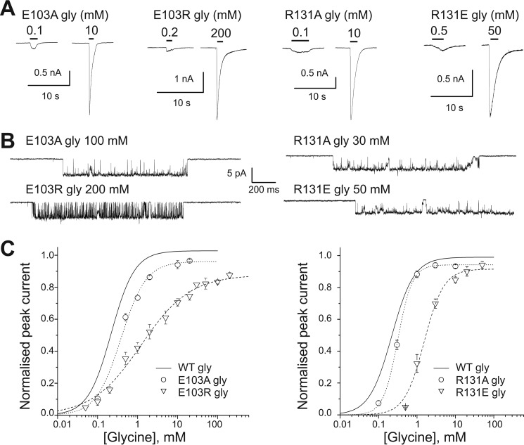
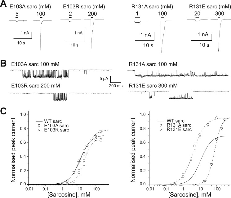
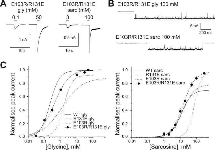
Glycine receptors (GlyR) belong to the pentameric ligand-gated ion channel (pLGIC) superfamily and mediate fast inhibitory transmission in the vertebrate CNS. Disruption of glycinergic transmission by inherited mutations produces startle disease in man. Many startle mutations are in GlyRs and provide useful clues to the function of the channel domains. E103K is one of few startle mutations found in the extracellular agonist binding site of the channel, in loop A of the principal side of the subunit interface. Homology modeling shows that the side chain of Glu-103 is close to that of Arg-131, in loop E of the complementary side of the binding site, and may form a salt bridge at the back of the binding site, constraining its size. We investigated this hypothesis in recombinant human α1 GlyR by site-directed mutagenesis and functional measurements of agonist efficacy and potency by whole cell patch clamp and single channel recording. Despite its position near the binding site, E103K causes hyperekplexia by impairing the efficacy of glycine, its ability to gate the channel once bound, which is very high in wild type GlyR. Mutating Glu-103 and Arg-131 caused various degrees of loss-of-function in the action of glycine, whereas mutations in Arg-131 enhanced the efficacy of the slightly bigger partial agonist sarcosine (N-methylglycine). The effects of the single charge-swapping mutations of these two residues were largely rescued in the double mutant, supporting the possibility that they interact via a salt bridge that normally constrains the efficacy of larger agonist molecules.

Keywords: agonists; efficacy; glycine receptor; homology modeling; ion channel; mutagenesis; patch clamp; potency; site-directed mutagenesis.


Conflict of interest statement

The authors declare that they have no conflicts of interest with the contents of this article

Figures

FIGURE 1.
FIGURE 1.
Homology modeling reveals a salt-bridge between residues Glu-103 and Arg-131. A, location of the Glu103-Arg131 salt-bridge as predicted by a GlyR homology model (cyan)(7) built using GluCl as template (PDB code 2XYS) (16). The model is overlaid with the recent cryo-EM structure (green) of the zebrafish α1 GlyR (PDB 3JAE) (17). Note that the Glu-103 side chain is not fully resolved in the cryo-EM structure, but is certainly within distance to form the salt bridge as postulated by the model. B, an amino acid alignment of this region for nine members of the pLGIC superfamily showing the position of the salt bridge between Glu-103 (red) and Arg-131 (blue) and its conservation in glycine receptors. Other conserved positions are indicated by a gray background. C, the agonist binding mode and the Glu103-Arg131 salt bridge in a GlyR homology model based on the zebrafish α1 GlyR structure (PDB 3JAE) (17). Glycine (left) and sarcosine (right) are shown in the final frame after 500 ns simulation time. Orange, principal subunit; purple, complementary subunit. For both ligands, the carboxyl group is sandwiched by hydrogen bonds with Thr-204 and Ser-129 and is further stabilized by a salt bridge with Arg-65. The ammonium moiety interacts with a water molecule in the binding pocket, and the water is stabilized by hydrogen bonds to Glu-157 and to the backbone carbonyl oxygen of Ser-158 (as in Ref. 7). The ammonium group of glycine engages in an additional cation-π interaction with Phe-207 and forms a hydrogen bond with the backbone carbonyl group of Phe-159. This is sterically hindered for sarcosine by its additional N-methyl group.
FIGURE 2.
FIGURE 2.
The E103K startle mutation reduces the sensitivity of α1 GlyR to both glycine and sarcosine and impairs channel gating. A, representative whole-cell current responses evoked by U-tube agonist application to HEK 293 cells expressing WT α1 GlyR (upper panels, 2 different cells) or E103K α1 GlyR (lower panel, 2 different cells). Black bars above the traces show the timing of the applications. Panels also show the responses to a saturating concentration of glycine obtained in the cells used for the sarcosine concentration-response curves. B, clusters of single channel activity elicited by saturating concentrations of glycine (upper panel) or sarcosine (middle panel) on WT α1 GlyR or (glycine only) on E103K GlyR (lower panel; cell-attached configuration, channel openings downwards). Note the decreased single channel Popen in mutant receptors. C, whole cell concentration-response curves to glycine and sarcosine in WT (n = 7 and 10, respectively) and E103K α1 GlyR (n = 4 and 3, respectively). Curves are scaled to the maximum single channel Popen measured at saturating glycine or sarcosine; for sarcosine on E103K GlyR, we could not measure the maximum single channel Popen and the curve is scaled indirectly (e.g. responses to sarcosine are scaled to the macroscopic maximum response to glycine in the same cell, and then scaled to the maximum glycine single channel Popen).
FIGURE 3.
FIGURE 3.
Reversing the side chain charge in positions Glu-103 or Arg-131 changes the potency and efficacy of glycine. A, representative whole cell current responses to glycine applied to GlyR α1 mutants. B, clusters of single-channel mutant GlyR activity elicited in cell-attached patches by saturating concentrations of glycine. C, whole cell concentration-response curves for the effect of glycine on E103A, E103R, R131A, and R131E mutant GlyR (n = 3, 8, 5, and 4, respectively). Curves are scaled to the maximum Popen obtained from single channel recordings. The WT concentration-response curve for glycine is shown for comparison (solid line; the data for this curve is shown in Fig. 2C).
FIGURE 4.
FIGURE 4.
Removing or reversing the side chain charge of Arg-131 increases the efficacy of the partial agonist sarcosine on α1 GlyR. A, representative whole cell current responses to sarcosine in α1 GlyR mutants. B, clusters of single-channel mutant GlyR activity elicited in cell-attached patches by high concentrations of sarcosine (saturating except for the R131E mutant) C, sarcosine whole cell concentration-response curves for E103A, E103R, R131A, and R131E mutant GlyR (n = 4, 4, 5, and 6, respectively). Curves are scaled to the appropriate maximum Popen measured by single channel recording. The WT concentration-response curve for sarcosine is shown for comparison (solid line; the data for this curve is shown in Fig. 2C).
FIGURE 5.
FIGURE 5.
E103R/R131E mutation rescues α1 GlyR response to glycine and sarcosine. A, representative whole cell current responses to glycine or sarcosine in the double charge reversal mutant E103R/R131E GlyR α1. B, clusters of single-channel mutant GlyR activity elicited in cell-attached patches by saturating concentrations of glycine and sarcosine, respectively. C, glycine and sarcosine whole cell concentration-response curves in WT and double mutant GlyR (n = 5 and 8, respectively). Curves are scaled to the appropriate maximum Popen measured by single channel recording. The WT concentration-response curves and the single-mutation curves for both glycine and sarcosine are shown for comparison (solid line for WT; the data for this curve is shown in Fig. 2C).

References

    1. Lynagh T., and Pless S. A. (2014) Principles of agonist recognition in Cys-loop receptors. Front. Physiol. 5, 160. - PMC - PubMed
    1. Galzi J.-L., and Changeux J.-P. (1995) Neuronal nicotinic receptors: molecular organization and regulations. Neuropharmacology 34, 563–582 - PubMed
    1. Zhong W., Gallivan J. P., Zhang Y., Li L., Lester H. A., and Dougherty D. A. (1998) From ab initio quantum mechanics to molecular neurobiology: a cation-π binding site in the nicotinic receptor. Proc. Natl. Acad. Sci. U.S.A. 95, 12088–12093 - PMC - PubMed
    1. Beene D. L., Brandt G. S., Zhong W., Zacharias N. M., Lester H. A., and Dougherty D. A. (2002) Cation-π interactions in ligand recognition by serotonergic (5-HT3A) and nicotinic acetylcholine receptors: the anomalous binding properties of nicotine. Biochemistry 41, 10262–10269 - PubMed
    1. Pless S. A., Millen K. S., Hanek A. P., Lynch J. W., Lester H. A., Lummis S. C., and Dougherty D. A. (2008) A cation-π interaction in the binding site of the glycine receptor is mediated by a phenylalanine residue. J. Neurosci. 28, 10937–10942 - PMC - PubMed

LinkOut - more resources