. 2012 Jan 22;19(2):184-92.
doi: 10.1038/nsmb.2231.

Structural basis for autoinhibition and phosphorylation-dependent activation of c-Cbl

Affiliations

Structural basis for autoinhibition and phosphorylation-dependent activation of c-Cbl

Hao Dou et al. Nat Struct Mol Biol. .

Abstract

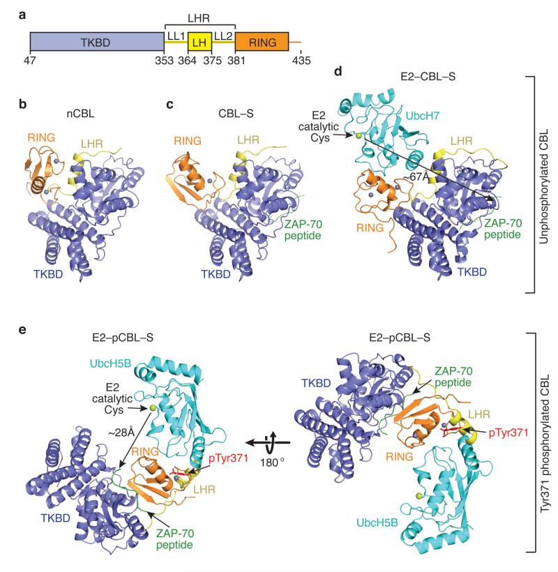
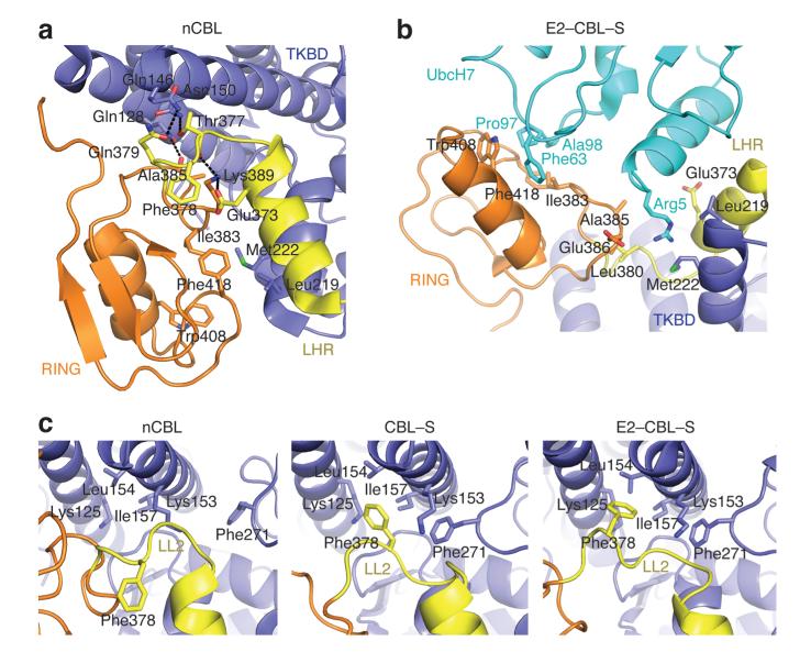
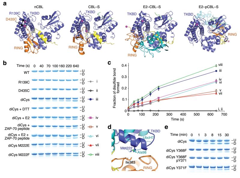
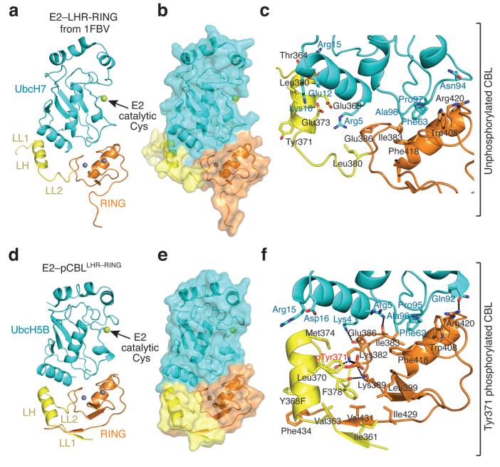
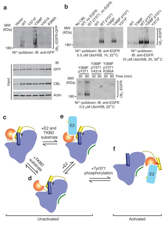
Cbls are RING ubiquitin ligases that attenuate receptor tyrosine kinase (RTK) signal transduction. Cbl ubiquitination activity is stimulated by phosphorylation of a linker helix region (LHR) tyrosine residue. To elucidate the mechanism of activation, we determined the structures of human CBL, a CBL-substrate peptide complex and a phosphorylated-Tyr371-CBL-E2-substrate peptide complex, and we compared them with the known structure of a CBL-E2-substrate peptide complex. Structural and biochemical analyses show that CBL adopts an autoinhibited RING conformation, where the RING's E2-binding surface associates with CBL to reduce E2 affinity. Tyr371 phosphorylation activates CBL by inducing LHR conformational changes that eliminate autoinhibition, flip the RING domain and E2 into proximity of the substrate-binding site and transform the RING domain into an enhanced E2-binding module. This activation is required for RTK ubiquitination. Our results present a mechanism for regulation of c-Cbl's activity by autoinhibition and phosphorylation-induced activation.


Figures

Figure 1
Figure 1
Ensemble of CBL structures. (a) Diagram of the crystallized CBL fragment: TKBD in blue, LHR (LL1, LH and LL2) in yellow and RING domain in orange. Residues encompassing each domain are indicated. All structures are colored as in a and oriented as in b. Zn2+ atoms are depicted as grey spheres, ZAP-70 peptide is green and E2 is cyan with its catalytic cysteine shown as a lime sphere. Phosphorylation state is indicated on the right. (b) nCBL structure. (c) CBL–S structure. (d) E2–CBL–S structure (PDB 1FBV). The measured gap between the E2’s catalytic cysteine and TKBD substrate peptide is shown by the arrows here and in e. (e) E2–pCBL–S structure. Right panel is in the same orientation as b and left panel is rotated by 180° about the x-axis. pTyr371 side chain is shown in red.
Figure 2
Figure 2
CBL autoinhibition via the E2-binding site. (a) Interactions in nCBL that stabilize the closed conformation. (b) Interactions in E2–CBL–S involved in CBL-E2 binding. (c) The LL2 conformations in nCBL (left), CBL–S (middle), and E2–CBL–S (right). All panels are colored as in Fig. 1.
Figure 3
Figure 3
The RING domain adopts different conformations in solution. (a) Structures of nCBL (left), CBL–S (middle left), E2–CBL–S (middle right) and E2–pCBL–S (right) colored as in Fig. 1 with TKBD’s Arg139 and RING’s Asp435 displayed as magenta spheres. Phe434 is displayed in E2–CBL–S because Asp435 is absent. (b) SDS-PAGE showing disulfide bond formation for unphosphorylated CBL47–435 variants in the absence or presence of E2, ZAP-70 peptide or both. Disulfide-bonded (D) and unmodified (U) species are indicated. The color symbols on the right with Roman numerals correspond to the chart legend in c. (c) Plots for fraction of disulfide bond formed over time for CBL variants in b. Curves are indicated with symbols and corresponding Roman numerals in b. Triplicate reactions were performed and the s.e.m. are shown. (d) Superposition of the RING domains from nCBL and E2–CBL–S. Coloring is as described in Fig. 1. (e) SDS-PAGE showing disulfide bond formation for diCys variants with or without Tyr371 phosphorylation. Labels are as in b.
Figure 4
Figure 4
pTyr371 alters LHR conformation and interactions. All coloring is as described in Fig. 1. Phosphorylation state is indicated on the right. (a) E2-LHR-RING portion of structure from E2–CBL–S (PDB 1FBV). (b) Surface representation of a. (c) Close up view of the interactions between UbcH7 and LHR-RING domain in E2–CBL–S. (d) Structure of E2–pCBLLHR–RING. (e) Surface representation of d. (f) Close up view of the pTyr371-binding site and UbcH5B-binding interface in E2–pCBLLHR–RING.
Figure 5
Figure 5
EGFR ubiquitination by CBL. (a) Immunoblots from in vivo EGFR ubiquitination by full-length CBL variants. Ni2+-pulldown products were Western blotted for GFP to detect His-Ub-EGFR-GFP. Protein input levels were assessed with the indicated antibodies. (b) Western blots from in vitro EGFR ubiquitination by CBL47–435 variants. Ni2+-pulldown products were Western blotted for EGFR to detect His-Ub-EGFR. (cf) Model for autoinhibition and phosphorylation-dependent activation of c-Cbl. c-Cbl exists in an unactivated state (ce) where its E2-binding affinity is reduced by a competitive RING autoinhibitory mechanism. Coloring of c-Cbl, E2 and substrate is as in Fig. 1. (c) In the absence of E2, c-Cbl adopts a closed conformation where the RING’s E2-binding surface associates with the TKBD. (d) TKBD substrate binding induces partial RING opening. (e) E2 binding causes the RING domain to adopt an open conformation. The TKBD competes against E2 for RING binding, reducing E2 affinity and E3 activity. In the unactivated state, Tyr371 (black ball-and-stick) secures the LH to the TKBD and limits the RING domain rotation to a region distal from the TKBD substrate-binding site. (f) pTyr371 (red ball-and-stick) activates c-Cbl by releasing LH from the TKBD, thereby abolishing autoinhibition, altering LH-RING-E2 interactions and promoting dramatic LHR conformational changes that bring the RING domain and E2 into proximity of substrate.

Comment in

  • Cbl exposes its RING finger.
    Kales SC, Ryan PE, Lipkowitz S. Kales SC, et al. Nat Struct Mol Biol. 2012 Feb 3;19(2):131-3. doi: 10.1038/nsmb.2241. Nat Struct Mol Biol. 2012. PMID: 22301873 Free PMC article.

References

    1. Hershko A, Ciechanover A. The ubiquitin system. Annu. Rev. Biochem. 1998;67:425–79. - PubMed
    1. Pickart CM, Eddins MJ. Ubiquitin: structures, functions, mechanisms. Biochim. Biophys. Acta. 2004;1695:55–72. - PubMed
    1. Dye BT, Schulman BA. Structural mechanisms underlying posttranslational modification by ubiquitin-like proteins. Annu. Rev. Biophys. Biomol. Struct. 2007;36:131–50. - PubMed
    1. Duda DM, Scott DC, Calabrese MF, Zimmerman ES, Zheng N, Schulman BA. Structural regulation of cullin-RING ubiquitin ligase complexes. Curr. Opin. Struct. Biol. 2011;21:257–64. - PMC - PubMed
    1. Deshaies RJ, Joazeiro CA. RING domain E3 ubiquitin ligases. Annu. Rev. Biochem. 2009;78:399–434. - PubMed

Publication types

Substances