. 2011 Sep 2;286(35):30691-30705.
doi: 10.1074/jbc.M111.247999. Epub 2011 Jul 6.

Structural and functional insights into DR2231 protein, the MazG-like nucleoside triphosphate pyrophosphohydrolase from Deinococcus radiodurans

Affiliations

Structural and functional insights into DR2231 protein, the MazG-like nucleoside triphosphate pyrophosphohydrolase from Deinococcus radiodurans

Ana Maria D Gonçalves et al. J Biol Chem. .

Abstract

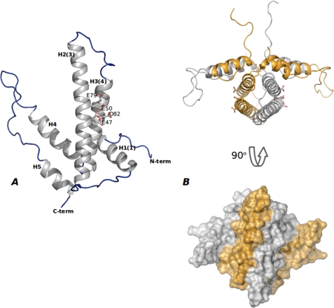
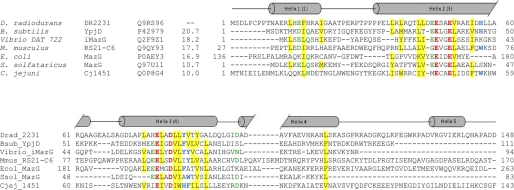
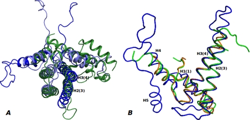
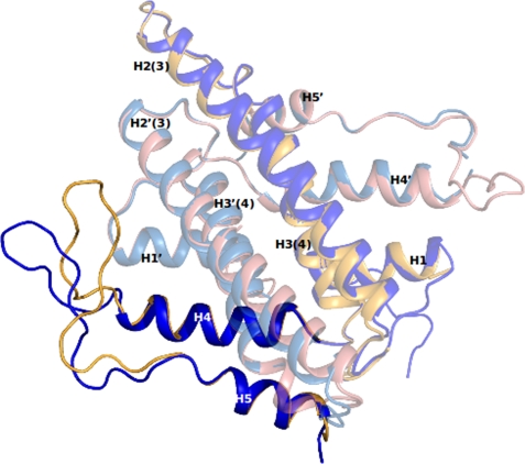
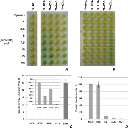
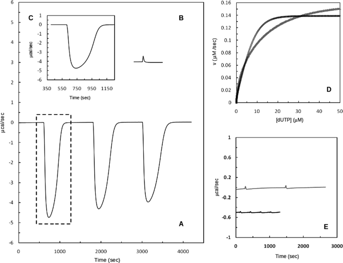
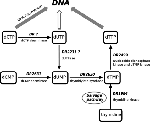
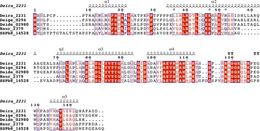
Deinococcus radiodurans is among the very few bacterial species extremely resistant to ionizing radiation, UV light, oxidizing agents, and cycles of prolonged desiccation. The proteome of D. radiodurans reflects the evolutionary pressure exerted by chronic exposure to (nonradioactive) forms of DNA and protein damage. A clear example of this adaptation is the overrepresentation of protein families involved in the removal of non-canonical nucleoside triphosphates (NTPs) whose incorporation into nascent DNA would promote mutagenesis and DNA damage. The three-dimensional structure of the DR2231 protein has been solved at 1.80 Å resolution. This protein had been classified as an all-α-helical MazG-like protein. The present study confirms that it holds the basic structural module characteristic of the MazG superfamily; two helices form a rigid domain, and two helices form a mobile domain and connecting loops. Contrary to what is known of MazG proteins, DR2231 protein shows a functional affinity with dUTPases. Enzymatic and isothermal calorimetry assays have demonstrated high specificity toward dUTP but an inability to hydrolyze dTTP, a typical feature of dUTPases. Co-crystallization with the product of hydrolysis, dUMP, in the presence of magnesium or manganese cations, suggests similarities with the dUTP/dUDP hydrolysis mechanism reported for dimeric dUTPases. The genome of D. radiodurans encodes for all enzymes required for dTTP synthesis from dCMP, thus bypassing the need of a dUTPase. We postulate that DR2231 protein is not essential to D. radiodurans and rather performs "house-cleaning" functions within the framework of oxidative stress response. We further propose DR2231 protein as an evolutionary precursor of dimeric dUTPases.


Figures

FIGURE 1.
FIGURE 1.
Overall shape of DR2231 protein. A, monomeric DR2231 protein structure. DR2231 is an all-α-helix structure with an overall hairpin-like fold. Secondary structure elements are labeled both according to sequence and to the convention of Moroz et al. (7) (in parenthesis) for facility of comparison with other dUTPase/MazG proteins. The EEXX(E/D) motif is presented (Glu47, Glu50, Glu79, and Asp82); Glu47 appears in a double conformation when in the absence of the divalent metal cation. B, DR2231 dimer in open conformation, presented also in orthogonal view. A surface representation illustrates extension of the interface in the dimer.
FIGURE 2.
FIGURE 2.
Sequence alignment of DR2231 with members of the dUTPase and MazG subfamilies. The alignment was constructed on the basis of a ClustalW 2.0.12 multiple sequence alignment (58) with minimal manual editing. Experimentally determined crystal structures are available for all listed entries: Bsub_YpjD (PDB code 2GTA), Vibrio_iMazG (PDB codes 2Q5Z and 2Q73), Mmus_RS21-C6 (PDB code 2OIE), Ecol_MazG (PDB code 3CRC), Ssol_MazG (PDB code 1VMG), and Cjej_1451 (PDB code 1W2Y). The proteins are listed by name of the source organism, followed by the protein name; the third column corresponds to UniProt identifiers, and the fourth column shows the percentage of identity with DR2231 sequence. Sequence numbers preceding and following matched segments are indicated. The structural elements shown above the alignment correspond to DR2231 determined in this work. Cylinders indicate α-helices, and the numbering of helices in parenthesis corresponds to the convention established by Moroz et al. (7). The Mg2+-binding residues are shown in red boldface type, and conserved residues are colored as follows: hydrophobic (yellow background), aromatic (blue boldface type), Asp/Asn/Glu/Gln (green boldface type). The E. coli MazG alignment is performed only on the enzymatically active C-terminal domain (residues 136–263); the C. jejuni dUTPase sequence was truncated at residue 143.
FIGURE 3.
FIGURE 3.
Overlay of DR2231 structure with known dimeric dUTPase and MazG structures. A, DR2231 dimer (monomer A (dark blue) and monomer B (light blue)) superimposed with C. jejuni dUTPase monomer (green). B, DR2231 structure (blue) overlay with S. solfataricus (PDB code 1VMG; orange) and B. subtilis (PDB code 2GTA; green) MazG structures.
FIGURE 4.
FIGURE 4.
Open (blue) and closed (yellow) conformations of DR2231. Changes are confined to the “loop” region encompassing residues Lys112 to Pro131.
FIGURE 5.
FIGURE 5.
A, stereo view of the putative binding site of DR2231 in the closed conformation bound to dUMP. The active site shows the contribution of residues from both monomers to substrate binding and coordination. When crystallized in presence of Mg2+, DR2231 presents only one divalent metal-coordinated equivalent to Mn1; a water molecule was modeled in the position of Mn2 (not shown). B, close-up of the binding pocket highlighting the role of structured waters in catalysis (W1 and W2), based on homology with C. jejuni dUTPase and substrate binding (W3, W4, and W5). W6 participates in the hexacoordination shell of Mn2.
FIGURE 6.
FIGURE 6.
Enzymatic activity of DR2231. A and B, screening of purified DR2231 for NTP-PPase activity against (d)NTPs in the presence of 0.623 μm (A) and 0.623 nm enzyme (B). The top row (labeled PPase -) in both panels corresponds to screening of each substrate at 10 μm in the absence of inorganic phosphatase. Hydrolysis of ATP is representative of all NTPs. C, specific activities of DR2231 (0.623 nm) at 5 μm substrate; the inset corresponds to determination of specific activities of substrates at 0.623 μm. D, divalent metal preference of DR2231 for 5 μm dUTP hydrolysis; depicted is the percentage of specific activity of the enzyme in the presence of a divalent metal relative to the specific activity in the presence of Mn2+. Enzymatic activity in the absence of divalent metals or in the presence of 20 mm EDTA was not detectable. For details, see “Experimental Procedures,” NTP Pyrophosphatase Assays, Error bars, S.D.
FIGURE 7.
FIGURE 7.
DR2231-catalyzed hydrolysis of dUTP in 20 mm Tris-HCl, 5 mm NaCl, 25 mm MgCl2 at pH 7.0 and 25 °C. A, representation of typical calorimetric trace (μcal versus time) obtained after the addition of three injections of 3 mm dUTP (20-μl injections) to the calorimetric cell containing DR2231 (12.5 nm). Change in instrumental thermal power was monitored until substrate hydrolysis was complete, returning to the original base line. B, thermogram of the dUTP dilution experiment. C, calorimetric trace (first injection) resulting after subtraction of the first peak of the dilution experiment. D, net thermal power (from B) was converted to rate and substrate concentration and fit to the Michaelis-Menten equation (black curve); the gray curve corresponds to a similar assay but in the presence of 25 mm MnCl2 (see “Experimental Procedures”). E, raw data representing the calorimetric trace (gray line) of 20-μl injections of 3 mm dTTP into the calorimetric cell containing enzyme (12.5 nm) and the equivalent dilution experiment in the absence of enzyme (black line). The trace of the dilution experiment is shifted for clarity.
FIGURE 8.
FIGURE 8.
De novo and salvage pathways for dTTP biosynthesis. Identifiable homologous loci from D. radiodurans are represented beside their respective arrows.
FIGURE 9.
FIGURE 9.
Sequence alignment of DR2231 with other putative subfamily members: Deira_2231 (D. radiodurans, Q9RS96), Deige_0294 (Deinococcus geothermalis, Q1J1N7), Deide_02980 (Deinococcus deserti, C1CZ79), Haur_2379 (Herpetosiphon aurantiacus, A9AYP1), and SSPb78_14528 (Streptomyces sp. SPB78, UPI0001B553EB). Identical residues are shown in white on a red background, and similar residues are shown in red on a white background. For the secondary structure assignment, α-helices are represented as helices, β-strands are represented as arrows, and β-turns are marked TT. This figure was prepared with ESPript (57). The multiple-sequence alignment was performed with ClustalW (version 2.0.12) (58).

References

    1. Kamiya H. (2003) Nucleic Acids Res. 31, 517–531 - PMC - PubMed
    1. Bessman M. J., Frick D. N., O'Handley S. F. (1996) J. Biol. Chem. 271, 25059–25062 - PubMed
    1. Koonin E. V., Aravind L., Galperin M. Y. (2000) in Bacterial Stress Responses (Storz G., Hengge-Aronis R. eds) pp. 417–444, American Society for Microbiology Press, Washington, D. C
    1. Makarova K. S., Aravind L., Wolf Y. I., Tatusov R. L., Minton K. W., Koonin E. V., Daly M. J. (2001) Microbiol. Mol. Biol. Rev. 65, 44–79 - PMC - PubMed
    1. Makarova K. S., Aravind L., Daly M. J., Koonin E. V. (2000) Genetica 108, 25–34 - PubMed

Publication types

MeSH terms

LinkOut - more resources