. 2009 Aug;37(14):4787-98.
doi: 10.1093/nar/gkp506. Epub 2009 Jun 10.

Functional importance of crenarchaea-specific extra-loop revealed by an X-ray structure of a heterotetrameric crenarchaeal splicing endonuclease

Affiliations

Functional importance of crenarchaea-specific extra-loop revealed by an X-ray structure of a heterotetrameric crenarchaeal splicing endonuclease

Shigeo Yoshinari et al. Nucleic Acids Res. 2009 Aug.

Abstract

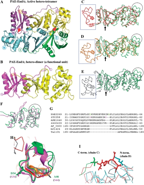
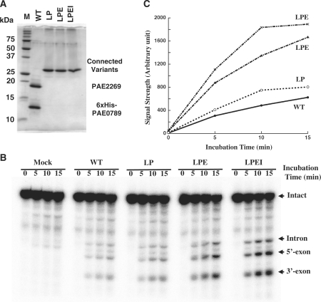
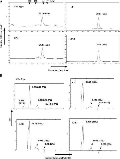
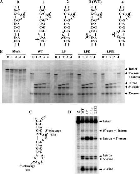
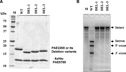
Archaeal splicing endonucleases (EndAs) are currently classified into three groups. Two groups require a single subunit protein to form a homodimer or homotetramer. The third group requires two nonidentical protein components for the activity. To elucidate the molecular architecture of the two-subunit EndA system, we studied a crenarchaeal splicing endonuclease from Pyrobaculum aerophilum. In the present study, we solved a crystal structure of the enzyme at 1.7-A resolution. The enzyme adopts a heterotetrameric form composed of two catalytic and two structural subunits. By connecting the structural and the catalytic subunits of the heterotetrameric EndA, we could convert the enzyme to a homodimer that maintains the broad substrate specificity that is one of the characteristics of heterotetrameric EndA. Meanwhile, a deletion of six amino acids in a Crenarchaea-specific loop abolished the endonuclease activity even on a substrate with canonical BHB motif. These results indicate that the subunit architecture is not a major factor responsible for the difference of substrate specificity between single- and two-subunit EndA systems. Rather, the structural basis for the broad substrate specificity is built into the crenarchaeal splicing endonuclease itself.


Figures

Figure 1.
Figure 1.
Crystal structure of PAE-EndA. (A) Schematic representation of a crystal structure of PAE-EndA. The unit is composed of two catalytic subunits (cyan and yellow) and two structural subunits (green and magenta). Red arrows indicate L10-loop-like interactions between the structural and the catalytic subunits to form a hetero-tetramer. The positions of the N-termini and C-termini of the proteins are shown as dotted circles. (B) A functional hetero-dimer unit of PAE-EndA (chains A and B). The positions of the N-termini and C-termini of the proteins are shown as circles. (C–E) Superimposed structure of the PAE-EndA (PDB ID: 2ZYZ, chains A and B) on; (C) AFU-EndA (PDB ID: 1RLV, chain A); (D) TAC-EndA (PDB ID: 2OHC, chain A); and (E) MJA-EndA (PDB ID: 1A78, chains A and B). (F) Catalytic subunits of crenarchaeal origin, STO-EndA (PDB ID: 2CV8, chain B), and PAE-EndA (PDB ID: 2ZYZ, chain D) were superimposed. Through (C) to (F), color-assignments are as follows: dark green for PAE-EndA structural subunit; light green for PAE-EndA catalytic subunit; red for AFU-EndA; orange for TAC-EndA; pink for STO-EndA; gray for MJA-EndA. Positions for L10-like loop are indicated with black arrows. The EndA N-subdomain missing in the PAE-EndA structural subunit is enclosed by blue-dotted square. The extra loop missing in the euryarchaeal EndA catalytic subunits is circled with black dots. (G) Part of an amino acid sequence alignment for EndA catalytic domains or subunits. Amino acid sequences from catalytic subunits of crenarchaeal origin [PAE2269 (P. aerophilum, residues 1–183. GenBank accession number AAL64075), ST0358 (S. tokodaii, residues 1–180. Accession number BAB65337), APE1646 (Aeropyrum pernix, residues 1–187. Accession number BAA80647), and SSO0439 (S. solfataricus, residues 1–182. Accession number AAK40764)], catalytic domains of euryarchaeal homodimeric EndAs, [AF_0900 (A. fulgidus, residues 150–305. Accession number AAB90338), and Ta1191 (T. aciophilum, residues 156–289. Accession number CAC12316)], and a subunit of a euryarchaeal homotetrameric EndA, MJ1424 (M. jannaschii, residues 1–179. Accession number ABW02570) were aligned with CLUSTALW version 1.83 (43) on DNA Data Bank Japan (DDBJ) server (http://clustalw.ddbj.nig.ac.jp/top-j.html) with default parameters. (H) Magnified view of crenarchaeal EndA-specific extra loop. Residues for D34 and A46 of PAE-EndA (green) and S34 and S44 of STO-EndA catalytic subunits are presented as sticks. Color assignments for the structures are the same as in Figure 1C–F. (I) Magnified view of the superposed structure around the linker-loop region. Green, C-terminal region of the PAE-EndA structural subunit; cyan, N-terminal region of the PAE-EndA catalytic subunit; red, interdomain loop region of AF-EndA. The C-terminal residue of the PAE-EndA structural subunit (valine), the N-terminal residue of the PAE-EndA catalytic subunit (methionine), and the reference residues (-LPEI-) in the loop region for linker variants are shown as sticks.
Figure 2.
Figure 2.
Engineered homodimeric PAE-EndAs retain splicing endonuclease activity. (A) GelCode-Blue-stained 15% SDS–PAGE gel. In each lane, 2 μg of the fraction eluted from TALON resin was loaded. (B) Splicing endonuclease assay. Sulfolobus tokodaii tRNATrp precursor was used as the substrate. Lane assignments are; M, molecular mass marker in (A); mock, mock incubation with enzyme solvent; WT, wild-type 6xHis-PAE-EndA; LP, variant LP; LPE, variant LPE; LPEI, variant LPEI. (C) Accumulation of 5′-exon on the time course.
Figure 3.
Figure 3.
Subunit assembly of the wild-type and engineered variants of PAE-EndA. (A) Gel filtration profiles. Arrowheads at the top of the figure indicate the elution position of the marker with corresponding molecular weights. Horizontal axis; retention time (min), vertical axis; Potential difference proportional to A280. (B) Analytical centrifugation profiles. The analysis were conducted at Research Institute of Biological Science, Katakura Industry Co. Ltd. Horizontal Axis; sedimentation coefficient, [S]; vertical axis, sedimentation distribution function, c(s). Abbreviations for the variants are the same as Figure 2.
Figure 4.
Figure 4.
Engineered homodimeric PAE-EndAs cleave substrates with noncanonical BHB motif. (A) Secondary structural representation of bulge number variants. The molecules were designed by deleting (variants 0, 1 and 2) or adding (variant 4) nucleotides at the 5′-exon–intron boundary of the S. tokodaii tRNATrp precursor. Arrows indicate cleavage sites of PAE-EndA and its variants. (B) Methylene blue-stained gel for the splicing endonuclease assay. The amount of protein used for the assay was 0.5 μg. (C) Secondary structural representation of a Mini-BHL molecule. Arrows in the figure indicate expected cleavage sites for the heterotetrameric splicing endonuclease. (D) Autoradiogram of a splicing assay gel. The reaction was carried out for 20 min. Abbreviations for the variants are the same as in Figure 2.
Figure 5.
Figure 5.
Extra-loop deletion variants lose the splicing endonuclease activity. (A) GelCode-Blue-stained 15% SDS–PAGE gel. In each lane, 2 μg of the fraction eluted from TALON resin was loaded. (B) Splicing endonuclease assay. Sulfolobus tokodaii tRNATrp precursor was used as the substrate. Lane assignments are; M, molecular mass marker for (A); WT, wild-type 6×His-PAE-EndA; DEL-1, variant DEL-1; DEL-2, variant DEL-2; DEL-3, variant DEL-3. The amount of the protein used for the assay was 0.5 μg.

References

    1. Li H. Complexes of tRNA and maturation enzymes: shaping up for translation. Curr. Opin. Struct. Biol. 2007;17:293–301. - PubMed
    1. Abelson J, Trotta CR, Li H. tRNA splicing. J. Biol. Chem. 1998;273:12685–12688. - PubMed
    1. Trotta CR, Miao F, Arn EA, Stevens SW, Ho CK, Rauhut R, Abelson JN. The yeast tRNA splicing endonuclease: a tetrameric enzyme with two active site subunits homologous to the archaeal tRNA endonucleases. Cell. 1997;89:849–858. - PubMed
    1. Di Nicola Negri E, Fabbri S, Bufardeci E, Baldi MI, Gandini Attardi D, Mattoccia E, Tocchini-Valentini GP. The eucaryal tRNA splicing endonuclease recognizes a tripartite set of RNA elements. Cell. 1997;89:859–866. - PubMed
    1. Paushkin SV, Patel M, Furia BS, Peltz SW, Trotta CR. Identification of a human endonuclease complex reveals a link between tRNA splicing and pre-mRNA 3′ end formation. Cell. 2004;117:311–321. - PubMed

Publication types

Associated data